-
Title
-
Acta Medica Philippina
-
Issue Date
-
Volume XV (Issue No. 1) July-September 1958
-
Volume XV (Issue No. 2) October-December 1958
-
Volume XV (Issue No. 3) January-March 1959
-
Volume XV (Issue No. 4) April-June 1959
-
Publisher
-
University of the Philippines, College of Medicine and the Institute of Hygiene
-
Year
-
1958
-
Language
-
English
-
Rights
-

-
extracted text
-
AN EPIDEMIC OF ACUTE RESPIRATORY TRACT INFECTION AMONG CHILDREN CAUSED BY HA VIRUS: I. SEROLOGY• VERONICA F. CHAN, B.S.HYG., C.P.H . ..• LOURDES ESPIRITU-CAMPOS, M.D., M.P.H. Department of Medical MicrobWlogy, Institute of Hygiene University of the Philippines In late July through mid-September, 1957, the Children's Memorial Hospital (CMH) in Banawe, Quezon City, recorded a total admission of 142 infants and young children with acute respiratory illness given the diagnosis of acute bronchiolitis. The iUness was associated with sudden onset of high fever, restlessness and even sleeplessness. Among younger patients, the manifestation of respiratory distress was observed to bC' more severe while the febrile reaction was slight. On the other hand, the older patients experienced more severe febrile reaction and less respiratory distress. The frequency of cases was highest in the one-month to one year age group (1). ThP CMH and the Virus Laboratory, Depa1tment of Medical Microbiology of the Institute of Hygiene, embarked on a collaborativ(' effort to study the illness. The plan was to study the diseasP simultaneously from the clinical as well as the laboratory points of view. A paper on the clinical aspects of the riisease ha..<; already been published ( 1). Workers abroad have associated a number of rccognizerl viruses as responsible for respiratory illnesses among humans. Aside from the various influenza viruses, agents that have already been cited in reports are the adenoidal-pharyngeal-conjunctival (APC) group of ,viruses (2, 3, 4), hemadsorption <HA) viruses (5), and with less certainty, the crouP-as:sociated (CA) virus (6), respiratory syncytial (RS) virus (7,8), •Th.11 RUlb wa1 aided by u in-ant hum the Nnturnl ScienC'O Keoenreb ··~nt"• .,r th~ llnlvenlty nl the l'hlllpnlneo. lte"'8reh altotmenr No. 1r.. YN•• 1~~~-1%~. 199 2-00 ACTA MBDICA PIDLIPPINA Johns llopkins (Jll) virus (9), and 2060 virus (10). With the ••backdoor approach" (2) as a guide, work has been done employing some of the viruses mentioned. The availability of seed virus in our laboratory dictated the use of the viruses in our investigation. The purpose of this paper is to present a preliminary report of the immunological responses of the cases studied. Isolation of virus will form the basis of succeeding reports. MATERIALS AND METllODS Serum collection: Blood specimens were extracted aseptically, early in illness and approximately two weeks later, from acute bronchiolitis patients at CMH. In both instances, blood samples were iced soon after extraction and while in transit to the virus laboratory. After allowing the blood clot to retract at 4°C., the sera were separated and stored in a -20°C. freezer. Out of the 29 patients ill with acute bronchiolitis that were bled during the acute phase of illness, onJy 17 were subsequently extracted for the convalescent sample. Hence, these 17 paired sera comprised our study group. Comp/£m<nt fU;ation (CF) test: (a) Preparation of antigen: Lyophilized adenovirus tYJ>e 4 was kindly sent to us by Dr. Garrison Rapmund of the Walter Reed Army Medical Research Center, Washington, D.C., U.S.A. In our laboratory, the adenovirus type 4 was grown in bottles of HeLa cells. When the cells showed sufficient degree of cytopathic changes, the bottles were frozen and thawed alternately, and this procedure was performed twice. Fluids were pooled and centrifuged in a refrigerated International Centrifuge ·(Model PR-2) at 2,500 rpm for 20 minutes. The clear supernate was ampouled, sealed and shell frozen and stored at -60°C. This preparation served as CF antigen. Uninfected bottles of HeLa cetls were similarly treated and served as control. (b) Immune serum: Rabbits used for the preparation of immune serum were pre-bled. A series of ten biweekly intravenous injections were scheduled and ten days after the last, SEROLOGY IN ACUTE BRONCHIOLITIS 201 the rabbits were bled by cardiac puncture. Serum was stored at -20°C. (c) Method: Briefly, the method employed 2 units of antigen, 2 full units of complement, 2 units of hemolysin and overnight fixation at 4°C. (11, 12). The highest dilution of serum which exhibited 75% or greater fixation of complement was considered the endpoint. Antibody rise in titer of fourfold or greater was considered significant. Hemaggluti11,Q,tion Inhibition (HI) Test: (a) Preparation of antigens: HA viruses type I and II were sent to us through the courtesy of Dr. R.M. Chanock of the National Institutes of Health, Bethesda, Maryland, U.S.A. Mon.key kidney cells grown in blake bottles prepared at our laboratory according to the technique of Younger et al. (13) were used to propagate the viruses. The monkey kidney tissu~ culture fluids simi1arly treated as that of the adenovirus type 4 were used as antigens. Uninoculated monkey kidney cells prepared in like manner served as controls. Infected chorioallantoic fluids of 12-day old embryonated eggs were sources of antigens for mumps virus and the influenza viruses: A'/FMl/47, A/PHIL/57, A/PR8/34, A/Swine-16 1976/31, A/Denver/57, B/Lee/40 and D/Sendai/52. (b) Immune serum: Immune sera were prepared in big roosters that were pre-bled. Ten days after a series of intravenous injections, the roosters were test-bled. If satisfactory titers were obtained, the roosters were exsanguinated by cardiac puncture. Sera were stored frozen at -2-0°C. without preservative. ·(c) Method: The sera were pre-treated with trypsin in phosphate buffer pH 8.2 and inactivated at 56°C. for 30 minutes (14). Guinea pig erythrocytes were used in the HI test of the HA viruses allowing the erythrocytes to sediment at 4°C. and the test read by the pattern method after 2 hours (5). With the influenza viruses, fowl erythrocytes were employed, sedimentation taking place at room temperature and the test read by the pattern method after one hour. As in the CF test, a fourfoJd or greater rise in antibody titer was considered signific&nt. 202 AcrA. llEDICA PmLIPPINA RESULTS Complement Fim.tion Paired sera obtained from 17 children with acute bronchiolitis were tested for development of complement fixing antibodies against adenovirus type 4. Results are sununarized in Table 1. Only one out of 17 cases showed a significant rise in CF antibody titer. Hemagglutination Inhibition: For the purpose of economy on serum samples, only the convalescent phase of the 17 paired sera were tested by HI test against the following influenza viruses: A'/FMl/47, A/PR8/34, A/Swine-16 1976/Sl, A/Denver/67, and B/Lee/40. In no single instance was an antibody titer greater than 1 :8 of serum diJution. A/Phil/57, an Asian influenza strain isolated in our laboratory and found to be closely related to A/Jap 305/57 was used in HI test with the 17 paired sera. Table 2 presents the data of HI tests performed with the 17 paired sera. In all instances, appreciable antibody titers were shown to exist starting at a serum di1ution of 1 :16. Out of the 17 pairs tested, 1 case showed significant increase in HI antibody titer. HI antibody levels against HA-I (para-influenza 3) and HA-II (para-influenza 1) ·(15) were determined from the 17 cases of acute bronchiolitis. Because of the relation that exists between the HA viruses and Sendai and or mumps virus ( 5). the latter viruses have also been included in the tests. Table 2 summarizes the data. As may be seen from Table 2, out of' 17 cases. 3 showed a significant rise in HI antibody titer against HA-I, and another against HA-II. Of interest to note is the presence of a significant rise in HI antibody titer against A/Phil/57 and HA-II in the same patient ·(No. 10 C.R.). Very low antibody leve1s have been exhibited in all cases against mumps and Sendai viruses. SEROLOGY IN ACUTE BRONCHIOLITIS 203 DISCUSSION With the adenoviruses sharing extensively complement fixing antigens (16) the CF test employing only one type of adenovirus as an antigen proves a serologic test of value in the detection of the existence of an infection caused by any of the other lmown types (2). Studies made by Heubner et al. (2) showed that more than 70% of persons infected with any given strain showed fourfold heterotypic rises against other antigenic types. The likelihood that a type of an adenovirus is the probable etiologic agent of this epidemic of acute bronchiolitis is not great since only one out of 17 cases manifested a significant antibody rise. It must be mentioned that cases of acute bronchiolitis aPpeared at a time when the new A/ Asian influenza/57 strain was circulating. One patient (No. 10) with significant antibody rise against the Asian strain may very well have been a true case of influenza. The HI antibody titers of all cases studied reflect recent exposure to the Asian strain of influenza epidemic of 1957 which occurred before the outbreak of the acute bronchiolitis epidemic. Five out of 17 cases showed a significant rise in antibody titer against the HA viruses, one of the 5 showing a significant rise to both Asian strain and HA-II and is probably a case of double infection. Chanock et al. (5) in a recent publication, incriminated the HA viruses, with HA-I significantly more prevalent among infants and young children with respiratory illnesses such as febrile pharyngitis, acute bronchiolitis and pneumonias, inasmuch as half of the cases studied yielded HA-I virus in their throat swabs. In reexamining the paired sera of cases obtained, we found that not all were ideal specimens for testing as far as tiffie of collection is concerned. In )!Orne of the cases, there is tendency to manifest a significant antibody rise but this probably did not become evident because the acute phase sera were extracted too late in the course of the disease. Because of this limitation, the authors with calculated optimism entertain the possible relation of the HA-I virus as t>tiologic agent in this epidemic of acute bronchiolitis. 204 ACf.A MEDICA PBILIPPINA Further and more extensive controlled studies to corroborate our findings regarding the HA viruses will be the subject of. a future work. SUMMARY A rough immunological screening was performed on 17 acute bronchiolitis cases. Tests employed were CF against adenovirus type 4 and HI against the various influenza viruses: viz., A'/FMl/47, A/ PRB/34, A/Swine-16 1976/31, A/Phil./67, B/Lee/40 and D/ Sendai/52; the mumps virus, HA-I and HA-II. , The likelihood that a type of an adenovirus as the probable viral etiologic agent of this epidemic of acute bronchiolitis is not great since only one of 17 cases gave a significant antibody rise in titer. The possible role of the various influenza viruses have been eliminated. The possible etiologic relation between the HA-I virus and acute bronchioJitis is suspected based on the observation that four (4) of the seventeen (17) cases tested or about one-fourth showed positive results, but further and extensive controlled study is needed. ACKNOWLEDGMENTS Grateful acknowledgment is due to Dr. Fe del Mundo, Director, Children's Memorial Hospital, who made possible the collection of clinical specimens. The authors wish to thank Dr. Potenciano R. Aragon, Head, Department of Medical Microbiology, Institute of Hygiene, for his suggestions in writing this paper. The technical assistance of Mrs. Gorgonia T. Ocampo is greatly appreciated. SEROLOGY IN ACUTE BRONCHIOLITIS 205' REFERENCES 1. MUNDO, F. de!, SORIANO, L., AFRICA-JULIANO, L., MATAWA· RAN, A., and SIAZON P.: An Epidemic of Bronchiolitis in Infants in 1957, Jour. Phil. Med. Ass., 12:729-735, Dec. 1958. 2. HEUBNER, R.J., ROWE, W.P., WARD, T.G., PARROTT, R.H., and BELL, J.A.: Adenoidal-Pharyngeal-Conjunctival Agents; a Newly Recognized Group of Common Respiratory System Viruses, Nev.England Jour. Med., 251:1077-1086, 1954. 8. PARROTT, R.H., ROWE, W.P., DEUBNER, R.J., BERNTON, H.W., and McCULLOUGH, N.B.: Outbreak of Febrile Pharyngitis and Conjunctivitis Associated with Type 3 APC Virus Infection, New England Jour. Med., 251:1087-1090, 1954. 4. HILLEMAN, M.R., and WERNER, J.B.: Recovery of New Agent from Patients with Acute Respiratory Illness, Pro. Soc. Exper. Biol, and Med., 85:183-188, 1954. 6. CHANOCK, R.M., PARROTT, R.H., COOK, K., ANDREWS, B.E., BELL, J.A., REICHELDERFER, T., K.APIKJAN, A.Z., MASTROTA, F.M., and HEUBNER, R.J.: Newly Recognized Myxoviruses from Children with Respiratory Diseases, The New England Jour. Med., 6:207-213, Jan. 30, 1958. 6. CHANOCK, R.M.: Association of a New Type of Cytopathogenic Myxovirus with Infantile Group, The Jour. Exper. Med., 4:555575, Oct. 11, 1956. 7. CHANOCK, R.M., REIZMAN, B., and MYERS, R.: Recovery from Infants with Respiratory lllness of a Virus related to Chimpanzee coryza Agent (CCA) I. Isolation Properties and Characterization, Am. Jour. Hyg., 63:281-290, Nov. 1957. 8. CHANOCK, R.M., and FINBERG, L.: Recovery from Infants with Respiratory Illness of a Virus related to Chimpanzee Coryza Agent (CCA). II. Epidemiologic aspects of Infection in Infants and Young Children, Am. Jour. Hyg., 63:291-300, Nov. 1957. 9. PRICE, W.H.: The Isolation of a New Virus Associated with Respiratory Clinical Disease in Humans, Proc. Nat. Acad. Sci. 42:892896, 1956. 10. PELON, W. MOGABGAB, W. J., PHILIPS, I.A., alid PIERCE, W.E.: A Cytopathogenic Agent Isolated From Naval Recruits with Mild Respiratory Illnesses, Proc. Soc. Exper. Biol. and Med., 94:262~267, 1957. 111.: BENGTSON, I.A.: Complement Fixation in the Rfokettsial Diseases Technique of the Test, Pub. Health Rep., 59:402-406, 1944. ACTA -MEDICA PBILIPPINA 12. BEEMAN, E.A., HEUBNER, R.S., and COLE, R.M.: Studies of Coxsackie Viruses. Laboratory Aspects of Group A Viruses, Am. Jour. Byg., 66:83-107, 1962. 13. YOUNGER, J.S.: Monolayer Tissue Cultures. I. Preparation and Standardization of Suspensions of Trypsin-Dispersed Monkey Kidney Cells, Proc. Soc. Exper. Biol. and Med., 86:202-206, 1964. 14. KEITH, E.J.: "Influenza", American Public Health Diagnostic Pro· ceduree for Viruses and Rickett8ial Diaeases, 2nd ed. New York, 1966. 10. ANDREWES, C.H., BANG, F.B., CHANOCK, R.M. and ZHDANOV, V.M.: Para-Influenza Viruses 1, 2, and 3: Suggested Names for Recently Described Myxoviruses, Virology, 8:129-130, 1969. 16. ROWE, W.P., HEUBNER, R.J., HARTLEY, J.W., WARD, T.G. and PARROTT, R.H.: Studies on the Adenoidal-Pharyngeal-Conjunctlval (APC) Group of Viruses. Am. Jour. Byg., 61:197-218, March 1965. SEROLOGY IN ACUTE BRONCBIOLITIS 207 TABLE 1 RESULTS OF CF TESTS WITH 17 BRONCHIOLITIS CASES TO ADENOVIRUS TYPE 4 1. J. c. (Male) 2. G. C. (Male) 3. H. C. (Female) 4. c. c. (Female) .'i. J. c. (Male) 6. L. H. (Female) 7. A. J. (Male) 8. M.L. Jr. (Male) 9. L. R. (Female) w. C.R. (Female) 11. J. s. (Male) 12. E. SJ. (Female) 13. L. SJ. (Female) 14. B. S. (Male) 15. JM. S. (Male) 16. BB. V. (Male) 9.4.57 23 8-25-57 9. 1-57 11 9- 9.57 11 8-20-57 12 8-27-67 II 9- 1-57 8-27-67 12 8-31-67 8-24-67 9- 1-57 8-30-57 11 8-28-67 8-25-57 8-21-57 "-·~?~"~,:;~~)~~- _ _l_~--25-~7. J_ 0 - Less than 1 :8 dilution. • 20 • 15 8 13 18 18 6 11 8 13 3 10 10 19 8 15 • 11 • 15 • 18 • ..!_2_ 1:266 1:256 0 1:64 0 1:8 1:16 1:8 0 __o_ x -The patient was first admitted at the CMH June 15, 1957, and re~~em~~~!n~i~asthbie~i8'roer l~'rt11~~dtss~!~~~b{in 4Se~~~b:~. is~i~'9stt: second blood sample was withdrawn. 208 . AC?A HIIDICA PBILIPPINA TABLE 2 m ANTIBODY TITERS TO INFLUENZA ASIAN STRAIN A/PHIL/67, HA VIRUS TYPE I AND BA VIRUS TYPE II Patient No. A/PhiV67 HA-I HA-ll 1:286 1:8 1:266 1:16 1:18 1:82 1:18 1:16 1:82 1:16 1:82 1:64 1:82 1:32 1:64 1:16 1:266 0 1:128 1:32 1:64 1:128 1:128 1:612 1:82 0 1:32 0 1:266 1:64 1:32 1:128 1:266 1:82 1:128 1:128 1:266 1:128 1:16 1:8 1:16 1:16 1:8 1:16 10 0 • 0 1:82 0 1:64 11 1:266 1:64 1:82 1:128 1:82 1:32 12 1:64 1:128 1:32 1:64 1:128 1:64 18 1:64 1:8 1:16 1:32 1:8 1:16 " 1:32 1:8 1:8 1:32 1:128 0 .. 1:64 1:8 1:8 1:64 1:8 1:8 •• 1:64 1:128 1:32 1:64 1:128 1:32 17 1:64 1:32 1:16 1:64 1:16 1:16 0-Leea than 1:8 dilution.
ANOREXIA, WEAKNESS, RESTLESSNESS AND PARKINSONISM ASSOCIATED WITH HYPOKALE· MIA FOLLOWING RADIOACTIVE IODINE THERAPY PAULO C. CAMPOS, M.D., IRINEO LAWAS, B.S.Chem. VISITACION MANIPOL, B.S.Chem. and ALICIA 0. CLEMENTE U. P. - P. G. H. Medical Cente,. 111 a series of 52 hyperthyroid patients treated with varying doses of radioactive iodine, we have observed in 10 of the patients symptoms very suggestive of a thyrotoxic aggravation - a worsening of the exophthalmos, tremors, irritability, increased weight loss and hyperhydrosis. This aggravation usually appeared within the first two weeks following therapy and generally associated with elevation of the protein bound iodine values. Exacerbation of the hyperthyroid state following radioactive iodine therapy has been described in the literature and has occasionally been rel'errl'<I to as the "thyroxine release syndrome." This paper, however, deals with a set of symptoms observed in four of the 52 patients - symptoms which we feel are apart from those in the thyroxine release syndrome. The uniformity of the signs and symptoms Jlresented by the four patients, the consistency in the time of appearance and the correspondence of the biochemical observations in all the patients excited our curiosity. It is possible that the four cases are those of a delayed, severe or at~rpical thyroxine release syndrome. They seem more likely to be what many would call cases of thyroid storm or thyroirl crisis - a vague anrl ill rlefined syndrome which has defied undel'stanrling. CASES CASE No. 1 : L..L\l., -17 years old, female, Filipino, marrierl, developed t:>xophthalmos, itritabilitr. loss of weight and bluning· of vision of six weeks' dunit1on. She also had all the signs and symptoms of hyperthyroidism. She was given 6.8 millicuries of RAI. There was marked improvement after the treatment. 12:l 124 ACT A MEDI CA PBILJPPINA Three weeks after the treatment, she developed anorexia, marked weakness, restlessness, insomnia, dizziness, intention tremors, mask-1ike facies and Parkinson-like movements. Laboratory results: Blood sugar (2-10-58) - 112 mg. per cent Cholesterol (2-10-58) -180 mg. per cent Protein bound iodine (12-10-57) - 4.0 gamma per cent ( 2- 8-58) - 2.9 gamma per cent ( 9-26-58) - 3.3 gamma per cent Sodium (2-7-68) -141 meg/L Potassium (2-7-68) -3.1 meg/L Blood pressure - normal. CASE No. 2.: P.B., 47 years old, male, Filipino, married, businessman, had an the typical signs of toxicity (palpitation, hyper-irritabi1ity and fine tremors). He was seen by an outside physician and was given 9 millicuries. There was immediate improvement after the treatment with marked diminution of the signs an<l symptoms. Four weeks later, he developed anorexia, marked weakness, coarse tremors, sweating and insomnia. This weakness was first noticed in the lower extremities, so much that the patient could not stand up. It later progressed to complete paralysis of the upper and lower extremities. Auricular fibrillation which disappeared two weeks after RAI therapy recurred and the patient showed Grade V failure on admission. Laboratory results (10-7-58) : Sugar ---------------- 104 mg. per cent NPN ----------------- 34 mg. per cent Uric Acid ------------- 3.5 mg. per cent Cholesterol ____________ 226 mg. per cent Protein-bound iodine ____ 5.1 ganuna per cent Sodium ---------------144 meq/L Potassium ------------- 2.2 meq/L Chlorides -------------- 99 meq/L Blood pressure ---------normal CASE No. 3.: B.B., 44 years old, male, Filipino, married, lawyer, had an enlarged thyroid, exophthalmos and all the typ. ieal signs of toxic goiter for the past 6 years. A month ago, he HYPOKALEMIA AFl'ER RADIOIODINE THERAPY 125 was given RAI therapy - 6.8 millicuries. There was marked improvement immediateJy after the treatment, both symptomaticaUy and objectively. There was regression of the exophthalmos; paJpitation, restlessness and irritability were markedly diminished. Three weeks after the treatment he cleveloped auricular fibrillation, marked weakness, anorexia, restlessness ancl insomnia. In addition, he showed Parkinson-like movements of the upper and lower extremities. He was admitted with a temperature of 37.5°C.; Blood pressure - 166/90; Pulse rate - 120; Respiration rate - 24. Laboratory results : 1or, m,,. per "'nt 47 •• 2.2" Pro!cin·hnuruli<>dine. l!hJOd p..,....,.,._normal 9 m11. l"'r cent 9 mir. Pf!• cenl 127 ..-i/L .... 127 me<r/L ... CASE No. -:1.: E.P .. 53 years old, female, Filipino, nurse, marrierl, has been having toxic symptoms for the past 6 months. Exophthahnos was nokd 2 months ago. She received 7.62 milJicuries of RAJ. Four weeks later, she developed anorexia, marked weakness, restlessness and intention tremors, particularly of the lower extremities. She was anoretic and unable to sleep. Tremors progressed to classical Parkinsonism with typical masklike facies. This set of symptoms subsided after 10 clays. The patient is now obviously well. Laboratory results: (10-22-5~) 13S meq/I. 148 """l/L .... 163 meq/L (11·'·58) rH hlood 7.4! Pot.. .. ium • Chloride~ •.. Su.irnr Cho!erterol . Ph01phoro111 P.R.!. '·' •• 3.2" Pf!•<'<!•I 111-11-681 Uriner~ l7·kelo9terlod1 . BIPf"I n ....... ure-nnrrnal ••• Ill m(I" •• ..,,. <'<1111 . ... 263 •••• l!.2"' 3.2 .... 12 J'tftmmn. 1>er«nt f!J.13·6~) 7.6 mfl",/U hr•. .1.2~ mg,/24 hr• 126 ACfA MEDICA PHILIPPINA DISCUSSION These four cases have been investigated with particular emphasis on various biochemical parameters among which were: protein-bound iodine, cholesterol, blood suPr. calciumphosphorous, pH (blood), uric acid, serum electrolytes, urinary 17-KS and 17-0H steroid excretion, anrl electrocardiog.raphic examinations. Calcium-phosphorus values seem to be perfectly normal and no physical signs (Chvostek's, Trousseau's, Erb's) suggestive of hypoparathyroidism were demonstrable in any of the patients. Blood sugar values in all the patients were completely normal. Cholesterol values were perfectly within the normal range in all the cases. Protein-bound iodine was elevated within the first two weeks in two cases with progressive diminution within the 3rd week. The values at the time the symptoms were observed, however, ranged from elevated to low values. The serum electrolyte results were interesting. Sodium was normal in all the cases, but potassium values were persistently low, ranging from 2.2 to 3.5 meq/L at the period when the symptoms were most evident. Curiously, the lo\\'est potassium value observed (2.2 meq/L) was in the patient with quadriplegia. Chlorides were perfectly normal. The blood pressure was normal in all the cases. COMMENTS Aside from the obvious background of a severe thyrotoxic disease and equall:i.• obvious measures to suppress it, we have no definite idea what produces this set of symptoms and how it is produced. We can only suggest that whatever it is, it seems to be intimately or remotely related with potassium metabolism. One instinctively wondt>rs if the adrenal cortex might bt: the culprit. There are, however, no clinical signs that may suggest an increased or decreased arlrenocortical activity; 17-KS and 17-0H steroid excretion was normal. Moreover, we wonder why all these symptoms have not been more frequently obHerved after surgical removal of the gland. There is also the possibility that the development of the symptoms depend on the retention in the body of substances resulting from the destructiori of the HYPOKALEMIA AFI'ER RADIOIODINE THERAPY 127 gland rather than from the sudden removal of such substances from the body. Another possibility is that some of the radioactive iodine introduced may produce radioactive metabolic by.products which act on liver cells and result in the production of the symptoms. The level of radioactivity alone, however, does not seems to be a very important factor, for two reasons; ( 1) we have given much bigger doses for patients with cardiov111.scular complaints without observing this phenomenon and (2) the syndrome appears when radioactivity detectable in the liver is almost negligible. The marked anorexia observed with these patients suggest that the liver is somehow involved. It is also possible that these patients may have from the very onset some form of sub-clinical Parkinsonism. Even in such a situation, however, one can not ignore the fact that some biochemical event occurring in these patients has contributed to making this Parkinsonism clinically manifest. We feel justified in describing the set of symptoms as a syndrome for a number of reasons: 1. If it is a thyroid storm, then we may have here some biochemical data relative to a heretofore very vague syndrome. 2. In view of the almost classifical Parkinsonism that is observed, a stud:t• of the syndrome, may help considerably in elucidating the heretofore obscure problem of Parkinsonism. 3. In view of the consistent hypokalemia, one wonders at the wide possibilities for investigation. SUMMARY This paper covers our observation on four cases which we feel might represent some lmusual observations in connection with thyrotoxicosis and radioactive iodine therapy. They present principally in the form of anorexia, marked weakness occasionally progressing to complete quadriplegia, restlessness, tremors later on progressing •Parkinson-like movement, and auricular fibrillation. These symptoms come on ·or about the 3rd or 4th week after therapy and are especially observed in the severely thyrotoxic cases, regardless of whether or not they have received previous antithyroid therapy. The salient features of these cases can be summarized as follows: 128 ACTA MBDICA PBILIPPINA 1. All the cases uniformly presente<l marked weakness. Two cases showed classical Parkinson-like movement and masklike facies. 2. The syndrome appeared uniformly on the 3rd or ·Ith week after therapy. 3. All the patients presented significant hypokalernia corresponding to the severity of the manifestations. 4. AU the cases were severely thyrotoxic. Two were rendered euthyroid with Tapazole whi1e the others did not have any antithyroid preparation. 5. Calcium-phosphorus values were normal in all. 6. Cholesterol values were normal in all. 7. Protein-bound iodine values showed no rdation to the symptoms. Some presented elevated, others normal, while still others depressed PBI values. 8. Blood pH in a few was normal; this makes the possibility of alkalosis and acidosis coming into the picture quite unlikely. 9. Blood sugar was normal in all. 10. Serum sodium and chlorides were normal. 11. 24 hour 17-KS and 17-0H steroid excretion \nts within the normal range. 12. All the symptoms lasted from one to four weeks with spontaneous improvement in an cases. ACKNOWLEDGMENTS Acknowledgment is hereby made to Dr. Mario Gutierrez of the Department of Medicine, P.G.H., who helped take care of the cases; to Dr. Angel Florentin, also of the Department of Medicine, who did the urine 17-KS and 17-0H studies; to Mrs. Serapia Roque-Rubio, who did the serum electrolyte studies; and to the numerous patients of the Thyroid Clinic who wholeheartedly cooperated in the investigations. HYPOKAl,EMIA AFTER RADIOIODINE THERAPY 129 .. . .. l t LU EEKS Fiv. l A Casi' of Thyrnxi1w Rt'le-ast' S.\"IHlrnnw. WEE.KS Fig. 2 Case No. 1. 13-0 ACTA MEDICA PHILIPPINA WEEKS Fig. 3 Case No. :!. wt t Ks Fig. 4 Case No. 3. IL4' ... _ BYPOKALEMIA APfER RADIOIODINE THERAPY 131 ... l i WEEKS Fig. !i Case No. '1. -.. , ~. ~ ~--l :rt: .. :1 ! - . ~ :i ! t ~ •' I, .. ~· Fig. 6 Changes in Laboratory Findings.
ARTHROGRYPOSIS CONGENITA: REPORT OF A CASE AMBROSIO F. TANGCO, M.D., MANUEL T. JUVERA, M,D. and ROMAN S. JBAY, JR., M.D. College of Medicine University of the Philippines Arthrogryposis congenita is a medical curiosity. It is rare and authors who have written on it count with their fingers the cases they have seen. We are reporting a case which was confined in the nursery ward of the Philippine General Hospital CASE REPORT S.J., a 35 yr. old G3P2, Filipino housekeeper from Malate, Manila was admitted in the Obstetrical ward of the Philippine General Hospital on June 20, 1957. She had moderately severe preeclampsia but with no signs of hydramnios or oligohydramnios. After 24 hours of labor, she deJivered spontaneously a baby boy weighing 27 40 grams with a head and chest circumference of 33 cm. and 30.5 cm. respective1y. His cry was like the soft crow of a rooster. He had slightly narrowed fonta· neUes but the suture lines in the skull were not fused. The deformities presented in the upper extremities were dorsiflexion of the wrists, flexion contracture of the elbows and slight bowing of the right arm due to fracture of the humeral shaft. The lower extremities showed characteristic bilaterial hip abduction and outward rotation, flexion contrac· ture of the knees and the heels almost touched the buttocks. The extremities had a firm muscular tone. The X·ray skeleta1 survey revealed a thin right humerus fractured at the middle and slightly underdeveloped right ra· dius and ulna. The rest of the skeleton was within normal limits except for the malpqsition of the lower extremities. Corrective casting of the lower extremities with gradual wedging at the knee and ankle was started on the 4th week. The upper extremities were placed in corrective sp1ints, changed weekly to produce extension of the elbow and the wrist. 185 186 ACTA MEDICA PHILIPPINA II~w~yer, the course of the baby in the wal'd was stormy. Feedidg·Wa.!r always by gavage aa tlie baby never sucked. He frequently became cyanotic and febrile. There was no trache~~ageal fistula on barium examinatian of the upper gut. On the 4th month, he developed persistent rises of temperature and died ten days after its onset. Autopsy reyealed acute bilateral pneumonia. DISCUSSION : : There- has been much conjecture about the etiology of this col\i:HtlOrt. The theory of direct intrauterine compression has beeii · 8dv0Cated because some had a history of hydramnios or oligohydramnios. On the other hand, there is also a good reason to accept primary germ variation as the immediate cause.·· , It Was Otto in 1841 who first described this entity and termed it" congenital myodystrophy. Since then, it has acquired many· Syrionyms, namely: amyoplasia congenita, myodystrophia fetalis; multi(>le congenital articuh1r rigidity and arthrogryposis congehita. The most widely used name is the last, meaning arthi-Os-~oint and grypos-crooked. The' sYnonyms have been given on account of the involvement of both the skeleton and soft tissues, but primari1y the latter~ Th.ere are multiple jOiht contractures and periarticular chariges but there is no bony aplasia nor any evidence of primary 'malformation of the bones from errors of suppression or differentiation . . Th# significant pathological feature is the aggregation of fat and degeneration of muscle fibers, resembling myodystrophies of .later life. This muscle atrophy is not due to neurogCnre ·invcilVement and' seems to occur rather late in embryonal life when the muscle fibers are already fully differentiated. Some·, ·ho\vever, may present degeneration of cells of the anterior atid. posterior horns of the cord and changes in the white matter· of ·the brain. The outstanding clinical feature i.S the rigidity of one or more JOints, usually a number of the larger joints. This contracture "never gives the impression of an absolute bony block but is always stringy, no matter how greatly the motion may ARTHBOGRYPOSIS CONGENITA 187 be restricted. The contracture may be of the flexion type as in this case or extension type or a combination of the two. The extension of the lmees may be of such severity sometimes that it resembles extreme genu recurvatum. Pain is characteristically absent. Sometimes the overlying skin is tense and shiny due to lack of motion producing lymphangiectasis. Among the conditions to be differentiated from it but rarely with difficulty are congenital c1ub foot, scleroderma and spastieity due to central nervous system disorders. MANAGEMENT There can be no generalization made as to the form of treatment as cases differ so much in degree and type. However, by far the best results are obtained by conservative treatment. In general, the success of nonoperative management is much greater in the very young than in the older infant. The flexion contractures are more resistant than the extension contractures. The clubfoot deformity yields better to corrective measures than the ordinary congenital talipes because of the atrophy of the muscles and ligaments. The conservative means employed are traction, splints and corrective casts. Gradual wedging of the cast is done till the desired result is obtained. The obstacles in the correction of the flexion contracture as in the case discussed are the danger of subluxation of the knee; the tension produced by the corrective efforts upon the sciatic nerve and popliteal vessels should also be considered. Operative treatment calls for operations upon the soft tissues as muscle lengthening, tenotomies and capsulotomies and on the bones as osteotomies. Postoperatively, traction devices are necessary and should be continued until .the correction is absolutely stable. To prevent deformities from recurring as much as possible after operation, the position of correction must be secured by braces. Exercise should also be done to provide a powerful stimulus for the development of the greatly weakened muscular apparatus. 188 ACTA llEDICA PmLIPPINA However, in conclusion, no treatment is satisfactory and when the deformities are corrected, they have a tendency to recur. Some improvement may be expected but even after the deformities are lessened, the muscle power is more often so slight that joint function cannot be anticipated. REFERENCES CHRISTOPHER, F.: Textbook of Surgery, 6th Ed., 1949, Saunders, page 86'. COLONNA, P.: Regional Orlhopedie Surgery, 1960, W. B. Saunden;, pp. 103-104. MERCER, W.: Orthopedic Surgery, IV Ed., 1960, Williams & Wilkini; Co., page 64. HOWORTH, B. M.: Textbook of Orthopedics, 1962, Saunders, pp, 460-462. SMITH & KNIGHT: Campbell& Orthopedic Surgery, 1966, Mosby & Co .. page 2116,
BACTERIAL AND SENSITIVITY STUDY OF LOCAL CASES OF CHRONIC SUPPURATIVE OTITIS MEDIA NAPOLEON C. EJERCITO, M.D .. LOURDES ESPIRITU-CAMPOS, M.D. and FELIPE TOLENTINO, JR .. M.D. U.P- P.G.H. Merlical Center and Jnstit·ute of Hygiene Univer.'iity of tl1e Phili'Pfl'ines From available records in the dispensary of the Philippine General Hospital, during the period starting from May 1, 1957 and ending in April, 1958, there were 1,026 cases of otitis media seen and treated. Of these, 659 were of the chronic exudative type. There were 9 cases of tympano-mastoiditis with subperiosteal abscess and 2 with intracranial complications admitted in the hospital. In 1957, there were 779 radical mastoidectomies performed in the Philippine General Hospital. These do not include various tympanoplastic procedures done. Accorrling to Dench (1). "We may assume that a discharge from the middle ear, which has failed to yield to proper therapeutic measures at the end of three months, constitutes a symptom of a chronic inflammatory process." Chronic exudative otitis media usually results in impaired hearing and/or grave complications. Various predisposing factors, such as congenital or acquired anatomical barriers to drainage, allergy, systemic disease an(j low resistance of the patient, play a big rule in its production. The exciting factor is usually bacterial in origin. This paper is chiefly concerned with the determination of the kind of organisms, and their scmlitivitr reactions to various drugs, that mar be encountered in cases of chronic cxudative otitis media. For this purpose, cultures were made of material taken from the middle ear of patients with chronic otitis media. 139 140 ACI' A MEDlCA PBILIPPINA METHOD The external auditory canal was carefully wiped off with a sterile cotton applicator. The middle ear was then swabbed with a fresh, slender sterile cotton applicator and this was immersed in a tube of sterile nutrient broth. Each of the swabs was immediately streaked on one blood agar plate and oD.e plain agar plate. A tube of thioglycolate medium was also inoculated with each swab. All media were incubated at 37°C and examined after 24 hours. Colonies growing in blood agar and plain agar plates wc>re isolated and identified by microscopic, cultural and biochemical characteristics. Growth in thioglycolate medium was examined microscopically, after 24 hours incubation, and again a week later to determine the presence of anaerobic bacteria. The sensitivity of each organism isolated and identified was made, using Bacto-aensitivity discs of low, intermediate, and high concentrations. This was done by preparing pour plate cultures of each organism and arranging 6 to 7 discs equidistally on the surface of the medium. The plates were incubated for 24 hours at 37°C, after which the resul~ were read as follows: Zone of inhibition of bacterial growth in all concentrations - (low, middle, high) - Very Sensitive. Zone of inhibition of bacterial growth in intermediate and high concentrations - Sensitive. Zone of inhibition of bacterial growth in high concentration only - Slightly sensitive. No zone of inhibition of bacterial growth in any of the three concentrations - Resistant. RESULTS The material was obtained from 98 cases of chronic suppurative otitis media with drum perforations. In two of the cases, no organism was recovered, and in three, the organisms could not be identified specifically by smear and by culture. This left only a total of 93 cases with culture results. The recovered organisms are as follows: BACTBBIAL STUDY OF OTITIS MEDIA 141 Organism Number of Ca.ses Proteus ------------------------ 32 Staphylococcus albus ------------- 16 Staphylococcus aureus ----------- 14 Pyocyaneus --------------------- 10 Diphtheroides ------------------- 8 Bacillus subtilis -------------- --- 5 Alpha Streptococcus ------------Yeast ---------- ---- -- ---------Escherichia coli ----------------Sarcina lutea -------------------Staphylococcus citreus ----------Streptococcus hemolyticus _______ _ Total ---------- 93 In 6 cases, staphylococcus aureus was isolated together with other organisms; namely, with diphtheroides in 3 cases, pyocyaneus in another 2 and staphylococcus albus in the remaining case. The results of the sensitivity tests are sununarized in the table below. The figures in the body of the table represent the number of cases in which each isolated organism gave the indicated reaction to the various drugs . PROTEUS Very sensitive Sensitive Slightly iu•n;iith•t• Resistant z . \I ;:: zw ~Ez -" "" rg ~ ~ .... "'0 .... _, 0 .. ~~g Wo .. _, "',. z "'"' - .. w <" "w .. 31~1~ ~I lz WI w w-' 3 < zO " .. -" ~ 5 ~ N !! Z "T g~§ ",.,. ~ 2:: ~ "'~I;] ~~ ~ ~ ~ ~ ;:;:Ii: >o"P:: p:: _, .... _,_, z 553 ii: 0 w w :i ra1~ "'"' < .. .. .. "'"'" ., .... 142 ACTA MEDICA PBILIPPINA STAPHYLOCOCCUS ALBUS Very sensitive Sensitive Slightly sensitive Resistant STAPHYLOCOCCUS AUREUS Very sensitive Sensitive Slightly sensitive Resistant PYOCYANEUS V~ry sensitive Slightly sensitive Resistant In 1 case with diphtheroides, the organism was found slightly sensitive to chloromycetin, dihydrostreptomycin and polymixin, but resistant to the other drugs. In 2 cases with alpha streptococcus, the organism was very sensitive to aureomycin, chloromycetin, erythromycin, penicillin, terramycin and tetracyline; anrl sensitive to rlihyrlrostreptomycin and matromycin. DISCUSSION Walz, Donnellr and Babbit (2) made a bacteriologic surve)· in chronic middle ear infection. Most of the cases showed mixed infection. The prevailing organisms were Strept.ococcus hemoJyticus and Staphylococcus aureus. However, in those cases where the cultures were made by intratympanic aspirations, the prevailing organisms, in order of frequency were the Hemolytic streptococcus, Staphylococcus aureus and Staphylococcus albus. Mortimer and Watterson (3), on the basis of their studies, doubt the importance of Staphylococcus as a primary etiologic agent in otitis media. They considered pneumoeoccus, betaheBACTERIAL STUDY OF OTITIS MEDIA 143 molytic streptococcus and Hemophilus influenzae as the chief offending agents. Their study was made on infants and children of the lower age group. Material was taken from cases of acute exudative otitis media without perforation or prior treatment. Mention was made of the 1950 studies of Bjuggren and Tuneval on children 15 years of age and younger, where each patient had a myringotomy or was discharging pus from the ear when seen. The results in 178 children were as follows: Per cent of Total Beta-hemolytic Streptococcus _ _ _ _ _ _ 48 Pneumococcus -------------------- 21 Coagulase positive Staphylococcus __ 12 Hemophilus influenzae ------------ 8 No p·athogens -------------------- 11 Total ---------- 100 Dysai·t (4) mentions the work of Friedmann (London) who studied experimentally otitis media in guinea pigs to determine whether the principal infecting bacteria had changed in virulence. The animals infected with Pseudomonas pyocyanea and Proteus vulgaris suffered most consistently, Infection frequently caused bony destruction of the mastoid, and sometimes there was cholesteatoma formation. Pneumococcus frequently caused death from meningitis but the clinical symptoms were less frequent. Two strains of Staphylococcus albus (Coagulase negative) caused no infection. None of the strains of streptococci produced fatal infections; most of them caused little or no suppuration. One should note that in our series, the cases involved those with perforations of the ear drum. We could not ascertain which were the actual causes of the infection and which were the contaminants. Such organisms as Bacillus subtilis, Sarcina lutea, diphtheroides and very often Staphylococcus albus are non-pathogenic and therefore they can hardly be considered as the causal agents in the infection. The mere presence of the others, although they are potentially pathogenic, is no assurance that they are responsible for the infection. No efforts wer.e made to look for acid-fast organisms. 144 ACTA MEDICA PHILIPPINA There were only 4 cases in which Staphylococcus aureus showed sensitivity to the various sulfa compounds. The rest of the organisms appeared to be resistant to the sulfas. The patients in this series were out-patients and although they were suffering from a chronic disease, this was the first time they were seen in the Philippine General Hospital Dispensary. Most of them denied any previous medication. 1t would appear from our series that the most effective drugs for Staphylococcus albus infections are Chloromycetin and Tetracyc1ine. For proteus infections, the effective drug appears to be Chloromycetin. For pyocyaneus infections, dihydrostreptomyein and polymixin seem to be the drugs of choice. One must bear in mind though that these assumptions are based on in vitro studies. However, they may provide clues in _the management of chronic suppurative otitis media. Perry (5) mentions that some topical medicament such as ointments containing penicillin, sulfonamides, a Jocal anesthetic or an antihistamine have a high index of skin sensitization. Senturia ( 6) states that "-in the treatment of ear disease, we have been pouring into the ear canal every variety of antibacterial and anti-inflammatory agents. As a consequence of the bactericidal mania, Lo and behold, we have produced for ourselves a vast number of cases of Otomycosis". SUMMARY In the order of frequency, the organisms recovered in this series were mostly Proteus, Staphylococcus albus, Staphylococcus aureus and Pyocyaneus. Although this does not appear to conform with the surveys made abroad, further study is indicated and comparison with the results of other local researchers should be made. The majority of the recovered organisms were resistant to the various sulfa compounds and to some antibiotics. REFERENCES t. DENCH, E.B.: Diseases of tl1e Ear, 1903, D. Appleton & Co., New York, page 197. 2. JACKSON, C. and JACKSON, C.L.: Diseases of the No~e, Throat and Ear, 1947 W. B. Saunders Co., Philadelphia and London, page 290, 3. MORTIMER JR., E. A. and WATTERSON JR., R.L.: A Bacteriologic Investigation of Otitis Media in Infancy, Pediatrics. li: 359 (March). 1956. BACTERIAL STUDY OF OTITIS MEDIA 145 4. DYSART, B. R.: Otitis lledia and Complications, A. M. A. Archives of Otolaryngology. 64: 415-416 (Nov.), 1956. 5. PERRY, E. T.: The Treatment of External Otitis. The Dermatologist's View Point. Transactions of the American Academy of Ophthalmology and Otolaryngology. 6J: 345 (May-June), 1957. 6. SENTURIA, B. H.: What's New in the Treatment of Otitis Extema. The Otologist's View Point., Transactions of the American Academy of Ophthalmology and Otolaryngology. 61: 347 (May-June), 1957.
BOOK REVIEW THE CHEMICAL PREVENTIOri OF CAltDIAC NECROSES- By Hans Selye. New York: The Ronald Press Co., 1953. 194 pp. $7.50 The common feature of all the different forms of cardiac necroses is the replacement of cardiac muscle fibers first by inflammatory cells and eventually by scar tissue. Hans Selye has coordinated and integrated in this monograph many isolated clinical and experimental observations on cardiac necroses scattered throughout the literature in the light of newly acquired knowledge about the role played by electrolytes and steroids in cardiopathies. Although this monograph deals mainly witli cardiac diseases, Selye points out that treatment with electrolytes and corticoids many times results in lesions in other organs outside the heart such as the skeletal muscles, cerebral edema, hepatic necroses or nephrocalcinosis. Recent observations in the author's laboratory have led to the conclusion that stress, electrolytes and steroids play conditioning roles in the response of the body to normally inoffensive agents resulting in morbid changes. The work in the author's laboratory now under wa~· deals mainly on the chemical production and prevention of cardiac necroses. More than 400 papers in the current literature as well as unpublished observations by the author have been included in the preparation of this monograph. It is hoped that the systematization of our knowledge will bring about a better insight into the complex relationships between electrolytes, steroids and stress which the author believes to be fundamental for the understanding and prevention of many diseases, in the same manner that the concept of the general adaptation syndrome has clarified many riddles in the past. - A.B.M. Sison
EVALUATION OF RADIATION HAZARDS IN THE PHILIPPINE GENERAL HOSPITAL F. A. MENDIOLA Ra.di<>isotope Laboratory U. P. - P. G. H. Medical Center In most of our hospitals today, very little attention has been given to radiation hazards resulting from scattered x-rays and ganuna-rays from medical isotopes. We have no knowledge of how much dosage our x-ray personnel have been exposed to in the past. The consciousness of radiation protection led us to survey the radiation level in and about the Radioisotope Laboratory and the Radiology Department of the Philippine General Hospital. The Radiology Department of the Philippine General Hospital has three exposure rooms. In Room I, there are three x-ray machines for therapeutic purposes, each with its own eave and operated at 100 kvp, 5 ma.; 200 kvp, 5 ma.; and 260 kvp, 10 ma., respectively. The nurse's table is separated from the exposure caves by a wooden wall. In Room II, there is a machine usually operated at 100 kvp, 200 ma., for diagnostic purposes. Built in this room is a cave where about 250 mg. of radium is kept. In Room III, there is an x-ray machine usually operated at 80 kvp, 50 ma., also for diagnostic purposes. The cave in the Radioisotope Laboratory keeps a maximum of about 30 me. of radioactive iodine shielded with lead blocks. In a situation like ours where radiation monitoring is not so well equipped yet, measurement of radiation becomes a problem. Tlie radioisotope laboratory of the Philippine General Hospital has a scintillation coO.nter for iodine suitable for our purposes. The method was to count the incident·x-ray or gamma photons per second, and from this number and from the t>nergy of the radiation, deduce the corresponding dosage rate in r-units. 165 166 ACTA MEDICA PHILIPPINA APPARATUS The scintillation counter utilizes a Nal (Tl) c1·ystal, %. inch in diameter and %. inch long, incorporating a DuMont phOtotube no. 6292. The counter was hooked into a linear count rate meter. The time constant of the circuit was chosen so as to yield an overall maximum percentage error in the meter reading of about 1 % . After testing the phototube charact.erietics, the operating voltage was set at 900 volts. There was a background of about 160 counts per minute. Assuming that this background is due to cosmic radiation, this number would correspond roughly to 0.1 mr/hr. Evidently, the backgroun<l reading is not due to electronic noise. Calibration of the instrument was done by computing for the effective area of the crystal for different photon energies, using Klein-Nishina's formula ( 1) for the absorption coefficient in the Compton range. Based on our experience, the theoretical effective area was multiplied by a factor of 1.26 to aJlow for scattered radiation in the shield. The effective area of the crystal for iodine gamma rays which was determined experimentally by the manufacturer was then checked against the calibration formula. METHOD Radiation level at various spots in and about the three exposure rooms in the radiology department and the radioisotope laboratory was counted. The incident intensity is given by NE I = ---X-where I is the incident intensity in ergs/cm2.; sec., N the count rate reg:istered by the counter in counts/sec., A the effective area of the crystal in cms2., and E the energy in ergs of the incident radiation. In the absence of information on the voltage waveform of the power supply of the x-ray machines, the energy of the x-radiation was based on the peak voltage. It is to be noted that this voltage does not always indicate the photon energy of the scattered beam which is usuall;t' of lower photon energy than the direct beam. HOSPITAL RADIATION HAZARDS 167 The dosage rate was computed from I on the basis of .th~ definition of the roentgen equivalent physical (rep), namely: 1 rep is that amount of x- or gamma-radiation which causes an absorbed energy of 93 ergs per gram of tissue. Tissue is assumed to have more or less the absorption characteristics of water. RESULTS Table l shows the calculated dosage rate a person would be exposed to at the corresponding places indicated. When the measurements were taken, 25 me. iodine was kept in the radiation cave of the Radioisotope Laboratory. The dose rate of about 10 mr/hr is simply an estimate since this level was found to be beyond the range of the counter used. A good estimate is obtained by considering the scattered radiation to be one-thousandth in intensity compared to the primary beam. On this basis, the values in Table 1 check with the measurements (2) of the usual direct x-ray dosages involved in medical applications. Table 2 shows the maximum permissible level of dosage per week for personnel. These values are determined to safeguard radiation personnel against immediate physiological effects. For the public, the limits shown should be divided by 10 to minimize severe genetic effects. These values are based on the latest recommendations of the U.S. National Committee on Radiation Protection and Measurements. In evaluating occupational hazards, conservatism is always a safe guide. The "hottest" dose rate of 10 mr/hr in Table 1 lasts only for less than a second for every x-ray shot. There are about 70 cases handled in room III every day, giving the personnel an accumulated dm;e of about 12 mrep per day or 66 mrep per 10-hour week. The therapeutic exposures in room I last in longer intervals. Based on a continuous 40-hour week, the nurse would receive a dose of 80 mrep per week. This nurilber is to be compared with the 90 mrem limit in Table 2. At the side corridor by room II, the disinterested public can acquire a dose of 13 mrep per 8-houi- day. The conservative .Ymit of 9 mrem per week for critical organs of the public Iii! 4Cl'A HBDICA. PBILIPPIHA renders the comdor an Wlhoalthful waiting place for tho patianla. Howeva, this doaage of 18 ~P ia negligible OOllQland to tho d.._ of alM>ut 1 to 10 rep that a patient receivee in tQ aommon x.-rv ~tions. There are no hazards in the Radioisotope Laboratory. COMMENTS Calculation or radiation survey does not give us a record of dosage a particular individual has been exposed to. A more faithful reproduction of individual exposure is rendered by a film monitoring system. One way to encourage a film badge monitoring service is to have one central film badge service for the different interested x~ray and radioisotope laboratory units in our country. It is hoped that with our consciousness of radiation hazards, greater care will be taken in utilizing radiation for medical purposes and that a record of accumulaterl dosage be kept for each individual. ACKNOWLEDGMENT The help extended by Drs. Paulo Campos and Paterno Chikiamko in this work is greatfully acknowledged. REFERENCES 1. KLEIN, 0. and Y. NISWMA.: Z, Phyeik 52: 853 (192~). 2. PLOUGH, B.: Radiation Tolerances and Genetic Effects, Nucleonic~ 10 (1962). HOSPITAL RADIATION HAZARDS 169 TABLE 1 llADIATION LEVEL IN AND ABOUT THE llADIOISOTOPE LABOllATOllY AND X-llAY DEPAllTMENT OF THE PHILIPPINE GENEllAL HOSPITAL. Place R<Ulioisotope Laboratory, 25 me. iodine. Closest approach of person to shield of Dose Rate in mrep/hour. isotopes inside cave. 0.5 Outside cave by the glass window. 0.3 Outside cave by the concrete and leadlined walls. background X-ray Roorn I, 200 kvp, 5 ma. By the nurse's table when the machines are on. X-ray Room II, 100 kvp, :WO ma. Inside x-ray room when machines are off (radium background) 0.5 At 2 meters from the door of radium caVl' when machines are off. Behind personnel shield when the machine is on. X-ray Room Ill, 80 kvp, 50 ma. Machines off. At 3 meters from the machine when it is on. 0.6 background 10? 170 • .\.CTA MEDICA PHILIPPINA TABLE 1 (Continued) Place Side Corridor by X·ray Room II (Public Place) By the waiting bench just outside the Dose Rate in mrep./hour. glass window of the radium cave. 1. 7 By the same waiting bench just outside the concretE' waU of the radium cave. 0.5 TABLE 2 MAXIMUM PERMISSIBLE LEVELS FOR PERSONNEL. Critical organs. Whole body skin. Hands and forearms (fractional exposure). Level per week in mrem. 90 190 14-00
HEMATOCRIT AND MEAN CORPUSCULAR HEMOGWBIN CONCENTRATION IN NORMAL ADULT FILIPINO STUDENTS" l\IANUEL P. i\IACAPl~LAC, 1\1.D., AllEl.IA ;u. ALBINO, ;u,n. and SOLITA F. CAMARA-BESA. 1\1.D., :U.S. (Biochem.) Department of Biochemistry, College of Mnlicine Unirersity of the Philivpine8 In a previous paper, Camara-Besa and Macapinlac ( 1) prl'· sented hematocrit values of normal Filipino stuclents obtained by the copper sulfate specific gravity method. Macapinlac, 1·r al. (2) showed that the hematocrit values of normal subjects obtained by this method were comparable with those obtainerl by the method of Wintrobe (3), but emphasized that the fom1e!' method was found by Van Slyke, et al. (4) to give accurak values for pathologic samples. For clinical purposes, therefore, the utility of the copper sulfate specific gravity metho<l of hematocrit determination becomes limited. The method of Wintrobe has not only been accepted as a reference method but i" probably still the one most commonly used in many clinical laboratories. To provide better values that can be considered normal for Filipinos, determination of hematocrit was e:,tended in the present study to include a larger number of subjects usin~ the standard method of Wintl'obe. It was deemed wortlmhilt' also to determine the mean corpuscular hemoglobin concentration since this value as stressed by Wintrobe (:j), is important in the study of anemias. EXPERIMENTAL METHODS The subjects were 155 medicine, nursing, dentistry, and B.S. Hygiene students, considered normal following till' eritcria previously used; to wit, 1. Absence of signs and symptoms of disease. •This study was supported in part hy a grant-in-aid fnm1 tlw l·.1·. J; Natu1·al Science Research Center. 66 ACTA MEDICA PHlLIPPINA 2. Normal blood pressure, not over 135/85 mm. Hg. 3. Normal urine findings by routine examination. 4. In the presence of gingivitis, subjects with vitamin C blood levels below 0.4 mg. per 100 ml. were excluded. The subjects consisted of 74 males, aged 18 to 30 years with a mean of 20.1 years, and 81 females, aged 18 to 26 years with a mean of 19.7 years. The blood samples were obtained by venipuncture and prevented from coagulating by delivery into test tubes previously lined with dried heparin (0.2 mg. heparin per ml. of blood). Hematocrit values were determined shortly after blood withdrawal. Standard Wintrobe tubes were filled to the mark, and the volume of packed RBC per 100 ml. of blood was determined after centrifuging the tubes for one hour in a No. 2 International Centrifuge at 3,000 rpm, following the procedure recommended by Wintrobe (3). An aliquot of the heparinized blood samples, in 35 males and 31 females, was analyzed for hemoglobin conw centration, following the iron method of Wong as modified by Ponder (7). The mean corpuscular hemoglobin concentration (M.C.H.C.) was calculated from the obtained hemoglobin and hematocrit values, using the following equation: Hemoglobin, grams per 100 ml. M.C.H.C. = ---- --~ - -- ---- x 100 Vol. packed RBC per 100 ml. RESULTS The mean hematocrit values obtained were 46.97 ± 2.79 (S.D.) volume per cent (cells) for males, and 41.59 :!: 2.82 (8.D.) volume per cent (cells) for females. Figures 1 and 2 show the frequency distribution of the values obtained. ·" .. , ... ~ 10 u 0 z 6 HE~IATOCRIT A:'IJD COHPL'~(TLAH HE:'!IOGLOBI~ 67 so §2 >S'l Hematocrit (vol. pel' cent of cells) Mean = 46.97 S.D. = 2.97 Actual Range = 35.0 - il 1.0 FiJ!:Ul"l' 1. Hl'matu('t it n1lurs in normal adult mall' Filipino !<tudC'nt~. 68 "' .. .. .. v "O 0 :2. 18 14IO ' 2. ACTA :mmICA PHILIPPl:.IA Hematocrit (vol. per cent of cells) Mean = 41.59 S.D. = 2.82 Actual Range = 34.0 - 52.0 Figure 2. Hematocrit values in normal adult female Filipino Students. The mean corpuscular hemoglobin concentration obtained for males was 33.57 + 1.81 per cent (S.D.), and those for females was 32.68 + 2~24 per cent (8.D.). The difference in the mean corpuscultll- hemoglobin concentrations obtained ( 0.89 per cent) in the two groups is not statistically significant (t = 1.7). HEMATOCRIT AND CORPUSCULAR HEMOGLOBI~ 69 Table 1 presents the hematocrit and mean corpuscular hemoglobin concentration obtained in our subjects anrl those obtained by Wintrobe (7) from a compilation of previously reporterl values for adult subjects. COMPARISON OF HEMATOCRIT AND MEAN CORPUSCULAR CONCENTRATION IN THIS STUDY WITH VALUES REPORTED IN THE LITERATURE Vol. Packed RBC i Mean Corpuscular Hemoglobin (ml. per 100 ml. Blood) Concentration ( % ) SEX - I -- ----- - - - - - - Filipinos Wintrobe's Filipinos Wintrobe's This Series I Series• This Series Seriesl Males 46.97 + 2.79 I 47.0 + 7.0 33.57 + 1.81 34 :': 2.0 I Feinales 41.59 + 2.82 42.0 + 5.0 I 32.68 + 2.24 I 34 :': 2.0 I Wintrobe, M. M. (6) Note: All figures given are means ~ standard deviation. In both male and female subjects in the present study, the hematocrit values agree very well with those given by Wintrobe. Stransky and Aragon (8) obtained a lower mean hematocrit value (37.2 vol. per cent cells), in a study of 130 non-pregnant Filipino women. The mean corpuscular hemoglobin concentration of our subjects are only silghtly lower than those given by Wintrobe. SUMMARY In 74 male Filipino students from 18 to 30, averaging 20.l years old, judged clinically healthy by criteria set forth in the text, the mean hematocrit value was 46.97 + 2.79 (S.D.) volume per cent cells and the mean hemoglobin c~ncentration in 35 of the subjects was 33.57 ± 1.81 (S.D.) per cent. Selected under the same set of criteria as normals, 81 female Filipino students from 18 to 26, averaging 19.7 years old gave a mean hematocrit value of 41.59 ± 2.82 (S.D.) volume per bent cells, and 31 of them had a mean corpuscular hemoglobin concentration of 32.68 + 2.24 (S.D.) per cent. 70 ACTA llEDICA PHILIPPINA As expected, the male subjects had higher hematocrit values than the females. On the other hand, the mean corpuscular hemoglobin concentration did not show any significant sex difference. ACKNOWLEDGMENT The authors are grateful to the members of Class 1962 of the College of Medicine, Class 1962 of the College of Dentistry and B.S. Hygiene and to Class 1968 of the School of Nursing for their cooperation as subjects in this study. REFERENCES 1. CAMARA-BESA, S. F. ond MACAPINLAC, M. P.: Studies with the Copper Sulfate Speeifie Gravity Method of Blood Analysis. I. Hemogglohin, HematoC'rit and Plasma Prott'in Va\UC'S of Normal Filipino Students. Acta :\led. Philippines, 13:71 (1956-1957). 2. :\IACAPINLAC, M. P., CAMARA-BESA, S. F. and ALBINO, A. i\I.: Studies with the Cl')pper Sulfate Specific Gravity Method of Blood Analy:;is. III. Compal'ison of Hematocrit Values with Wintrobe's llethod and a Micromcthod of Determination. Acta l\led. Philippina, JJ:9l ( 1956-1957). B. WJNTROBF., M. :!\.I.: A Simple and Accurate HematoC'rlt. ,J. Lab. ard Clin. J\lcd., J.'i::t.~i (Hl2U). ~. VAN SLYKE, D. D., PHILLIPS, R.A., DOLE, V.P., HAJ\IILTON, P.B. ARCHIBAID, R.M. and PLAZIN, J.: Calculation of Hemoglobin from Blood SpeC'ifk Gravities, J. Biol. Chem., JR:/:819 (1950). Wl~TROBE, M. ll.: Clinical Hematolo!l'Y· 31·d Ed. l!Vil, Lea and Febigcr, Pa., U.S.A. pp. 32!l-33l. G. Ibid. pp. 94-!l!i. i. PONDER, E.: The Relation netween Red Blood Cell Densit>· and Co!'pUsC'ular Hem.,::-lobin Concentration. J. Biol. Chem., JU:333 (HM2). 8. STRANSKY, E. and ARAGON, G. T.: Anemia in Pl'egnancy in the Philippines. Acta :\led. Philippina, .5:25 (1949).
ISOLATION OF THE PATHOGENIC FUNGUS, MICROSPORUM GYPSEUM, FROM PHILIPPINE SOIL ALEJA:"llDHO C. ltEYES, M. D., :\I. P.H. Dept1rfment of Medical Mic1·obWl-Ogy, Institutr. of Hyg·i.e'IU' Unfrer."ity of the PhiUppines During the past few years, there has been a growing interest in the search for the natural habitat of the human pathogenic fungi. Current investigations are directed towards seeking an answer to the question of whether the pathogenic fungi are obligate parasites of man and lower animals or saprophytes that possess ability to infect susceptible individuals under certain conditions. A perusal of the literature on tht' subject, points to the soil as the natural habitat of many pathogenic fungi (1-12). Interest has been focused on the search for pathogenic fungi in soil as a result of the pioneering studies of Enunons (1). He was the first to point to the role of the soil as a reservoir of pathogenic fungi including those causing systemic and superficial infections. Subsequent investigations done by other workers resulted in the isolation from soil of several pathogenic fungi including AUescherUi boydii, Sporofrichum .<Jchenckii, Candida albicans. Cryptococcus neoformans, Coccidioides immites, Hi.stovlmmw. capsulaf.wm. Mic1'0Sf)()rllm l!YPseum and T1·ichop/1yton menfaf/l'O}Jhytes. The saprophytic existence of the dermatophytcs, the etiological agents of ringworm infections, was suggested by the ease with which these fungi were grown in soil under laboratory conditions. Early attemp~. howeyer, to isolate the dermatophytes directly from test soils ended in failure due to overgrowth of the culture tube by saprophytic molrls. The introduction by Vanbreuseghem (12) of a selective procedure for isolating kcratinophilic fungi has made possible the i!;;olation 148 ACTA MEDICA PHILIPPINA of the dermatophyte fungi from soil. By placing hair filam6nts on the surlaee of moistened soil in which Trichophyton mentagrophytes, Trichophyton rubrum and Epidermophyton floccos-um had been grown, Vanbreuseghem observed that the bait became visible overgrown by mycelium which in some cases were seen to be penetrating the hair shafts by means of "perforatfog organs." Vanbreuseghem examined a number of soil samples from Belgium by this hair-baiting technic and isolated a keratinophilic fungus which is now known as Keratinomyces ajelloi, but none of the known dermatophyte fungi. His method, however, was successfully employed in the isolation from soil of Miaosporum gypseum by Ajello (4, 6, 6), Gordon (8), Frey and Durie (9) and Rodriguez ·(10). This paper is a report on the isolation of M. f/ypseum from Philippine soil using the hair baitin_g technic. MATERIALS AND METHODS The soil samples examined in this study came from various parts of Manila, Makati town, Rizal, Quezon City and the campus of the Col1ege of Agriculture at Los Bafios, Laguna. Soil samples were collected from the sides of streets, near the fence, near the house, under the house and in the woods. Specimens were collected directly in sterile Petri dishes by scooping up the top most layer of the earth with the bottom part of the Petri dish. The cover was replaced after enough soil was collected to half fill the container. The soil samples were processed the same <lay as collected. In isolating the dermatophyte, the technic described by Vanbreuseghem (12) and later employed by Ajello (4) was employed. To the Petri dish half filled with soil sample, sufficient sterile distilled water was added to moisten the soil thoroughly. About 20 to 30 ml. of water was required depending upon the nature of the soil sample. Short strands of autoclaved human hair were placed on the surface of the moistened soil. The baited Petri dishes were then kept in a drawer at room temperature and observed over a period of 8 weeks. Hairs that became covered with mycelium were examined microscopically and cultured on a selE!ctive medium introduced by MICROSPORUM GYPSEUM IN PHILIPPINE SOIL 149 Georg et al. ·(13) which contained 0.5 mg. cycloheximide (Actidione•), 20 units penicillin and 40 units of streptomycin per ml. of Sabouraud's dextrose agar. Animal inoculation was performed to determine the pathogenicity of the isolated fungus. On a shaved area approximately 3 cm. by 4 cm. on the flank of a guinea pig, a heavy suspension of 10-day old highly sporulating culture of the fungus isolate was rubbed in with sandpaper. The sandpapering was done very lightly so as not to cause bleeding. The animal was kept in a separate cage for observation. RESULTS Of 104 soil samples examined, 23 (22.1 % ) yielded cultures of M. gypseum. The time of appearance of a visible growth of this fungus on the soil plates was very variable. In some plates growth was visible as early as the third week, while in others as late as the sixth week. The fungus made its first appearance as a fine creamy down covering the hair filaments (Fig. 1). The color usually turned to tan after several days. The growth of the fungus upon the hair is luxurious enough to be easily detected with the naked eye. Microscopic examination of the hairs covered with mycelium showed abundant ellipsoid, rough, thin-walled macroconidia measuring 36----61 microns ·(ave. 51 microns) in length by 7.8-12.4 microns (ave. 9.9 microns) in width and containing from 4 to 7 cells (Fig. 2). Few singl1Xelled, oval to clavate microconidia attached to the sides of hyphae were also observed. Many hair filaments were seen with wedge-shaped perforations caused by penetration of the hair with cone-shaped masses of mycelium (Fig. 3). Pure culture of M. gypseurn was obtained by inoculating the mycelium covered hair into Sabouraud's dextrose agar with cycloheximide, penicillin and streptomycin. Growth of the fungus on this medium was fairly rapid, its colony measuring about 2 cm. in diameter after one week incubation. The colony 160 ACT A ~IEDICA PIDLIPPINA was powdery and almost cinnamon brown in color, while the reverse side was paJe orange yellow (Fig. 4). Microscopic examination of a young colony showed abundant ellipsoid macroconidia charactei·istic of this species and a few microconidia attached to the sides of hyphae (Fig. 5). Of the 2 isolates inoculated into the skin of each of two guinea pigs, only one produced a skin lesion from which the growing fungus was demonstrated microscopically and recovered by eulture. Inoculation of the fungus into the guinea pig was followed 2 days later by an acute dermatitis. This traumatic inflammatory reaction which subsided within a week, was replaced by a slight induration, crust formation and small, irregular, slightly erythematous areas. KOH mount of skin scrapings taken during the second week of infection, showed abundant branching hyphae in the scales. No infection of the hairs was noted. M. gypseum was cultured from the skin lesion using Sabouraud's dextrose agar with cycloheximide, penicillin, and streptomycin. A gradual clinical recovery was noted which was associated with the disappearance of the fungus from the skin scrapings, Gordon (8) has verified on a human subject the infectiveness of a culture of M. gypseuni isolated from one soil sample. DISCUSSION A survey of published literature revealed that M. gypseuni has been successfully isolated from soil by different workers in the following places; various parts of the United St.ates, Hawaii, Panama, Nigeria and Canada by Ajello (4, 6); Cuba by Fuentes (14); Australia by Frey and Durie (9); and Ecuador by Rodriguez (10). Ajello reported a recovery of 31.9% for soil samples from Tennessee and Georgia, Frey and Durie reporterl 12.5% from Australian soil and Rodriguez isolated the fungus from 4 out of 10 soil samples from Ecuador. The recovery of M. gypseum from 22.1 % of soil samples from the Philippines adds to the evidence now available which show that this fungus is prevalent in the soil throughout the world. The ease with which M. gypseum can be isolated from the soil strongly suggests a saprophytic existence for this fungus. MICROSPORUM GYPSEUM IN PHILIPPINE SOIL 151 Definite proof of sap1·ophytism, however, was furnished by the report of Gordon et al. (7) on the demonstration of the character. istie maeroconidia of M. gypseum, which never are produced on tissues of living anima1s, in a soi1 sample from Tennessee. Infections with M. gypseuni are rare and sporadic in occur· rence and distribution. A review by Ajello ( 4) of reported cases gave only 155 instances of human infections in the United States and 115 cases distributed among the following countries: Argentina, Brazil, Canada, Panama, Puerto Rico, Uruguay, Austria, Belgium, Denmark, England, Finland, France, Germ. any, Hungary, Italy, Ireland, NetherJands, Spain, Switzerland and Austra1ia. Among lower animals he found on record 61 instances of M. gypseum infections, 50 of which were in horses. -1 in monkeys, 1 in dog, 4 in cat.s, 1 in tiger and 1 in chicken. Jn our laboratory (16) only 6 human infections with this fungus were seen since 1950. Bocobo and Gutierrez (15) reported a case of M. gypseum infection in 1952. There is no local report of M. gyvseum infection in lower animals. Human infections by M. gypseum which are sporadic in occurrence and distribution can hardly be explained by trans· mission from one person to another. The rarity of infections in lower animals seems to minimize their importance as the primary source of human infections. The occurrence of M. ,qypseum in a large precentage of soil samples throughout the world has placed this natural habitat of the fungus as the more important source of infection of man and lower animals. This view is in accord with that of Ajello (4) who made the con· clusion-"that soil must be considered the main source of human infections. Lower animals, thus, can no longer be implicated as the prime source of M. gypseum. They, like man, are infecterl from soil. Only infrequently are infection~ transmitted from animal to animal." In spite of the prevalence of M. !IYJJsi~um in soil, it is signi. ficant to note that infections with this fungus are rare. Because of this, one is led to consider M. gypseuni a primarily soil inhabiting saprophyte, where-as suggested by other workers -it takes part in the breakdown of keratinaceous materia~ and only under certain special conditions can it bring about an Infection. i52 Aa'A MEDICA PBJLJPPINA So far only 2 species of dermatophytes have been isolated from soil, M. gypseum which is regularly obtained from soil and T. mentagrophytes which bas been rePorted isolated by Lurie and Borok (11) from soil of caves and by Rodriguez (10) from Ecuadorian soil. There are indications that some other species of dermatophyt.es may also exist as saprophytes in soil but have remained undetected probably because the methods presently employed in searching for them are inappropriate. SUMMARY The pathogenic fungus M. gypseum was isolated from 23 out of 104 soil samples (22.1 % ) collected from various parts of Manila, Makati town, Rizal, Quezon City and the campus of the College of Agriculture at Los Baii.os, Laguna. The method employed in isolating the fungus was described. The implication of the presence of M. gypseum in a large percentage of soil samples was discussed. REFERENCES 1. EMMONS, C. W.: The Isolation F'l'Om Soil of Fungi Which Caus,.. Diseases in Man, Trans. New York Acad. Sc. 14:51-54, 1951. 2. EMMONS, C. W.: Isolation of Cryptococcus neoformans from Soil, J. Bacteriol. 62:685-690, 1951. 3. EMMONS, C. W.: The Significance of Saprophytism in the Epi· demiology of the Mycoses, Tmns. New York Acad. Sc. 17:157-166, 1954. 4. AJELLO, L.: The Dermatophyte, Microsporum gypseum, a8 a Saprophyte and Parasite, J. Invest. Dermat. 21:167-171, 1963. 5. AJELLO, L: Occummce of Histoplasma capsulatum and Other Human Pathogenic Molds in Panamanian Soil, Am, J, Trop. Med. Hyg. 3:897-904, 1954. 6. AJELLO, L.: Soil as Natural Reservoir for Human Pathogenic Fungi, Science 123:876-879, 1956. MICROSPORUM GYPSEUM IN l"BILIPPINE SOii~ 153 7. GORDON, H. A., AJELLO, L., GEORG, L. K. and ZEIDBERG, L. B.: Hicrosporum gypaeum and Bistoplasma capsulatum Spores in Soil and Water, Science 116:208, 1962. 8. GORDON, M.A.: The Occunence of the Dermatophyte, Mic1-osporum gypseum as a Saprophyte in Soil, J. Invest, Dermat. 20:201-206, 1963. 9. FREY, D. M. and DURIE, E. B.: The Isolation of Keratinophilic Fungi, Including Microsporum gypseum, from Australian Soil, Austral. J. Exp. Biol. & Med. Sc, 34:199-204, 1956. 10. RODRIGUEZ, J. D.: Aislamiento de Hongos Patogenos del Suelo, Rev. Eeuator. Hig. Med. Trop. 15:5-12, 1958. 11. LURIE, H. I. and BOROK, R.: Trichophyton mentagrophytes Isolated from Soil of Caves, Mycologia 47 :506, 1955. 12. VANBREUSEGHEM, R.: Technique Biologique pour L'solement des De1matophites du Sol, Ann. Soc. Beige Med. Trop. 32:173-178, 19!)2. 13. GEORG, L. K., AJELLO, L. and GORDON, M. A.: A SelE>ctive Medium for th<' Isolation of Cocddioides imn1ites. S<'iE'DCE' 114::!8'(. 389, 1951. 14. FUENTES, C.: Cited Uy Ajello (6) 15. BOCOBO, F. C. and GUTIERREZ, I'.: Dermalo11hytosis among Filipinos, Acta Med. Philippina 8:l:ll (Jan.-March), 1952. JG. REYES, A. C.: UnpubJigherl data. 164 .ACl'A llRDICA. PBILIPPINA Figure 1 Soil Jllal•· showing a11J)C1&tam::e of Mic1·oi;1mrum gypseum on h:i.ir rilamrnt~ uHNI n~ hi1it. Firure 2 Profuse production of macroconidia by Microeporum gyp1eum on hair filament expoeed to soiL (x 150) MICROSPORUM GYPSEUU: IN PHILIPPINE SOIL 166 Figurt> a Perforatiou of hair rilamo?rit hy mycelium of Microsporum gypseum. (x 200) Figvre4 Cultunl appearance of Microsporum ppseum on Sabour.ud's slucoae agar 1 week old. (x 314) 106 It.CU. IUDICA PBIUPPJNAFigure 5 Mat'J'OCOnidia or MiCTOBJ)OMlm gypseum (x-500)
LABORATORY STUDIES ON THE INFLUENZA EPIDEMIC OF 1958 LOURDES ESPIRITU-CAMPOS, :\l.D., M.P.H. and VERONICA F. CHAN, B.S.HygiE'lll', c.r.H. Department Of Med-ical Microbiology, Institute of HYftiene University of th~ Philippines Sometime in mid-June, 1958, an unusually large number of cases of an acute febrile disease associated with upper respiratory tract infection, headache, muscle and joint pains was reported in Pangasinan. An increasing number of cases of a similar nature was subsequently noted in other provinces and cities, including Manila. The disease which eventually reached epidemic proportions was reported as influenza, the diagnosis based solely on the clinical picture presented. Unequivocal diagnosis of influenza is based on laboratory tests aimed at isolating the influenza virus from the respiratory tract and demonstrating a rise in antibody titer against the agent in the patient's blood. The opportunity for conducting such tests presented itself late in July when inquiry from the Directors of the University Health Service in Diliman and in Los Bafios revealed that there were a number of students confined in these units with illnesses similar to what had already been described. MATERIALS AND METHODS Patients studied A total of 94 patients with illnesses clinically resembling influenza were included in ,this study. Eighty of these were students confined at the Infirmary of the College of Agriculture in Los Bafios. Fh·e were confinerl at the Uniwrsity Health Service in Diliman. Four others "·ere medical students, and one was a B.S. Hygiene student. The rest consisted of a visiting lecturer at the Institute of Hygiene and two patients conAned at the San Lazaro Hospital. 3.1 ACT A l\IEDICA PHILIP.PINA Materials obtained Throat washings in brain heart infusion broth were obtained from four of the patients confined at the U.P. Health Service, from two patients at the San Lazaro Hospital and from the visiting professor at the Institute of Hygiene. A total of seven throat washings were thus obtained. These specimens were collected with'.n the first three days of illness when the patients were still febrile and acutely ill. At this time a blood sample was also collected from each of these seven patients as well as from ei!fhtv-two others from whom throat washings were not obtained. These specimens are referred to as the acute phase blood sample. Another blood sample, called the convalescent phase blood sample was collected from these patients 10-14 days following the first extraction except from one of the students from whom throat washings was obtained. The four medical students as well as the B.S. Hygiene student were seen only after they had fully recovered from their illnesses; hence, only the convalscent phase blood sample was extracted from them. Blood collected from these five students at the beginning of the school year was used in place of the acute phase sample. There were therefore a total of 93 paired blood samples. All throat washings obtained were iced in transit to the laboratory where they were kept in a Revco freezer at approximately minus 60°C. The blood samples were allowed to clot and the serum was separated and stored in an ordinary freezer at minus 10°C until the time when they were tested. Serological Test To determine whether a strain of influenza virus was the etiologic agent involved in this epidemic, a test run employing the hemagglutination-inhibition (HI) test (1) on 3 paired serum samples was performed. The antigens used in the test were A/FMI/47; A/PRS/34; A/Formosa '303/57; a strain closely related to Swine strain isolated here in 1957; and B/Lee/40. None of the 3 paired serum samples tested showed a significant antibody increase against any of the antigens used. It was felt that failure to obtain a significant antibody increase in a11 of the three paired serum samples might have been due to the improper selection of antigens used. INFLUENZA EPIDEMIC OF 1958 35 Another run on 3 other paired serum samples was done, this time using all of 15 strains of influenza virus available in our laboratory. In addition to those already mentioned, the following strains were included. A/WS/33 A/Conley /54 A/Valley Forge/57 A/Rhode Island/57 A/AA A/Denver/57 B/Great Lakes/54 C/1233/49 D/Sendai/52 A strain similar to Japan 305 isolated here in 1957. Except for the two strains isolated locally, all other strains were obtained from the United States. Two of the 3 paired samples showed a significant increase in antibody titer against strains of influenza belonging to Type A. One of the two samples showed significant increase in antihemagglutinins for WS, FMl, PRS and Japan 305 strains. The other paired sample gave a significant antibody increase for FMl and Japan 305 strains. The third sample did not show any significant antibody increase against any of the antigens used. With these results, we felt justified to continue the HI test on all remaining serum samples including the 3 samples used in the first run. To save on materials and to simplify the work, only 5 strains of influenza virus belonging to Type A were used; namely, WS, PRS, FM1, Japan 305 and Denver. These 5 strains were chosen on the basis of the results of the preliminary HI test run which showed significant antibody response to these strains. Virus Isolation Attempts to isolate virus from all seven specimens of the throat washings were maae employing a method similar to that used in a previous study (2). All amniotic fluids obtained were tested for the presence of an hemagglutinating agent with guinea pig red blood cells as well as chicken red blood cells. Parallel tests at room and refrigerator temperature were done. Results \Vere considered negative upon failure to recover an hemagglutinating agent after three successive amniotic passages. Amniotic fluid pools from which an hemagglutinating 36 ACTA :\IEDICA PHILIPPINA agent was recovered were subsequently passed into the allantoic cavity of embryonated eggs. Further al1antoic passages were undertaken until the virus had reached a titer sufficiently high for identification of the specific strain. Typing serums from roosters or large hens were prepared according to the method described by Jensen ( 1). Identification of the agents obtained from embryonated eggs was done by the hemagglutination-inhibition test (HI). Immune serums prepared against PRS, FMl, Japan 305, Denver and Swine strains were used. Two-fo1d serial dilutions of each serum in saline, starting with a l :50 dilution and ending with a I :25,600 dilution, were employed in the test. RESULTS Out of a total of 93 paired serum samples tested, 31 or one-third showed a significant increase in antibody titer against one or more of the five strains of influenza virus Type A. As shown in Table 1 there were 14 serum samples registering significant antibody increases against each of the newer Type A influenza viruses - A/Japan 305 and A/Denver - than against the older strains of Type A. There were only 4 paired samples showing significant antihemagglutinin increase against A/WS; 9 against A/PRS; and 10 against A/FMl. It is also to be noted that the antibody increase against the newer Type A strains were generally higher than against the older strains of the same type. Seventeen of the 31 individuals with significant HI results registered significant antibody increase against more than one of the antigens used while 14 showed a monotypic response. The frequency of significant responses to the different antigens used in the test can be seen in Table 2. Four out of the 7 throat washings inoculated in embryonated eggs yielded an hernagglutinating agent. As shown in Table 3 one of the individuals from whom an hemagglutinating agent was recovered had a significant increase in antihernagglutinins against A/FMl; another against Il"FLUENZA EPIDEMIC OF I958 37 both A/Japan 305 and A/Denver strains; the third against A/FMl, AJPRS and A/Japan 305, Antibody response of the fourth individual was not determined because there was no paired blood sample. Table 1. DEGREE OF ANTIBODY RISE IN 31 PATIENTS WITH SIGNIFICANT HI TEST RESULTS (July 8- September 15, 1958) Patient -Age in WS PRS Fi\H 305~ Code No. Years-------· _ _ J_a~--4-fold 8-fold 20 14 4-fold 8-fold 32-fold 1as1° 2a 4-fold 1370 20 4-fold 4-fold 1371 12 4-fold 1377 20 4-fold 1380 21 4-fold 16-fold 4-fold 1381 17 1388"'.. 19 4-fold 4-fold 16-fold 1393 22 4-fold 1395•*"' 17 1396 20 1398 18 1402 16 1403 22 1405 21 1407 17 4-fold 4-fold 4-fold 4-fold 4-fold 4-fold 8-fokl 4-fold 1409 30 1410•.. 17 1413 17 8-folrl 1444 21 1423 20 8-fold 1445 24 4-fold 4-fold 1453 15 1456 23 1460 19 147o•n 22 1473 1fi 1476 26 1489 21 Total -4 • Included in first test run •• Included in second test run ·1-fo\rl 4-fold 8-fold Sl.-fnl-1 4-fold f:l_t,,ltl 4-folrl lfl-folrl -:q- - 10 14 ••• With history of influenza in 1957 4-fold 16-fold 4-fold 4-fold 4-fold 16-fold 16-fold 8-fold 4-fold 4-fold 8-fold G4-fold 4-fold 8-fold 14 38 ACTA MEDICA PHILIPPINA Table 2. DISTRIBUTION OF HEMAGGLUTINATION-INHIBITION TEST TO RESULTS IN 93 INFLUENZA SUSPECTS ACCORDING TO THE ANTIGENS AGAINST WHICH SIGNIFICANT RISE IN ANTIBODY TITER WAS DEMONSTRATED (July 8 - August 10, 1958) Antigena Japan Number of WS FMI PRB 305 Denver aubjeda + - ----+ - __ + ____ +_ --- --,-+ + + 1 + + 1 + + + + + + + + + + + + + + + + 3 + 2 + 7 62 Tot.al 4 10 14 14 93 Table 3. ANTIHEMAGGLUTININ RESPONSE OF INDIVIDUALS FROM WHOM ISOLATION OF VIRAL AGENT WAS ATTEMPTED (July 8 - August 10, 1958) HI response to antigens -J.~--- Patient--Re8ult-of Code No. isolation F~ll PR8 305 Denver WS + + + 1370 1371 1361 1357 1359 1360 1350 Positive Posith·e Positive Positive Negative Negative Negative + + + No paired blood sample + Significant antibody increase - No significant antibody increase Identification of the isolates by HI test gave the results shown in Table 4. INFLUENZA EPIDEMIC OF 1958 39 Table 4. IDENTIFICATION OF THE VIRUS ISOLATES BY HI TEST USING CHICKEN ANTISERUM"' Virus Isolate Japan Code Number PRS/34 FMt/47 305/57° Denver/57 1357 500* 50""""' 400 1361 50"'0 50""""' 800 1370 50"'0 500• 400 1371 50"'""" 50°"' 1600 "'Titel'S expressed as recip1·ocals of initial serum dilutions. u Strain isolated loeally in the epidemic of 1957. • 0 Less than 50. DISCUSSION 60 200 50 100 The results obtained by the various tests employed in this study leave no doubt that there was an epidemic of influenza. The results of the HI test alone was sufficient evidence of the existence of the epidemic. The information obtained by this test showed that Type A influenza virus was responsible for the epidemic since significant antibody rise was noted only against this type and not against types B, C or D. It was not easy, however, to define with certainty the precise immunologic strain involved in this outbreak from the results of the HI test alone since more than half of the individuals included in this study showed a heterotypic antibody response to antigenically related strains. The relatively small proportion of individuals showing a positive serological response is not surprising since a similar observation was noted among cases studied abi·oad <luring the epidemic of influenza in 1957 (3). The use of the complementfixation test woul<l have probably enabled us to <letect more paired blood samples with significant increase in antihemagglutinins but the lack of antigens at the time this sturly "·as con<lucted precluded the use of such a test. The heterotypic responses noted in a number of individuals can be ascribed to a recapitulation of experiences in the past <vith antigenically related strains resulting in the reinforcement of the primary antibody as well as the broadening of the antiACTA MEDICA rHILirrISA body spectrum. Such an occurrence can be summarized by the exoression "the doctrine of original antigenic sin" so called by Davenport (4). While the results of the HI test does not permit one to draw definite conclusions regarding the precise immunologic str&in of influenza virus involved in this epidemic, the result of the study on the isolation of etiologic agent gives more information in this respect. Identification of the 4 hemagglutinating agents obtained show that they belong to the Far East strains of influenza virus and antigenically similar to A/Japan 305/67 and to a slight extent to A/Denver/57. As suggested by Dr. C. H. Andrewes (5) of the World Influenza Center, we propose to call these 4 strains A/Philippines 1-4/58. SUMMARY Ninety-three paired blood samples collected from cases with clinical illnesses resembling influenza were tested for evidence of antihemagglutinin response against WS, PRS, FMl, Japan 30!') and Denver strains of Type A influenza virus. Thirt:ir-one paired samples showed significant antibody increase to one or more of the antigens usect. In general, there were mOre significant and higher antibody responses against the newer Type A strain - Japan ·305 and Denver - than to the older WS, FMl and PRS strains. Significant antibody increase to the older Type A strains can be ascribed to effects on the antibody-forming mechanisms of antigenically related strains during childhood. Four hemagglutinating agents were isolated from 7 throat washings. HI test with chicken antiserums reveal that these are closely related to the Far East strains A/Japan 305/57 and to a lesser extent to A/Denver/57. These 1-4158. isolates have been designated A/Philippines ACKNOWLEDGMENTS We are indebted to Dr. Josefina Ayuyao and Dr. Padlan of the U.P. Health Service, Diliman, and to Dr. Antonio Tan I:SFLUENZA EPIDE~IIC OF 1958 41 of the Infirmary in the College of Agriculture, Los Bafios, for their assistance in the collection of specimens. We are also grateful to Dr. Rosalina B. San Juan of the Bureau of Health for sending us some specimens. Thanks are due Dr. Potenciano R. Aragon, Head, Department of Medical Microbiology, Institute of Hygiene, for his invaluable suggestions in the preparation of this paper. The technical assistance of Mrs. Gorgonia Ocampo of the Manila Health Department is also acknowledged. REFERENCES 1. JENSEN, K. E.: Influenza. In Diagnostic P1'0cedu1"es fol' Virus and Rickettsial Diseases, (2nd ed.), 1956, American Public Health As· sociation, New York, l'P· 241-260. 2. CAMPOS, L. E., ZARCO, R. M. and CRUZ DE LA, L.: Viral studies on the influenza epid<'mk in and al'OU!Hl the City of Manila, J. Phil. Med. Asso<". 39:751·75:j, May, 1957. 2. JE~SE~. K. F.. and HOGA:\, R. B.: Public Health lteports, ;./:1-10144, 1958. 4. DAVENPORT, F. M. and HENNESSY, A. V.: A scrologic recapitulation of past experiences with influenza A; antibody response to t' monoqtlrnt \'ar<."ine, J. Expel'. Med. JO.\ :S::.-97, rn;,6. 6. ANDREWES, C. H.: Personal communication.
LYMPHOMAS AND LEUKEMIAS Pathological and Histogenetic Aspects ADELAIDA E. DALJ\IACIO-CRUZ, 1\1.D. U. P. - P. G. H. Medi.cal Center Lymphomas and leukemias are both malignant neoplasms of the hematopoietic system. Lymphomas form a group of malignant tumors arising usual!}• in multiple foci, from the lymphoid-reticular system. As everr organ in the body normally has l:i,'mphoid and reticular cell components, each can be a primary site for these tumors. The different organs are affected roughly in the following frequencies: lymph nodes are involved in 90 per cent of cases; spleen, 50 per cent; bone marrow, 20 per cent; other viscera as the liver, lungs, kidneys, and gastrointestinal tract, 10 per cent; and central nervous system, 0.5 to 1 per cent. Lymphomas may start as a solitary 1esion and may remain as such for a long time. As a rule, however, the lesions are multiple from the beginning. The multiplicity of lesions is due to the systematic nature of the disease and usually not to metastases. Occasionally, however, dissemination by me· tastases mar occur. Lymphomas are composed primarily of immature and/or mature cells of the lymphoid-reticular system. For a clearer concept of the origin of cells forming the lymphomas, let us review the histogenetic graph below: I + LYMPHOBLAST I I + LYMPHOCYTE HISTOGJ<:NETIC GRAPH STEM CELL --1 + IMMATURE Rl':TlCULUM I CELL I + HISTIOCYTfo: I + FIBROBLAST 184 ACTA MEDICA PBILIPPINA It is widely accepted that lymphoid and reticular cells arise from a common cel1 - the lymphoid-reticular cell. Lymphomas may arise from these cells at any stage in their development. The type of lymphoma depends on the degree of immaturity of the cells and on the direction of differentiation. If the tumor cells arise from the stem cell stage, a stem-cell lymphoma develops; if from the lymphoblast, lymphoblastic lymphosarcoma results; if from the lymphocyte, lymphoeytic lymphosai:coma; if from the immature reticulum cell, reticulum cell sarcoma, if from the histiocytic -stage, clasmatocytic type of reticulum-cell sarcoma. Where then is the origin of Hodgkin's disease? For a diagnosis of Hodgkin's disease, the Dorothy-Reed Sternberg cell must be present. This polymorphous giant cell is but an atypical form of the reticulum cell - a peculiar reaction to the unknown etiologic agent of Hodgkin's disease. Aside from this pathognomonic cell in Hodgkin's disease, there is much histoJogic pleomorphism in this type, especially in Hodgkin's granuloma. Based on the histology, lymphomas are placed under two main groups: I. Monomorphous 1. Stem-cell lymphoma 2. Reticulum cell lymphoma 3. Lymphoblastic lymphoma 4. Lymphocytic lymphoma 5. Giant follicular lymphoma. II. Polymorphous 6. Hodgkin's disease 7. Mycosis fungoides The monomorphous group includes all the types which are composed wholly of tumor cells. Polymorphous group includes the types which have inflammatory elements aside from the tumor cells. These inflammatory elements may include inflammatory cells of neutrophils, eosinophils, plasma cells, histiocytes, and fibroblasts, at times to the extent of large areas of fibrosis. It is believed that these inflammatory cells and fibrosis repreLYMPHOMAS AND LEUKEl\llAS 135 sent a favorable reaction on the part of the host to the disease. In Hodgkin's paragranuloma, the inflammatm·r elements, except for a few, scattered, isolated Reed-Sternberg cells may chieflr comprise the tumor. The relationship of Hodgkin's paragranuloma to Hodgkin's granuloma may be analogous to the relationship of a primar~· tubercle to a fibrocaseous type of tuberculosis. H0<\gkin's sarcoma is reticulum cell sarcoma plus Reed-Sternberg cells. .Mycosis fungoidcs, a malignant lymphoma of the skin has also a polymorphous pictm·c, similar to Hodgkin's granuloma. It has been observed by many that cases of malignant lymphoma sometimes present features of more than one \'aricty or exhibit changes in type during the course of the disease. Actually, this has also been the observation in the U.P.-P.G.H. Medical Center based on autopsies and/or biopsies of patients with malignant lymphomas. The following is a g1·aph basec\ on the study of Custer and Bernhard on 1,300 cases of malignant lymphomas in the A1·med Forces Institute of Pathology, Washington, D.C. This study bascrl on biopsy and later autopsy or on sequential biopsies of the same case gi\'es (urtlwr evi<l~nce of the above observation. INTERRELATIONSHIPOF LYMPHOMAS 186 ACTA MEDICA PHILIPPINA The heavy lines indicate the most frequent transitions; the lighter lines, the less frequent; and the dotted lines, the unusual h·ansitions actually observed. Only about 20 per cent maintain a pure type lesion throughout their course. Fifty-five per cent show 2 or more types of lesion. Approximately 40 per cent of autopsied cases having previous biopsy display a complete alteration of histologic pattern from one type to another. Follicular Jymphoblastoma or giant follicular lymphoma may follow a benign clinical course for years, the longest being i7 years. At this stage, it should be differentiated from reactive hyperplasia of the lymph node. Hodgkin's paragranulorna may have the same benign clinical course for years as follicular lymphoblastoma, but with the passage of time one type undergoes transition into other types. In general, cases tended to progress towards greater malignancy, though this is not invariable. These observations are of gi·eat interest because they indicate an underlying unity in these groups of diseases. They are also a warning against any too rigid system of classification based on purely h istologic criteria. Nevertheless, in spite of these tl'ansitions, it is essential for the understanding of the clinical features to recognize the existence of each type. Abnormal reticulum cells in monocytic leukemia are sometimes indistinguishable from Sternberg-Reed cells. Repeated observations of this phenomenon have led to the conclusion that Hodgkin's sarcoma and reticulum cell sarcoma are identical and at·e closely allied to monocytic leukemia. At either end of the scale lymphosarcoma and hrmphatic leukemia are distinct clinical entities, but there is a very large numbe1: of cases in which a clear distinction is not possible. The mere presence of abnormal lymphoid cells in the circulating blood is not adequate definition because a considerable number of cases show characteristics of lymphosarcoma for a long time before developing a leukemia blood picture. Equally, the presence of tissue invasion does not exclude a leukemia blood picture. Histologically, neither the pattern of tissue nor the cell cytology assists in making a distinction though the presence of leukemic blood can be recognized in tissue sections. It is nnly by taking into consideration everything- the clinical history, complete blood and bone marrow examinations, repeatedly done during the course of the illnesS, and biopsy of different LYMPHOMAS AND LEUKEMIAS 137 organs as lymph node, spleen, or liver that a definite diagnosis can be given. At present, one can say that although two typical disease entities can be defined, the overlap is sufficient to suggest that they may well prove to be variants of one basic process. Dr. Khokhlova of the Pathologo-anatomical Laboratory in Lenin Institute of Hematology and Blood Transfusion conducted a series of experiments, wherein he injected benzol extracts of organs from leukemic patients into mice. He brought about the following results in the mice: leukemia alone, leukemia and tumor formation, or tumor alone. This again is an observation suggesting a close pathogenetic relationship between the two diseases. There seems to be a permeable boundary line between lymphomas and leukemias. In the former, there ma)' be a release of immature tumor cells into the blood stream; on the other hand, a true leukemia may present features of lymphoma when actual tumor formations are formed in the different organs. flJ•;FEREJ\"CES 1. JACKSON, H. and PARKER, F. Jr.: Hodgkin's DiS<'asr and Allied Diso1·ders, 19'17. Oxfol'd Uni\'ersit)• Press, New York. 2. LEVER, W. F.: Histop:itholog)· of the Skin, 1!149, J.B. LiJlpincott Compan)', Philaclel11hia aml l\lontl·eal. 3. MOORE, H.A.: A Tl•xthook of Pathology, 1951, W.B. Saunders Comf'any. Philach.J11hia and London. 4. KHOKHLO\"A, JI.I. P.: Pathologic Anatomy of Experin1ental Leukemia l'rnrllu-rcl hy Injc·c·tio•1s 0f B<'mwl extrarts from OJ'gans of ),('Ukemia [•atients, Bloml J:t: !117-!125 (Oct.), 1958. fi. HARIUSOX, C. \'.: Rl•li<-ulosis and Rcticulo-sarcorna, R(~(·ent Ad· vances in l'alhology, rn,;3, Geoffrey Hadfield; J. and A. Churchi!I Ltd., London. 6. CUSTER, R. P. and BERNHARD, W. G.: The Interrelationship of Hodgkin's Disease and Othl'r Lymphatir Tumo1·s, Am. J. Med. $!."., fJG: 62fi-642, 1!148. 7. GALL, E. A. and MALLORY T. B.: Malignant Lymphoma, Arn. J. Path., 18: 381-416. 1942.
NOTES ON SANITARY FACILITIES IN THE PHILIPPINES REYNALDO M. LESACA, DR. ENG'G. and WILFREDO L. REYES, M.P.B. Institute of Hygiene University of the Philippi.nu INTRODUCTION The last five years ushered in the Philippines an expanded rural development program which brought to the grassroots level, the barrio, sanitary facilities for the basic needs of the people. The sanitation phase of this program includes the country-wide development of rural water supply sources, such as drilling artesian wells, improvement and protection of springs, construction of piped water system and the initiation of campaigns to provide excreta disposal facilities on a national scale. These sanitary facilities are part of a total health program intended to bring to the people better medical care, improved disease prevention and health promotion. WATER SUPPLIES Records as of June 1958 showed that there are some 14,870 separate water supply sources developed, maintained and operated by the government, serving approximately 9,243,000 people or about 41.0 percent of the total population. Tables I to III give a breakdown of water supply sources classified into piped water systems (i.e. with distribution system), "artesian" wells and improved springs. These tables were compiled from records and reports of the National Watenvorks and Sewerage Authority (NAWASA). the Division of Sanitation and the Section of Epidemiology and Vital Statistics of the Bureau of Health. Data on other individual water sources which commonly include open dug wells, driven wells provided with open pitcher pumps and other types are not included because they are not available for reference. These latter sources however, 171 172 ACTA llEDICA. PBILIPPJNA are eousid~ llQt. llAtisf~'11 11!1-Qi <\rinking wat.e. 1lllless the water is previously bojJed 0.l' treated with some suitable disinfectant. TABLE I-STATUS OF ~~ED WATER SYSTEMS IN TBtl PBILIPRnll!S INDIVJDUAL SYSTEM POPULATION SERVED FISCAL YEAR Number Cumulative Number Cumu1ative Total (lOOO's) Total (1000'&) Before 1954-1966 427 427 2,'786 2.'186 1954-1966 61 "' 64 2,860 1966-1956 66 644 260 8,100 1956-1967 51 ••• 826 3,426 1957-1968 76 671 62 3,488 Note: Estimated population served is rounded to the nearest 1000, The Manila and suburbs waterworks system (formerly the Metropolitan Water District) is excluded from this tabulation. TABLE II-STATUS OF "ARTESIAN" WELLS IN THE PHILIPPINES• POPULATION SERVED INDIVIDUAL SYSTEMS FISCAL YEAR (lOOO's) Cumulative Cumulative Number Total Number Total Before 1954-1966ll 8,281 3,281 820 820 1954-1966 1,816 4,697 329 1,149 1955-1956 2,694 7,291 674 1,823 1956-1957 8,884 11,176 971 2,784 1957-1958 2,416 13,590 ... 3,398 Note: Estimated population served is rounded to the nearest 1000. SANITARY F4~1'8 IN. 'PBE PIULIPPINES 173 x Artesian wella in tbia U.bui!A\i.,D rei!IJ'< to frei?-fl11wiug wella ~ lfWI 1\8 non-free fi(t~I{ welJ,s. No. dietin~on is made between true artesian wells and deep or shallow wells, :u: Number of "artesian" wells before th,e fiacaJ. year 1954-1955 does not include some 4,700 dug and open-piteher pump wells constructed before the war 88 they are not considered satisfaetory drinking water SOUfC98 unJess treatment ia made. TABLE Ill-STATUS OF IMPROVED SPRINGS IN THE PHILIPPINES POPULATION SERVED INDIVIDUAL SYSTEMS (IOOO's) FISCAL YEAR - Number Cumulative Cumulative Tot.al Number Total Before 1954-1955 40 40 36 36 1964-1956 56 96 49 85 1955-1956 98 I 194 63 148 1966-1957 108 302 66 214 1!)57-1958 314 616 191 405 Note: Estimated population served is rounded to the nearest 1000. TABLE IV- YEARLY ESTIMATED POPULATION SERVED BY THE MANILA AND SUBURBS WATERWORKS (FORMERLY THE MANILA METROPOLITAN WATER DISTRICT)" YEAR POPULATION SERVED June 30, 191i4 - 1,664,000 June 30, 1956 - 1,736,000 June 30, 1956 - 1,808,000 June 30, 1957 - 1,880,000 June 30, i9&8 - 1,952,000 Note: Popu1ation served rounded to the nearest 1000. " n is estimated that 80% of the people within the wat:er district are ; ; served by the waterworks. 174 AClA MEDICA PHILIPPINA TABLE V-SUMMARY OF POPULATION SERVED BY INDIVIDUAL WATER SYSTEMS IN THE PHILIPPINES, 19M-1U68. YEAR Estimated Estimated p...,.., (June 30) Population Served Total Population Population Served .... 6,306,000 21,180,000 26.0 .... 6,820,000 21,518,000 2'1.0 1966 6,879,000 21,867,000 81.6 1967 8,314,000 22,196,000 87.6 1958 9,243,000 22,634,000 41.0 Note: E11timated populations rounded to the nearest 1000. TABLE VI-SUMMARY OF INDMDUAL WATER SYSTEMS IN THE PffiLIPPINES, 1958. TYPES NUMBER Piped Water Sys. 671 ''Artesian" wells 13,690 Improved springs 616 TOTAL 14,877 Tables V and VI give summaries of the total population served by the different types of water supply sources and the total number of individual systems under each type. Tables VII and VIII show the status of water quality of the individual water supply sources as determined by physical, chemical and bacteriological examinations. Records of the Bureau of Health (Water Analysis Station) show that more than one-fourth (26.6%) of all water samples submitted for bacteriological examination indicate positive results (up to completed test) for coliform, as of May 1959. Records as of June 1959 show that about one-fifth (18.2%) of all water samples submitted for physical and chemical examinations did not satisfy standards set up by the Bureau of Health. SANITARY FACILITIES IN TBE PHILIPPINES 175 TABLE Vil-RESULTS OF PHYSICAL AND CHEMICAL EXAlrDNA· TIONS OF WATER SAMPLES, MANILA WATER ANALYSIS STATION, JULY 1958 - JUNE 1959, Pereent of Source of Water Total Samples Samples Found Sam.plea Found Sample Examined Unsatisfactory Unsatisfactory Piped Wat.el' System 295 12 4.1 Springs • 1 12.5 Artesian wells 27 • 33.3 Deep wells 104 •• 44.2 Shallow wells 28 16 6'1.1 TOTAL 462 .. 18.2 Note: Individual water systems usually examined for physical and chemical properties. TABLE VIII-RESULTS OF BACTERIOLOGICAL EXAMINATIONS OF WATER SAMPLES, BUREAU OF HEALTH WATER ANALYSIS STATIONS, JULY 1968 - MAY 1969. Souroe of WateJ' Tot.al Samples Samples Positive Percent Sample Examined Coliform Positive Piped Wat.el' System 7,613 1,666 22.8 Springs 187 10'1 57.2 Artesian wells 1,881 '" 29.6 Deep wells .,. • •• 50.3 Shallow wells 1,067 ... 27.8 TOTAL 11,611 3,063 26.6 Note: Piped wateJ' systems have about ten samples each; otheJ'8 usuall~· sampled fol' bacteriologice1 exo.mination once a year. EXCRETA DISPOSAL Compiled reports of ProvinciaJ and City Health Officer:o illi. the Bureau of Health show that there are some 1,996,65(1 ei:Creta disposal units in the Philippines (excluding the cit:.· 176 ACTA IWBDICA 'PBILIPPINA of '.Ma!llla) as of tit• end of 1958. It is """"'8ted that tlll!re are some 3,853,560 families in 1958 (except '.Manila), so that there about 61.8 percent or a little more than one out of every two families with a reported excreta disposal system. Said excreta disposal s;vsmms are classified as septic tanks, antiPolo system, bored-hole latrines, or other sanitary systems. Septic tanks usually do not have any further treatment (secondary treatment) for the effluent and the reported other sanitary systems may not be satisfactory at all, so that the actual percentage of families with sanitary means of excreta disposal ie probably much less than 50 percent. Table IX gives the breakdown of excreta disposal systems according to type from 1964 to 1968 in the Philippines. TABllE IX- SUIUIARY OF EXCRETA DISPOSAL SYSTEM'S, PHILIPPINES, 1964-1958.:.: - T Y P E · 1191>4 1966 1966 1967 1958 Septic tanks 113,953 74,446 89,HR> 92,268 97,l'i10 Antipolo S78'ems ! 467 ,083 492,370 620,208 636,468 561,549 Hored-ho1e Latrine 6,234 8,893 10,131 10,267 ..... Other Sanitary 1.a'9;.41 ......... 1,088,141 1,176,392 1,377,100 1,431,243 ------- -----------TOTAL 1,625,411 1,761,100 1,996,629 2,070,231 1,996,660 ----------~-----------Estimated Total No. of 8,622,170 3,680,020 3,737,860 8,796,710 3,863,660 Families ----·----1----1---- -------Percent of Families with Excreta Dis- 44.9 posal Facilities 47.6 ~ Excladlng the City of lflanll•. 53.4 54.6 61.8 SANITARY FACIL1TIES IN THE PHILIPPINES 177 REFUSE COLLECTION AND DISPOSAL The collection and disposal of refuse in the different towns in the Philippines is generally a function of the Mayor's Office, the local health unit or the public works department. In the city of Manila, a City Department of Public Services undertalces the collection and disposal of refuse. The cities of Iloilo and Tacloban handle their refuse by contractual ser,•ices. Refuse in cities and the larger towns is usually disposed of by the landfill or open dump method whenever low, spacious and cheap land suitable for dumping is available. The garbage portion of refuse is commonly used for hog and other animal feeding. This is especially true in the smaller towns of the country where refuse disposal is the responsibility of the individual household. Combustible materials are either burned or buried in the ground. The Bureau of Agricultural Extension has initiated a program of small scale composting in rural areas with the help of school teachers. So far, available information on refuse collection and disposal in the different t.Owns is very scanty. REFUSE COLLECTION AND DISPOSAL IN 'ril:E CITY OF MANILA The city of Manila produces an average of 850 cubic meters or 280 tons of refuse daily. The collection and disposal of this refuse is undertaken by the City Department of Public Services. For purposes of collection, the city is divided into fifty-one (51) districts and each district is of such size th.at one truck with a complement of four ( 4) personnel could cover the whole area in eight hours. Collection of refuse is done daily in residential districts and two to three times during the <lay only in market places. As a means to improve the efficiency of collection and to instill civic cleanliness into the city inhabitants, an anti4 1itter campaign and other communities in the country have followed suit. However, the. collection of :;weepings from some minor streets is complicated by the presence of animal-drawn vehicles. A landfill method is used for the disposal of the refuse of ?4.anila. The present dumping ground is located in Tondo, a loW area near Manila Bay. The refuse is covered with saw· 178 ACTA MEDICA PBJLIPPINA dust, handsprayed daily and power-sprayed twice a week with DDT to control flies and other vermin. The refuse disposal problem of the city of Manila appears to be the inavailabilicy of suitable dumping grounds. The low areas within economical hauling distances, suitable for dumping refuse are being filled quite fast. The city administration therefore created an Advisory Committ.ee to study the refuse collection and disposal problem of the city and studies are underway to determine the economical and practical refuse disposal method for the refuse of Manila with the unavai1ability of dumping grounds. A pilot composting plant is being set up to assess its practicability a~ a disposal method for the refuse of Manila. SEWAGE DISPOSAL lN THE CITY OF MANILA The city of Manila is served by a sewerage system which was in operation since 1909. About one-third of the city is oonnected to this sewerage system. ·Financial considerations and topographical conditions in some areas however, delay the needed expansion of the city's sewerage services. Two-thirds of the city therefore, have to depend on individual septic tanks, septic wells and pail privies for the disposal of their sewage or excreta. The sewerage system of Manila disposes its sewage load into the Manila Bay without prior t1·eatment. The system is under the administration of the National Waterworks and Sewerage Authority (NAWASA). The septic tanks, septic wells and pail privies are however under the administration of the City Department of Public Services. Septic tanks may be constructed by homeowners in unsewered areas in accordance with standard plans prepared by the city. It is however to be noted that septic tank effluents are being disposed. into the streets gutters without prior· treatment and thereby pose potential contamination hazards to the public. Septic well type toilets may be constructed in unsewered areas when the assessed value of the building does not exceed P3000 and P5000 when the buiJding is located in sewered areas. The same public health hazard is encountered with this type of sewage disposal in the city. Records of the city Department of Public Services indicate that there are some 13, 720 septic well toilets serving approximately 68,600 people or about 5.5 percent of the city population, as of 1956. SA.NITAltY l?ACILITIES IN THE PHILIPPINES i79 BuildingS located in unsewered areas having . an· aiSessed value less than PlOOO are required by City Ordinance to have at least a private pail installation for excreta disposal, A charge of PG.00 per month is made for such service. Coliection of pail privies is done daily by coUectors who bring the pails to the nearest pail deposits to be transported to disposal stations at night. The contents of the pails are dumped finally into the sewers at the pumping stations. Records of the Department of Public Services show that as of 1956, there are about 3,388 pails serving approximately 16,900 people. PHILIPPINE STATISTICAL SURVEY OF HOUSEHOLDS In a surve~· of households undertaken by the National Economic Council and the Bureau of Census and Statistics on a national scale in 1956, part of the data collected inc1uded the sources of drinking water, types of toilet facilities and the methods of refuse disposal. Tables X to XII give the percentage distribution of households classified according to th<> above criteria. TABLE X- PERCENTAGE DISTRIBUTION OF HOUSEHOLDS BY SOURCE OF DRINKING WATER FOR THE PHILIP· PINES, URBAN AND RURAL: MAY 1956. Source of Drinking Water Philippines Urban Rural All Sounes (thousands) 3,812 1,269 2,643 Percent 100.0 100.0 100.0 Shallow well, unprotected 26.6 9.4 36.3 Shallow well, pi-ole<"ted 5.4 4.2 6.0 Springs 12.6 4.5 16.6 Drilled wells, free flowing 4.0 6.2 3.3 Drilled wells, pitcher pump 24.0 21.0 26.li Rivers, streams and Jakes 3.6 2.7 3.9 Stond rain wnte1· 3.0 :J.O 3.0 Munidp•l pipod ""'" 'Y'''ml 19.6 48.8 6.0 Othe1'!1 0.8 0.8 0.8 ~t ~ported 0.6 0.4 0.6 180 ACTA MEDICA PBILIPPINA The Bulletin of the Philippine Statistical Survey of Households states, "About one-third (32%) of all households etiU depe,nd on shallow wells for their drinking water, three-tenths (28%) on artesian wells, and one-fifth (19.6%) on municipal piped water systems. One out of 25 households still depend on rivers, streams and lakes as their source of drinking water. However, three-fourths (75%) of all households in urban areas have artesian wens or municipal piped water system as their ·source of drinking water compared with only one-third (33.8%) in rural areas. More than one-half (57.9%) of all households living in rural areas depend on shallow wells or sprinP · 8.s the source of drinking water." Are a TABLE XI- PERCENTAGE DISTRIBUTION OF HOUSEHOLDS BY TYPE OF TOILET FACILITIES, FOR THE PHILIP· PINES, URBAN AND RURAL: MAY 1956, Total Households Percent of Total by Type of Toilet Facilities I~~.:;;: ~ee;; 0ti~n ~::!- ~p~; Flush s:S~m None Others Not -Ph-il-ip-pi-n•-,----~--,,-,.-.-l--10-0-.0-l--27-.3- 13.0 &.< --.:61:3--..:6 o:1 o:7 Metropolitan Manila 292 ~--"4.7 O:S ---;:3 62.8 ~ ~ -=-o:t Proper 176 100.0 0.2 ~ ---U-- -"69.6 !U ""'16.9--=-- - Suburbs 116 100.0 11.4 8.3 16.9 43.8 6.6 12.8 - U.a Urban (Excluding Metro- - - - - - - - - - - - - - - - - - - politan Manila) 977 I 100.0 I 28.0 21.2 8.3 8.0 0.9 33.1 (x) o.• Rural 2,543 I 100.0 I 29.6 10.8 IO:SIO:Sl--.:2162.Slo:tl---.:9 (:II) Less than 0.1 percent 181 182 ACTA MEDICA PHILIPPINA The Bulletin of Philippine Statistical Survey of Households states, "Only a little more than one-fourth ·(26.0%) of all households are said to be using sanitary toilet facilities (Antipolo System, Sanitary Pit, and Flush System). About forty.five precent (44.5%) reported no fixed toilet facilities. More than one-half ( 52.3 % ) of the households living in rural areas were reported in this ci.tegory. Almost seventy percent (67.9%) of households living in Metropolitan Manila use Antipolo, Sanitary pit or Flush System, while about thirteen percent (12.Go/o) still use the pail system." TABLE XII- PERCENTAGE DISTRIBUTION OF HOUSEHOLDS BY REFUSE DISPOSAL FACILITIES, FOR THE PWLIPPINES, URBAN AND RURAL: MAY 1956, ·--· .. Refuse Disposal Facilities Philippine& Urban Rmal Bouaebolda (thousandsj 3,812 1,269 2,643 Percent 100.0 100.0 100.0 Burning 46.4 41.2 49.0 Compost pit 6.8 7.0 6.0 Throw-out I 34.'1 26.0 39.6 Collected I 9.8 23.6 2.2 Others 2.6 2.6 2,7 Not reported 0.6 0.7 •.. "About one-half ·(46.49'o) of all households dispose of their refuse by burning, one-third, by throwing it out, one-tenth have it co11ected and one-sixteenth collect it in compost pits. Almost one out of four households (23.5%) living in urban areas have their refuse collected while the same number just throw it out," the Philippine Statistical Survey of Households Bulletin concJudes. Tables XIII and XIV give estimates of cost of sanitary facilities in the Philippines for a family of five and for a group of five to ten households. Data on average cost of "artesian" wells were obtained from the National Waterworks and Sewerage Authority and from the Division of Sanitation of the Bureau of Health. SANITARY FACILITIES IN THE PHILIPPINES 183 TABLE XIIl-ESTIMATED COST OF SANITARY FACILITIES IN THE PIDLIPPINES, 1959. Sanitary Facility .\, SHALLOW DUG WELL with sixineh concrete casing at least lO ft. deep ••••.......•.. 1. Open without pump 2. Covered and provided with a self-priming hand pump B. TOILETS - 1. Septic tank 2. Antipolo Toilet ..... 3. Pit Privy •...... 4. Bored-hole latrine For a Family For a Group of Five of 6-10 Houses Pl50 .... P660-P660 P300 r •• PlSO .... p 160 Pl,500 p 600 p 750 p 160 p 460 TABLE XIV-AVERAGE COST OF COMMUNITY "ARTESIAN WELi.'' FOR 250 PEOPLE, 1959. Depth of Well in Feet 20- 30 ft. Driven 10- 60 ft. Drilled 60-100 ft. 100-200 ft. 200-300 ft. SOG---400 ft. Estimated Cost p 160 p 960-Pl,350 Pl,350--Pl,860 Pl,860--P3,050 P3,050-P5,700 P5,700-P7,100 The National Waterworks and Sewerage Authority (NAWASA) is the government agency charged with the construction, operation and maintenanoe of waterworks and sewerage systems in the Philippines. This government corporation was created by Republic Act 1383. It now owns and/or has jurisdiction, supervision and control over all territory formerly embraced by the Metropolitan Water District as well as all areas 3erved by existing government-o\vned waterworks, sewerage and 4lrainage systems in the Philippines. Its jurisdiction ex184 ACTA MEDICA PBILIPPINA tends to the construction, maintenance, operation and control of nonself-supporting and/or nonrevenue producing water systems and sanitary works whether undertaken at the expense of the Authority or through subsidy of the National Government. However, there are a number of cities that have contested the constitutionality of this Act and have refused to submit to its authority. REFERENCES 1. Records of the National Waterworks and Sewerage Authority, lllanila. 2. Annual Report of the National Waterworks and Sewerage Authority. 1965-1956, 1966-1967 & 1967-1968. 3. Philippines (Republic). Bureau of Health. Annual Report, 1954-1966, 1955-1966, 1966-1967, 1967-1968. 4. BRILLANTES, CONCEPCION, SUAREZ and LESACA. The Collection, Disposal and Utilization of Organic Wastes in the Philippines. (WHO Western Pacific Regional Conference Working Paper, tmpublished). 6. Philippines (Republic). National Economic Council and Bureau of Census and Statistics. The Philippine Statistical Survey of Househ0Td11 Bulletin. Series No. 2 vol. 1, May 1966.
OCCUPATIONAL DISEASES OF THE FISHERMAN'' E. W. BAADER. M.D. University of MunstriFirst of all I would 1ike to express my sincere thanks to the Co11ege of Medicine of the University of the Philippines and the Institute of Hygiene for the invitation to speak before this audience. The opportunity to do so is a great honor and a genuine pleasure to me. It was not easy to select an appropriate theme from the vast field of industrial medicine. I have chosen the occupation of the fisherman an<l the work m the fish industry because fish as a source of food is becoming increasingly important for Germany with its dense population amt small territory, and this industry is also very important for Asiatic countries such as the Philippines with its extensive coast line and growing population. The mortality of Eul'opean fishermen is statistically lowe1· than that of the general population, a fact which is attributed to the good physical condition and the work in pure air free from dust and microorganisms. The comparatively low susceptibility to tuberculosis is explained in the same manner by Germany's Nestor of industrial medicine, Franz Koelsch. On the other hand, however, there are a number of occupational diseases to which fishermen are exposed. The most well known of them is seasickness, in which a disturbance of the ,·egetative nervous system results from the rolling, rocking and stamping motion of the boat. This disturbance may be tracer! to an abnormal stimulation of the labyrinth by the displacement of the endolymph and to the tug on supporting tissues and Jigament!'.'i of the intestinal organs, which results in an irritation of sympathetic centers of the upper abdomen. Today we know that these vegetative irritations are conducted to the nuclei of the vagus nerves and vegetative centers of the central nervous system. This in turn leads ,to a derangement of adrenal function via the sympathetic and of pancreatic .s.ecretion via the vagus nerves. •Lecture given at the College of Medicine, University of the Philippines, on September 27, 1958. 113 114 ACTA MEDICA PHILIPPINA A general occupational lesion of fishermen is the so~lled "seamen's skin." Due to the influence of weather, solar radiation and sa1t water, the skin is usually severely tanned, the exposed parts of the skin are initially thickened, later atrophic, rich in pigment deposits or areas of depigmentation, with a tendency toward the formation of cancroids. Histologically, the typical alterations consist in severe hyperkeratosis, brOacl proliferation of epithelium into the deep layers of the skin, cleavage of the basal and lowest layers of prickle cells, multiform alterations of cell nuclei and inflammation of subepidermal connective tissue. Thus I was able to find an 80 times higher incidence of cancer of the face and lips at the Cancer Institute of Lisbon on the Portuguese seacoast than at the cancer institute of the inland city of Bel·iin. Cancer of the lips as an occupational disease of fishermen was described by Shambaugh among the fishermen of Massachusetts, who held the taned nets between their teeth while spreading them out or took tarred needles into their mouth. Cellulitis or furunculosis are further occupational diseases of fishermen, since the hygienic facilities are often inadequate in the narrow quarters of the fishing boats. This condition leads to more contact and smear infections. Deep stab injuries are often proclucecl by the ends of steel trusses and the ti1>s of wire roping. These injuries may result in secondary infection and phlegmonosis. Stormy weather, wind, colcl and rain produce rheumatoses (muscle rheumatism, arthritis, lumbago, sciatica); this group of disorders ranges high in frequency. ='Text in frequency are respiratory diseases. In the cold seasons, childblains and localized frost injuries constitute a hazard for fishermen. According to the Swedish author Sfrinrlberg, a typical occupational disease of fishermen is homosexuality, which he also designated as an occupational disease of European monks. This most likely applies especially to the fishermen out at sea for long periods, for instance in the Arctic or on whaling expeditions. Imbalanced diet and the consumption of large quantities of concentrated alcohol and tobac.co not infrequently produce disturbances in European fishermen. Gastritis and ulcers of the intestinal tract are fairly common. DISEASES OF FISHERMEN 115 Among the crew of two fish cutters, a total of 6 men, a curious acute disease was observed following the ingestion of tuna fish liver, which has been attributed to the unusually high intake of vitamin D and A. Tuna fish liver is used in the pharmaceutical industry for the production of vitamins; 100 grams of tuna fish liver contain approximately 400,000 units of vitamin D. The fishers had eaten 100-400 gm. per man. According to their reports, the severity of the disease had been dependent on the amount of liver which they had eaten. The first symptoms appeared 4 hours after the meal in the form of acute headache and severe nausea. Later, a totally adynamic condition, pain of the larger joints, neck pain and meningismus followed. In all cases edema of the face appeared. The fishermen named their disease "thick head." There was a loss of appetite. Urinary excretion was very low during the first hours of the acute condition. The urine had a grass-green color. Conspicuously, the s:i-•mptoms of the disease subsided rather quickly with the onset of strong diuresis. One of the patients admitted to the ships hospital exhibited an increase of all reflexes and very pronounced fatigue. The liver was painful on pressure and some,vhat enlarged. There was a slight increase of protein in the urine. Special examinations could not be performed on the high seas. The joint, head and neck pain subsided onlr slowly. The disease is obviously due to an acute hypervitaminosis. Goethe has also pointed out that in addition to the liver of tuna fish, the liver of turbot possesses a similar action. The eating of uncooked fish, for instance in fish salad, has led to infections with Bothrio('ep/w/11s latu.s, the larva of which occurs in fish. This infection leads to severe toxic anemia. A further danger resulting from the consumption of raw fish, especially the whitfish Leuciscus, is the infection with the liver leech Op~torchis felineus. Both of these parasites, the fish tapeworm and the liver leech: were found among German fishermen especially in East Prussia. The path~logist Askan2.zi saw 8 cases of liver carcinoma due to opistorchis which he correctl:i,r interpreted as an occupational cancer of fishermen. (Qpacities of the lens of the eye were produced by a trematod<' Jarva from the feces of sea1n11ls. This disorder was due either 116 to the contamination of 4riJl}c.ing water or to direct cOntact with the larva, which lives in the skin of fish. A similar infection well known in Japan is that due to Mefa!}onimus ynkoyawai.) Du1·ing the years of 1924 to 1933 there occu1Ted in Ea.st Prussia a curious disease among the eel fishers of the so-called Haff, the fresh water bay connected with the Baltic sea. This disease became internationally known as Haff disease. Approximately 1,000 fishermen became ill with severe back pain, muscle rigiditr and anemia. The bloodstained urine contained myoglobin. Volatile arsenic compounds from the sewage of a Konigsberg cellulose (paper) factory were at first considered responsible, later toxins from the eels. The probability of a causal relationship with arsenic received new support when in 1948 twelve patient& were admitted to the Kiel University Hospital with severe irritation of the mucous membranes of the respiratory and digestive tract, some of them with unquestionable lesions of the liver and kidneys. These patients had ingested cod liver oil obtained from fishermen from the Danish island Bornholm, where a short time previously large quantities of arsenic compounds of German origin intended for chemical warfare had been dumped into the sea. That the sea is not a suitable place for poisonous munitions or atomic bombs, which may produce terrible lesions in fishermen or in the people who eat the fish, is a fact which I surely do not need to stress in Asia, ,,·here people will immediately remember the tragedy of the Japanese fishing boat "Lucky Dragon." It is a noteworthy fact that especially among eel fishers conjunctivitis has bE!en frequently observed, and it has been believed that a toxin from the blood serum and bile of these fish may be the cause. Approximately 150 different species of poisonous fish are known. Most of them live in tropical waters and secrete their toxins, which circulate in their blood serum, from excretory organs, stings and quills, or teeth. For these poisons wounds of the skin may be the portal of enh'y into the human body. Following the catch, bloody lacerations and infections of the hands may be caused by the sharp fins in the sorting, slaughtering or preparation of the fish, and the handling and cutting at the fish markets. Jn German seaports the DISEi\BES OF FISB'BRllEN 117 most frequent injury is that due to the stings of haddock or red perch. Of 2,000 accidents reported to the sea trade association, 241 cases were due to stab injuries caused by these stings. After return to land a lengthy treatment of these injuries was necessary. Usually, an infection of the sting canal and surrounding tissue, in many cases an erysipeloid of the hands and forearms - often termed "fish rose" by doctors - sometimes cellulitis and phlegmonosis develop. This disorder was also seen quite often at fish markets and among housewives. Thus in Munich an extensive epidemic of erysipeloids due to the preparation of haddock occuned during the winter of 19-16-1947. The cause of this infection remained unclear for a long time. Today we know that there are two pathways of infection. At the Institute for Hygiene at the UniYersity of Hamburg it was found that the mucous from the body of the fish, i.e. not only the sting, has a toxic effect on the human skin and leads to inflammation with erythema, edema and pain. This toxic action is possessed not only by the mucous of haddock, but also of rayfish. On the other hand, the nterinar}' school at Hannover was able to isolate a bacterium from the swollen spleen of experimental animals injured by the sting of haddock. Morphologically, culturally and serologically this bacterium was found in the fish landed during the months of l\fay to October, not during the winter months, however. The erysipeloid bacteria were found most frequently in haddock, also in the Knurrhahn (grumbling fish), shellfish and other species. Whereas contact with the toxic mucous in sting injuries produces inflammation lasting only for a few days, the infection involving Rosenbachs bacillus seldom lasts less than 8 days. In addition to itching and fever, rheumatoirls of many weeks duration may develop. Several fatal cases of fish erysipelas in man with manifestations of toxemia have occurred. On autopsy, subendocardial hemorrhages werf found. Chronic diseases, one of them with a recurring erysipeloid of 9 years duration, are also <1n record. Specific serum therapy yielded ~ncertain results, penicillin is most effective. I must not withhold the fact, howe\•er, that other investigators did not succeed in finding erysipeloid bacterias. Thus Goethe, a Hamburg shipping doctor who to:ok part in expeditions of the fishing research boat "Anton 118 ACTA MBDICA PHILIPPINA Dohm" of the German ministry of nutrition, only i·arely saw erysipeloids, although skin inflammations with abscess formation were extremely numerous among the German fishermen sailing the waters around Iceland and Greenland ; he found staphylococcus pyogenes aureus in the purulent material from the skin and subcutaneous infections. The question, whether haddock are infected with erysipeloid bacteria of staphylococci while still at sea, or whether this infection takes place in the refrigerating rooms of the fishing boats, has not yet been clarified. The source of the bacteria in the native mucous of the fish is also still a matter of controversy. For the purpose of protection against the stings, backfins and gills of the haddock, the fish are not hanrlled with hare hands any more, but only with hooks. Another Jess serious, but rather painful affection of the skin occurs in mackerel workers not only in the fish in<lustr:r along the coast, but also in inland establishments. On the American anrl English sea coast this is known as "red feed dermatitis"; this <lisorder we have also seen in Germany during the months of July to September. It is characterized by an edematous erythema in the palm of the hands and flexor sides of the fingers, with hyperhidrosis. The cause is the intestinal content of mackerels (scomberoidae), with which the hands of the mackerel workers become soiled. When the mackerel cleaner firmly grasps the slippery mackerel with the left hand to s1it the ventral si<le with a knife helcl in the right hand, she presses out the contents of the wide cloaca of the mackerel. During the summer months this consists of red crustaceans (copepods) which stain the intestinal contents red. This has led to the name of the disease "re<l feed," ancl must be considered as the noxious agent. At other times of the year, when the food of mackerel is derh-ed from othet· sources, red feed dermatitis does not occur. Another fre<1uent lesion of the skin is found in the packe1·s of salt-conserved hetTing. The frequency of this disease is 96 per cent. The conservation of herring in salt anct the packing into bar1-els is carried out by hand ancl exclusively an occupation of women. Due to the salty brine, the fingernails break, sometimes with dissolution and partial destruction. The skin of the back of the hands. elbows and extensor sides of the DISEASES OF FISHERMEN 119 forearms swells. In packers who sometimes have to reach down deep int.o the barrels, the lower and middle thirds of the forearms are also afflicted. A popular name of this disease is "Salzfrass" (salt caries). Injuries due to the fms and fish· bones of herrings are frequent. As a reaction so-called "birds eyes" ai·e formed. These consist of a sting canal with inflammation in the epidermis, sometimes also the cutis, and the formation of an elevated ridge along the upper side. Closer inspection reveals a rlark hemorrhagic scab on a sharply delineated, rather deep ulcer, the edge of which, consisting of the remains of a bulla, appears slightly elevated. Some women are severely afflicted, with sometimes as many as 30 birds eyes on one arm. The packing takes place on the basis of piece work and is carried out as rapidly as possible, without regard for injury, in order to achieve a good weekly wage. The packing season begins about the middle of May anrl lasts until November. Occasionally deep ulcers reaching down to the ligaments and bones develop. Consequently, cicatl'ization and the lesions of the nails described above are the typical occupational stigmata of herring packers. The prevention of this disease is verr difficult. To prevent "birds eyes" on the tips of the fingers, steel thimbles are worn, which offer only partial protection, however. In therapy, cocl liver oil and sulfonamirle ointm<>nt have provecl effective. Fishers also frequently suffer from so-callecl "salt water festering" in the form of pyodermia of the hands and wrists produced primarily hr the rubbing of the creases of the oil coats against the skin and the irritative action of salt water. The condition has been called "salt boils" of the wrist. An occupational disease observed especially in the logger fishers is tenclovaginitis of the hands and arms due to overstrain. This is an occupational disease subject to compensation in Germany. The tendovagi1iitis 01· paratenonitis, resulting particularly during heavy catches with long slaughtering periods, occasionally may lead to a serious phlegmonosis of the forearms or hands with extremely hard infiltrations, severe paiu and complete immobilization. A remarkable finding was that baithing in cold water alleviated the pain and improvecl mobility. The disease often requires weeks of conservative therapy. 120 ACTA MEDICA PBILIPPINA After a discussion of the most important occupational lesions of fishermen, to whom we owe a considerable part of our food, I now would like to discuss the fishing industries and their occupational hazards. The loss of the German territories in the east and the overpopulation of we.stern Germany has seriously reduced the nutritional basis of our nation. An intensification of German agriculture was therefore inevitable. One of the rlecisive measures consisted in the cultivation of calorically high rootcrops. For animals, fish meal has proved to be a first rate feed, producing optimal weight increase when given in combination with 10--15 per cent rootcrops. The high demand for this type of feeding material led to the rapid development of industrial fisheries (fish processing industry) in Germany since 1951. From the end of 1\larch until the end of June, the source of fish meu.l is the Sandspierling (Ammodytes lanceolatus), an eel-like fish, 15-25 cm. in length, living at the bottom of the sea. The animals prefer sandy grounds, into which they can quickly squirm in case of clanger. In 1956 the German catch amounted to 5.000 tons, in 1957 it increased to 25,000 tons. The processing .vie\ded 6,400 tons of fish meal and 1,700 tons of fish oil valued at over six million marks. From the beginning of July until No\"ember oil herrings are caught. These are the immature 2 to ·3 year old herrings (Clupea harengus) of high fat content, averaging 19 cm. in length. The catch of these herrings has increaserl greatly since 1951 and now comprises approximately 80 per cent of the total catch of small boat fishing on the high seas. Since the herrings are industrially processed, they are not eventrated or refrigerated. The use of chemical conservatives such as formalin is prohibited by the sanitary police laws. A certain degree of putrefaction is therefore inevitable. On long fishing trips on hot summer days this may be considerable. During the summer of 1955 there was a concurrence of a very good catch and hot weather. Oil herrings which could not be processed immediately accumulated in the harbor docks and many ships had to remain before the piers because the unloading could not take place immediately. As a result of the bountiful catch many of the cutters also had a deck load of herrings, which rapidly became subject to putrefaction and disintegration. A foul stench pestered the longshoremen and inhabitant.a of the harbor regions. On this occasion we became acquainted DISEASES OF FISHERMEN 121 with a new occupational disease. The unloaders who had to enter the loading space of the boat to shovel the catch into the unloading machines often developed eye trouble. This illness begins with an inflammation of the conjunctivae and increases in severity to an acute painful irritation, with blepharospasm. A bilateral superficial keratitis with multiple defects of the corneal surface, often with opacities of the cornea, is found. This condition usually subsides in a few days. In persisting cases, however, "oil-herring keratitis" developed, with severe ulcerations leading to permanent opacities of the cornea, reduction of vision in some cases to the point of blindness. The number of men who became ill due to the work of unloading oil-herrings and sand eels rose to 80 per cent. In spite of the introduction of good prophylaxis and treatment, 56 per cent of the workers are still afflicted according to Ophthalmologist Prof. Friemann, Bremen. It seemed reasonable to assume that substances deriving from the herring juice were responsible. The caustic action of ammonia and lesions due to hydrogen sulfide were considered. The assumption seems more probable, that alkaloids from the animal cadavers were the cause. A parallel was seen in the fact that parenchymatous keratitis can be produced with the vegetable alkaloid colchicin. The cadaver alkaloids were studied intensively by my teacher Ludwig Brieger. The~' were produced under the influence of oxygen and moist warmth and are ammonia derivatives, most of them simple amines: mono-, di-, and trimethylamine. Trimethylamine has a characteristically pungent fishlike odor and also occurs as a normal product of metabolism in vaginal secretion. Even many years ago, Brieger succeeded in producing irritations of the cornea experimentally. Ratton saw painful conjunctivitis and chemosis in a cheesemaker, who handled ripe Gorgonzola and then wiped out his eye with one of his fingers. To clarify oil-herring keratitis, animal experiments were carried out by Friemann, Overhoff and Wolter. They put drops of the pressed.out juice of putrifying oil-herrings and various ptomaines into the eyes of rabbits. The et'fect achieved was Jess intensive, however, than the gases, for mono-, di-, and b·imethylamine quickly evaporate in the unloading and transporting of the fish. The fact, that both eyes were always affected, also supported the conception of the action of the gaseous substances and not of ehance lesions due to droplet.ci sprayed into the eyes. 122 ACTA 1'1EDICA PHILIPPINA In summary, the histological examinations in experimental trimethylamine keratitis of the rabbit's eye showed severe, primarily superficial degeneration especially of the nervous, but also of the cellular elements. It seems very fortuitous that the German industrial medical specialist and ophthalmologist Friemann was able to find that cortisone ointment facilitates the recovery from the keratitis and prevents severe lesions. This frequent illness of fishermen and unloaders of fishing boats with sand eels and oil-herrings was first studied intensive!)' in Germany. It seems very probable, however that thi!l. ciisorder will also occur in other fishing regions, for instance in the Philippines. Indian ophthalmologists along the east coast near :Madras, where the catch quickly deteriorates under the hot tropical sun, han already observed this illnes8. Not only ocular lesions have been observed. A number of unloaders in the freight space of a cutter lost consciousness on a hot day of August, 1955 and subsequently had severe circulatory disturbances. In the city of Glauchau a factory for the production of hexamethylenetetramine is located, During the manufacture of this product, trimethylamine is liberated. On a hot summer <lay of 1957, two workers became ill, one died of a subarachnoid hemorrhage, the other suffered a cardiac infarction with severe cerebral lesions (alternating hemiplegia, confused mental state). It has not yet been pL·oved definitely, however, that these casualties were due to trimethylamine. I would like also to point out, that the workers in fish meal factories are also threatened by an occupational disease, for these factories employ trichloroethylene for the extraction of fat from fish meal. During this procedure trichloroethylene intoxications with loss of consciousness and narcotic states, in one case apoplexy and retrobulbar neuritis have been observed. I hope that I have been able to show that the occupational diseases of fishermen in catching the fish, of unloaders of fishing boats and of workers employed in the processing of the fish are numerous, and that they deserve the increased attention of industrial medical specialists in cooperation with dermatologists, ophthalmologists and internists. I would be very happy, if my lecture has not tired you and if it wiU serve as a basis for the exchange of experience made in the Philippines and in Germany on the diseases of fishermen.
PRIMARY CARCINOMA OF THE LIVER AND ITS ASSOCIATION WlTH CIRRHOSIS A REVIEW OF 54 DEATHS BENJAl\llN BARRERA, J\I.D. and ADELAIDA E. DALl\fACIO-CRUZ, 1\.1.D. U .P. - P. G. H. MEDICAL CENT.ER It is a common observation that primary carcinoma of the liver is relatively rare among Westerners but is quite frequent among Orientals 1ike the natives of Malaya, Java, China, Japan, India and the Philippines, and also among the Bantu natives of South Africa (5, 6, 8, 9, 10, 11, 12, 13, 29, 31). This striking difference in racial and geographic distribution is of great interest and opens a field for further study that may lead to the elucidation of some factor or factors possibly concerned in the etiology of primary liver carcinoma. The purpose of this paper is to contribute what little it can to the vast knowledge that has already been advanced, to reemphasize the frequent occurrence among Orientals, the Filipinos in particular, of primary liver carcinoma, and to furnish additional evidence of its high frequency of association with cirrhosis. MATERIAL The necropsy files of the Department of Pathology, College of Medicine, University of the Philippines conring a four-year period from April 1953 to April 1957 were reviewed. Out of 4,539 autopsies of infants, children and adults, 54 deaths with primary carcinoma of the Jh·er were found, or a percentage of l.12. FREQUENCY IN VARIOUS GEOGRAPHIC AREAS The opening statement that liver carcinoma is more common among Orientals is attested to by the figures in Table 1. lli.ese autopsy data may not represent the actual occurrence in ACTA MEDICA PHlLlPPlNA living patients but they are more exact than those obtained from the clinics. Statistics from American and European countries give a range of 0.19 to 0.36 per cent frequency of primary liver carcinoma among necropsied cases. In Asia and Africa it is 0.98 per cent. Our study which consisted of 52 Fi1ipinos and 2 Chinese deaths yielded a higher percentage - 1.12. In 1926, Smith (33) reviewed 872 autopsies of Filipinos and found 12 cases of primary carcinoma of the 1iver; this gave a higher percentage of 1.4 compared to 1.12 of the present report. The percentage of 0.036 to 0.76 given by Lichtman is the usual range quoted by several authors who have made studies on both Occidental and Oriental populations. Table 1. COMPARISON OF FREQUENCY OF PRIMARY CARCINOMA, LIVER, AMONG NECROPSIES JN VARIOUS GEOGRAPHIC AREAS No. of neero1>sies No. with p1imal'y Ca, liver Percentage ~~~~~~~~~~-!-~~--~~--~~Edmondson and Steiner (12) (Los Angeles County Hospital) 48,900 100 0.20 Hoyne and Kernohar. (19) (Mayo Clinic) 16,303 31 0.19 Gustafson (16) (Bellevue Hospital) 24,400 62 0.254 Hermosilla et al (17) (South America) 4,337 16 0.36 Literature (American and European) cited by Hoyne and Kemohan (19) 159,144 339 0.227 Lichtman (23) (quotes numerous authors) 0.036-0.76 Literature (Asia and Africa) cited by Hoyne and Kernohan (19) 47,292 465 0.983 Pirie (29) (South Africa) 3,000 36 0.923 Smith (33) (Philippines) 872 12 1.4 Barrera and Dalmacio-Cruz (Philippines) 4,539 54 1.124 LIVER CARCINOMA AND CIRRHOSIS ASSOCIATION WITH LIVER CIRRHOSIS The association of liver carcinoma with a chronic degenerative and regenerative disease of the liver (cirrhosis) is definitely acknowledged and established by numerous authors (2, 5, 12, 19, 20, 21, 25, 28). Table 2 compares the statistics given by different investigators of the incidence of primary carcinoma of the liver associated with cirrhosis. Between the two types of Jiver carcinoma, the table shows that cholangiocarcinoma or bile duct carcinoma has less association with cirrhosis, the range given by various authors is 18.2 to 52. 7 percent in contrast to a greater association with cirrhosis of hepato-carcinoma or liver cell carcinoma, which ranged from 74.1 to 90 per cent. Our study conforms to the above pattern and gave 20 and 89.8 per cent for cholangiocarcinoma and hepato-carcinoma, respectively. Table 2. ASSOCIATION OF PRIMARY CARCINOMA, LIVER, WITH CIRRHOSIS I ~~~.~~~o::~~i::r ~:ie~f ;~~~:r~:=~-1 ce~~~e ---------, Berk and Lieber (5) (Collected from literature) Hoyne and Kemohan (19) (Mayo Clinic) Ande1son (2) Moore (25) IHepato Ca·351 l ·Cholangio Ca-131 Hepato Ca-20 1 Cholangio Ca-11 Cholang10 CaI Hopato C•Hepato CaCho\ang10 CaJaffe and Lafvendahl (20) I Edmondson and Steiner (12) Hepato Ca1cholang10 CaPell~1· (28). I Bal'rera and Dalmacio·Cruz 41 54 (Philippines) !~~:~~g~a~:.5 20 45 44 l 74.l 52.7 18.2 75 90 37-50 90 so 90 89.2 I za.5 ! 48.8 I:: ACTA MEDICA PHILIPPINA From the necropsies which were covered by our study, 155 were found with cirrhosis of different types - Laennec's postnecrotic, parasitic (due to schistosomiasis) and biliary cirrhosis. Table 3 gives a breakdown of the 155 necropsies with cirrhotic livers. It appears that postnecrotic cirrhosis is most clQsely associated with liver carcinoma. It is perhaps the greater tempo of proliferative and regenerative activity in this type of cirrhosis, exceeding the boundary line of physiological hyperplasia, which predispose to actual neoplastic growth ( 4, 24, 30). Table 3. FREQUENCY OF OCCURRENCE OF PRIMARY CARCINOMA OF THE LIVER IN 166 CASES OF CIRRHOSIS Type of cirrhosis No. of cirrhosis No. of cases Perwith cal'cinoma centage 1. Laennec's cirrhosis 71 32 46.07 2, Postnecrotic cirrhosis 19 10 62.64 3. Parasitic cirrhosis 41 7.31 (schistosomiasis) 4. Biliary cirrhosis 24 0.00 Total 155 45 29.03 Table 4 presents a comparison of the frequency of occurrence of carcinoma in cirrhotic livers, given by different investigators. It is interesting to note that our survey gives the highest percentage of 29.03 of cirrhotic 1iver associated with carcinoma. The range given by foreign authors is only from 3 to 7 .5 per cent. Why do a greater proportion of cirrhosis necropsies in the Philippines show liver carcinoma? Dr. Hans Smetana (32) of the Armed Forces Institute of Pathology, Washington, D.C., who came over for a visit to the Philippines, was struck by this significant finding. He examined the slides of several of the cases which were materials for this study and he commented that most of the cases of Laennec's cirrhosis here have some points of difference from Laennec's cirrhosis abroad; cases in the Philippines show more unrest of regenerating liver LIVER CARCINOMA AND CIRRHOSIS cells and larger regenerative nodules. The histopathological picture reminded him very much of the changes in the livers of experimental mice induced by the feeding of butter yellow. Can it be that a substance whose action is analogous to the effects of feeding butter yellow to experimental mice might possibly be invoked as the factor in the production of both the cirrhosis as well as the primary carcinoma of the liver in humans? This is just a casual speculation and exhaustive research is needed, especially about the diet of Filipinos. Sufficient casein and riboflavin in the diet of mice and rats have been found to prevent the development of the hepatic cancer which can be induced by a diet containing azo dyes such as butter yellow (11). Rice and fat in the diet seem to favor the production of hepatic tumors by butter yellow (27). Thus, certain dietary factors appear to predispose the liver to cancerous change, although the actual carcinogenic agent is unknown (11, 14, 18, 21). AlTable 4. ASSOCIATION OF CIRRHOSIS WITH PRIMARY CARCINOMA, LIVER, AS FOUND BY VARIOUS WORKERS Berk and Lieber (5) (Collected from literature) Sheldon (1935) (31) Binford, Laurence and Wollenweber (8) i\.nderson (2) Moore (25) Counsc\ler and Mcindoe (IO) Blumcnau (9) Peller (28) Barrera und Dalmacio-Cruz (Philippines) I No. of cases I of cirrhosis I 1,989 Cases of hemo· , chromatosis 387 cases of hemochromatosis 127 198 155 No. of cases I Perwith Ca, liver centage 90 4.6 7.1 29 7.5 3-7 3-. 3.5 3.29 45 29.03 ACTA MEDICA PHILIPPINA though the hepatomagen butter yellow is said not to be widely used in human foodstuff, other azo dyes have wider use and one of these, "oil orange E" (benzeneazo B naphthol), used in coloring margarine in Great Britain has induced hepatomas in mice (22). FREQUENCY BY SEX AND AGE The observation in different countries by different authors (2, 6, 10, 12, 18, 15, 16, 25) that liver carcinoma is more common in males than in females has also been seen in our survey and appears to be even more striking. Table 5 shows the sex distribution of liver carcinoma, as associated with the different types of cirrhosis and also the age distribution. The uncorrected sex ratio of male to female is 5.8 to 1. However, it should be noted that for liver carcinoma not associated with cirrhosis, the male to female ratio is reversed and is 1 to 2. Four out of the 6 females with liver carcinoma without cirrhosis, had cholangio-carcinoma, and only 2 had hepato-carcinoma. This is in agreement with the greater number of carcinomas of the extra-hepatic bile ducts and the gall bladder in women. Three of the 4 females with cholangio-carcinoma had a concomitant chronic biliary tract infection, in the form of chronic cholecystitis and/or choledocholithiasis. These factors bring about some etiological implications with regards to occurrence of cholangio-carcinomas. Primary liver carcinomas occur at all ages, from newborn infants to the very old. The recent review by Bigelow and Wright (7) concludes that 95 acceptable cases in infancy and childhood are now recorded in literature. Jn adults, the average age in a series of reported cases varies with the total frequency of liver carcinoma in the population. Where the frequency is low, as in Europe and North America, the average age is in the sixth decade as in many other carcinomas; in populations where frequency is high, the average age is lower (12). This observation also holds true in our material in which the average age in both sexes is 87.7 years. The highest average age is in those liver carcinomas associated with Laennec's cirrhosis which is 44.4 years: the lowest in those associated with parasitic cirrLIVER CARCINOMA AND CIRRHOSIS hosis, 26.6 years. The average age for females is 38.1 years. almost five years younger than the average age for males, which is 42.4 years. Table 5. SEX AND AGE DISTRIBUTION OF 54 NECROPSIES WITH CARCINOMA, LIVER Number of cases Sex incidence: Male Female Age range: Age distribution: Below 20 years 20-30 years 31-40 years 41-50 yeal's 51-60 years 61-70 years Above 70 years I Associated I Post-necrotic Parasitic! Without:~-w/Laennec's cilThosis I cil'rhosis cirrhosis No.) Percitthosis j j 1 centage 32 10 I "i 100 (1 Chinese) (1 Chinese) 30 10 I !61 85.2 2 14.8 22-72 yrs. 20-55 yrs. 116-42 yn;·24-67 yr 16-72 yrs. II 2 12 12 12 13 a -1 i _A_vo_,._•_• _••-'~-"-·•_Y_"_· __ 38.3 yr~ 26 6 yrs. n 3 y1 s. 37.7 yrs. In an analysis of malignant tumors among Filipinos seen in the U.P. - P.G.H. Medical Center during ten years, 1947 to 1956, primary carcinoma of the liver was 27.11 per cent (ranked No. 1) and 6.73 percent (ranked No. 6) of all carcinomas in autopsy materials in males and females, respectively (3). QUESTIONABLE CO-CARCINOGENS Many factors such as malnutrition, (11, 1-1, 18, 21), parasitism, (29), alcoholism, (11, 21), malaria (21), and viral 1.infections of the liver (24) have been invoked but have not been 'definitely established to be factors in carcinogenesis. However, ACTA l\IEDICA PHILIPPINA these factors undoubtedly contribute to the total integrity of the liver functionally and structurally, and are significant determinants of how the liver components will react to further injury of whatever sort. Table 6 gives the results of recorded inquiry into the personal and social histories, and past diseases of some of the 54 patients. A history of birth and/or residence for sometime in an endemic area for schistosomiasis, as Samar, Leyte, Sorsogon, and Mindoro have been recorded in 9 cases. In one of the 3 patients who were verified to have parasitic cirrhosis, a history of endemicity was not elicited, possibly due to failure of the historian to inquire. Inquiry into the history of alcoholism was made only in 18 cases. Twelve cases or 22.2 per cent of the 54 patients were alcoholics. Six patients were definitely non-alcoholics. The determination of whether one's diet is nutritionally sufficient or not is very difficult, especially on the part of the patients. Thus, even with detailed inquiry into the dietary regimen of every patient, data will not be re1iable. In this study, there were only 2 with strongly positive history of dietary insufficiency - one from a patient who was a guerilla for five years during World War II, and the other from a beggar. A positive history of malaria in the past, ranging from l to 37 years ago was present in 16 cases, or 29.6 per cent of the 54 cases. A history of probable liver disease, possibly hepatitis, sometime in the past, from 8 to 36 years age was elicited in 4 cases. In the 10 cases of postnecrotic cirrhosis, there were only 2 where positive history of probable hepatitis in the past was elicited. It is to be noted that no factor in Table 6 gives a percentage of 50 or more, allowing due consideration for the probable failure of inquiry into the histories of some of the cases. It is indeed accepted that the pattern of organization of the liver, like that of any other gland structure, is not automatically maintained but is dependent on a hierarchy of continuously operating, coordinated stimuli, including those arising from the diet, from the liver itself, and through its secretions, as well as from various endocrine glands (14). LIVER CARCINOMA AND CIRRHOSIS Table 6. PERTINENT DATA ABOUT PERSONAL, SOCIAL HISTORY AND PAST DISEASES OF 54 PATIENTS WITH PRIMARY CARCINOMA, LIVER I · Associated with /w· Total Pe< Portal Postnecrotie'Parasitic . l~o~t No. of :rnt -------l:':::in:ch::_••_is cirrhosis cirrhosis~~~ Number of Cases 32 10 3 9 54 100 in endemic area for Resided for sometime I schistosomiasis 16.7 alcoholism: 12 18 33.3 Inquiry into history of I ( +) for alcoholism 7 12 22.2 ( - ) for alcoholism 5 6 11.1 Definite evidence of · dietary deficiency ( +) history of mala- ( ria in past I 12 ( +) history of "liver disease" w i th t::~~-ice 6 or j I ,_, 16 7.4 COMMENTS The frequency of cirrhosis as an associated lesion and its role as an etiological factor in liver carcinogenesis is of considerable importance. It is the general impression by nearly everyone (2, 5, 12, 19, 20, 25, 28) who has written on the subject that the frequency of association of cirrhosis with hepatocarcinomas is 75 to 90 per cent and in cholangio-carcinomas, 20 to 25 per cent. The majority (5, 31, 8, 12) believe that cirrhosis precedes and influe,nces the development of carcinoma, while a few (35) believe that cirrhosis is se~ondafy to the carcinoma. In experimental production of hepatic carcinoma by selenium, cirrhosis has been found to precede the development of the cancer (26). Karsner (21) in his personal experience has never seen liver carcinoma without some degree of cirrhoi.sis. Roth and Duncan (30) reported a case of hepato-carcinoma in a two year-old infant following a "giant cell hepatitis" with 10 ACTA l\IEDICA PHILIPPINA subsequent postnecrotic cirrhosis. Wegelin (35), on the other hand, advanced the concept that cirrhosis follows cancer, explaining that as the result of the newgrowth, the liver parenchyma undergoes atrophy and necrosis, thus giving rise to scar tissue formation. A third group (15) believe that cirrhosis and liver carcinoma may have the same provocative factors. Could the high degree of association of liver carcinoma with cirrhosis in our series be explained by this third concept? It is very apparent that the high frequency of liver carcinoma, specifically hepato-carcinomas, in males is due to the fact that cirrhosis is also more frequent in males. Likewise, the fact that cholangio-carcinomas are more frequently found in females is because it is not as frequently associated with cirrhosis as hepato-carcinomas. However, although it is generally believed that cirrhosis regardless of its course antedates primary carcinoma of the liver, it must always be kept in mind that there are also cases of primary carcinoma which do not develop on a cirrhotic liver. SUMMARY 1. Fifty four cases of primary carcinoma of the liver were noted among 4,539 necropsies at the Department of Pathology, College of Medicine, University of the Philippines, a percentage of 1.124. Forty nine were hepato-carcinomas, and five were cho1angio-carcinomas. 2. Out of the 54 primary liver carcinomas, 45 were associated with cirrhosis, giving a percentage of 83.3 which is not in discrepancy with those reported by different authors abroad. Hepato-carcinoma has a higher frequency of association with cirrhosis than cholangio-carcinoma, 89.8 and 20 per cent, respectively. In 155 cases of cirrhosis of different types, 45 cases had superimposed carcinoma, giving a percentage of 29.03 per cent which is significantly high in comparison with the figures given by foreign authors. 3. The uncorrected ratio of males to females was 5.8 :1 for liver carcinoma in general, but is 1 :2 for those liver carcinoma not associated with cirrhosis; more than half of the latter cases LIVElt CARCINOMA AND CIRRHOSIS 11 were cholangio-carcinoma. This indicates differences in the etiological factors for these different varieties of tumor. 4. The average age of the patients with liver carcinoma was 37.7 years; this is about 15 years younger than those reported abroad, and is in accord with the observation that in populations where the frequency is high, the average age is lower. 5. No significant data regai·ding the roles played by schistosoma infection, alcoholism, dietary deficiency, malaria, and previous viral infection of the liver were obtained, although these factors are supposed to play predisposing if not exciting influences in liver carcinogenesis. 6. The possibility of a carcinogen in the diet of Filipinos, the effects of which in the liver are similar to the changes produced by butter yellow in experimental mice, is entertained. REFERENCES 1. ALLEN, R. A. and LISA, J. R.: Combined Lh•er Cell and Bile Duct Carl'inoma, Am. J. Path. 25:647-655, 1949. 2. ANDERSON, W. A. D.: Pathology, 1953, The C. V. Mosby Company, St. Louis, page 828. 3. BARRERA, B., CANLAS, B. D., and CUYEGKENG, J.: An Analysis of Malignant Tumors Among Filipinos Seen in the U.P.-P.G.H. Medical Center During Ten Years (1947-56), Phil. Jour. of Cancer 2:189-215 (July-Sept.), 1958. 4. BEGG, C. F. nnd BERRY, W. H.: Isolated Nodules of Regenerative Hyperplasia of the Liver, Am. J. of Cli. Path. 2J:5, 477-483 (May), 1953. 5. BERK, J. E. and LIEBER, M. M.: Primary Carcinoma of the Liver in Hemochromatosis, Am. J.M. Sc. 202:708·714 (Nov.), 1941. 6. BERMAN, C.: P1·imary Carcinoma of the Liver: A Study in Incidence, Clinical Manifestations, Pathology and Etiology, 1951, H. K. Lewis and Co., Ltd., London. 7. BIGELOW, N. H. and WRIGHT, A. W.: Prima1-y Carcinoma of the Li\•er in InfanC"y and Childhood, Cancel' 6:170-178, 1953. 8. BINFORD, C. H., LAURENCE, R. L., and WOLLENWEBER, H. L.: Hemochromatosis with Primary Carcinoma: of the Lh•er, Arch, Path. 25:527-533 (April), 1!.138. 9. BLU:i\IENAU, E.: Arch. of Verdnuungkr., 27:1-17, 1921. 10. COUNSELLOR, V. S. and MdNDOE, A. H.: Primary Carcinoma of the Liver, Arch. Int. l\led .. 1'7:363-387 tJ\Iard1), 1926. 11. CRAMER, W.: The Origin of Cancer in :\lan. J.A.J\LA., 119:311· 316, 1942. 12 ACTA MEDICA PHILIPPINA 12. EDMONDSON, H. A. and STEINER, P. E.: Primary Carcinoma of the Liver, A Study of 100 Cases Among 48,900 Necropsies, Cancer 7:462-503 (May), 1954. 13. FRIED, B. M.: Primary Carcinoma of the Liver, Am. J, Med. Sc., 1118:241-267 (Aug.), 1924. 14. GILLMAN, J., GILBERT, C., and SPENCE, I.: Some Factors Regulating the Structural Integrity of the Intra-hepatic Bile Ducts with Special Reference to Primary Carcinomas of the Liver and Vitamin A, Cancer 7:1109, (Nov.), 1954. 16. GUSTAFSON, E. G.: An Analysis of Sixty Two Cases of Primary Carcinoma of the Liver Based on 24,400 Necropsies at Bellevue Hospital, Ann. Int. Medicine 11:889-900 (Dec.), 1937. 16. HALPERT, B. and ERICKSON, E.: Carcinoma of the Liver, A Study of Twenty Eight Cases, Cancer 8:992-1002, 1955, 17. HERMOSILLA, I. M., RODRIGUEZ H.H., GU'M'MAN, B., and SAFFJE, F.: Cancer Primitivo del Hip:ado, Rev. Med. de Chile 78: 600-601, 1950, cited by Edmondson and Steiner (12). 18. HIGGINSON, J., GROBOELAAR, G. G., and WALKER, A. R. P.: Hepatic Fibrosis and Cirrhosis in Man in Relation to Malnutrition, Am. Jour. of Path. ,,0:29-54, 1957. 19. HOYNE, R. M. and KERNOHAN, J. W.: Primary Carcinoma of the J.ivf'r: A Study of 31 Cases, Anh. of Int. Med. 79:532 (May), 1947 20. JAFFE, R. H. and LIFVENDAHL, R. A.: Med. Clin. North America 1.9:1159, 1930. 21. KARSNER, H. T.: Morphology and Pathogenesis of Hepatic Cirrhosis, Am. Jour. of Clin, Path. U:569-606 (Nov.), 1943. 22. KIRBY. A. H. M., and PEACOCK. P. R.: Liver Tumors in Mice Jnjl'ctf'd with Commercial Food Dyes, Glasgow M.J. 30 :364-872, 1949 cited by Edmondson and Steiner. (12). 23. LICHTMAN, S. ft: Disease~ of the Liver, Gall Bladder, nnd Bile Ducts, 1942, Lea and Febiger, Philadelphia, pages 576-590. 24. LUCKE, B.: The Pathology of Fatal Epidemic Hepatitis, The Am. J. of Path. !0:471 (May). 1944. 25. MOORE, R. A.: A Textbook of Pathology, 1951, W. B. Saunders Company, Philadelphia and London, page 617. 26. NELSON, A. A., FITZHUGH, 0. G., and CALVERY, H. C.: Liver Tumors following cirrhosis caused by selenium in rats. Cancer Research 3:230-236 (April), 1943. 27. OPIE, E. L.: Influence of Diet on Production of Tumors of Liver by Hutter Yellow. J. Experimental Med. 80:219-231, 1944. 28. PELLER, S.: Malignant Tumors in Persons with CilThosis of the Liver, Am. J. l\l. Sc. i'o.;:798-807, 1943. LIVER CARCINOMA AND CIRRHOSIS 13 29. PIRIE, J. H. H.: Carcinoma of the Liver in Natives and Its Frequent Association with Schistosomiasis, Med., J. South Africa 17:87-97, (Dec.), 1921. 80. ROTH, D. and DUNCAN, P.: Primary Carcinoma of the Liver After Giant Cell Hepatitis of Infancy, Cancer 8:986-991, 1955, 31. SHELDON, J. H.: Hemochromatosis, 1935, Oxford University Press, London. 32. SMETANA, H.: Personal Communication, Feb., 1958, 33. SMITH, L. W,: Arch. Path. 1:365-380, 1926. 34. VON GLAHN, W. C. and LAMB, A. R.: Med. Clin, North Am. 8:29, 1924. 35. WEGELI!'J", K.: Vil'ch. Arch. of Path. Anat-Omy 179:95-153, 1905.
RADIOACTIVE FALLOUT IN THE MANILA AREA AND ITS PUBLIC HEALTH SIGNIFICANCE* REYNAJ,DO P.I. LESACA., Dr. Eng'g . ..• WIU'REDO I,, REYES, M.P.H. Department of P1tl1lic Health Engineeri11y Institute of Hy{/iene, Univei·sity of the Phili1minn; When a nuclear weapon is detonate<l, fission or fusion 1n·oducts are formed together with the various comJ)onents of the weapons assembly which are \'Olatilized by the intense heat generated. This mass of luminescent gas, popularly known as the fireball, sucks up varring amounts of dust from the gl'ound and forms eventually the familiar mushroom-shaped cloud so characteristic of these explosions. Th<' radioactin~ particulate matter that are formed are t-arried to great heights and are often carried br the atmospheric jet stream all O\'Cl' th<' '''01·Jd. These particles then begin their slow rlescent to ca1th, subject to existing meteorological conrlitions. Radioactive dust thus accumulated on the earth's surface following nuclear explosions are known as fallout. The increase in rarliation background obser\·ed following these rletonations are due primarily to the rleposition of these very fine materials, especially the relatively Jong-lived ones (1, 2, 3). Up to the present time the exact mechanism by \vhich these particles settle to the earth's surface is not too well understood. However, it is now \1·ell accepted that megaton yield weapons ( H-bombs) carrr rarlioactive material to the stratosphere (abon~ 50,000 ft.) and are almost entirely responsible for the world-wide fallout, while kiloton yield weapons carrr radioactive debris to the troposphere (below 40,000 ft.) and is responsible for localizerl fallout ('I). Historically, fallout began in 19,lfi when the firl'il atomic bombs were explorlerl in New l\'lexieo a1ul .Japa·n. Subsequent • Read at the regular monthly mcl'ting o( !hi' Philippinl' Public Health AS&Oeiation, Dttember 15. 1958. 87 88 ACTA MEDICA PHILIPPINA tests Of conventional bombs also produced fallout, buUn all thl!se cases it was small in amount and more or less localized in extent. Numerous studies have been and are being undertaken in other countries on the extent of this problem, and al1 indications point to the fact that nuclear detonations definitely cause an increase of radioactive debris in the upper layers of the atmosphere, the debris floating down very slowly to the earth's surface. This fact principally has led to the general agitation of peoples all over the world to ban further nuclear testing. In this country no such studies have yet been undertaken save perhaps a few samples examined every now and then as reported by the world-wide gummed film network of the U.S. Atomic Energy Commission (3). The purpose of this study then, is to obtain some data on the fallout in the Manila area as measured at the Institute of Hygiene building, resulting from the various nuclear explosions that have recently occurred. These detonations started on April 28, 1958 and lasted till about the first week of November when no more explosions were reported in the papers. It is hoped that this paper may stimulate others in undertaking or initiating further studies along this line. Fallout measurements are generally given in disintegration l'ate (curie) units because of technical difficulties and expense that would be involved in making continuous low-level rediation dose measurements. Actually it would have been better to give the results in dose rate units - rads, roentgen, rems - since these are the expressions that are of genetic and biological importance. Approximate indirect methods have been devised to express fallout measurements in terms of dosage, but the literature and equipment available at the present does not permit us to do this. SAMPLING AND MEASUREMENT Three methods of sampling were used. The first method consists of drawing air through an ordinary filter paper and the accumulated dust coming from the atmosphere is measured for radioactivity. Results are then expressed as disintegration per minute and converted to millicuries per cubic meter of air sampled. An ordinary baby milk glass bottle with plastic screw RADIOACTIVE FALLGUT .. 89 cap and inverted rubber nipple was connected to a suctioii pump through a gas meter as shown in Figµ.re A. The al·ea of the exposed filter paper was approximately 0.8 square inch. The second method makes use of filter paper of known area exposed to the air for 24 hours and the measured activity is then expressed in disintegration per minute transformed . into millicuries per square mile per 24 hours. This method has been lately improved by using the 1 foot square of gummed filin (similar to Scotch tape) provided by the Philippine Atomic Energy Commission. Whenever this supply is exhausted, use is made of a 10 inch x 10 inch ordinary filter paper coated with vaseline (petrolatum) in order for the settled particulate matter to adhere to the paper. The latter method is used by the Japanese (5). In both methods, the collecting media (filter papel' or gummed film loc~ted in the roof of the Institute of Hygiene building about 70 feet above ground level) are then ashed in an evaporating dish at 550°C. (about l000°F.), washed with distilled water to a I-inch stainless steel planchet, dried to constant weight and counted for activity by a Tracerlab GeigerMuller (GM) Tube of about 1.8 mg. per sq. cm. window thickness. A matched decimal Scaler records the activity of the sample which is placed about 8.7 mm. from the thin end window of the GM tube whose rliameter is 27 mm. Another method tried utilized the electrostatic precipitator to gather the airborne dust particles. These are collected by rlrawing air into a chamber where a potential of 12 kilovolts is applied. Once collected, the dust particles are washed into a containet· with distilled water and filtered through the membrane microfilter. The filter is then ashed and counted for activity as in the other two methods, the results being expressed in disintegrations per minute per unit volume of air. Use of this method in this study was limited to days when there were noticeably high counts in the other two methods and served mainly as a check. The above methods referred to collection of airborne particles directly. Mention should also be marle of the attempt to determine the radioactivity brought down by precipit.ation. Here, a measured amount of rainfall, 3li collected in a beaker, 90 ACTA llEDJCA PHILIPPINA Filter filtor cap (a) Filter Paper-Suction Pump @Board I film (bl Gummed Film Method (c) Electrostatic Precipitator FIG.A- DIAGRAM OF SAMPLING SET-UP RADIOACTIVE FALWUT 91 is slowly evapol'ated directly into a planchet and counted. Results are then expressed in disintegrations per minute and conve1ted to micromicrocuries per liter of water sample. RESULTS AND INTERPRETATION The results of the various measurements made are given in the tables and figures. Table 1 shows the air dust radioactivity in net counts per minute per cubic meter of air sampled. This activity is then divided by the efficiency of the counter (0.186 as determined by using 2 reference standards: simulated 1m with an activity of 2.30 x 10·5 microcuries and m2io with Table 1. RADlOACTIVITY, INSTITUTE OF HYGIENE, MANILA, PHILIPPINES, JUNE-NOVEMBER, 1958 in Net CPM/cu. m. AIR, FILTER PAPER-SUCTION PUMP SAMPLE Air Dust Air Dust Air Dust DATE Activity, Net. DATE Activity, Net DATE Activity, Net cpm/cu. m. c1>1n/cu. m. cpm/cu. m. -- - -- -- - -- --- - Tune ' :1.0 July 10 2.2 Sept. 1 0.1 ' 3.7 11 1.3 3 o.o 6 1.9 12 3.1 4 0.5 8 3.4 17 0.3 • 0.8 • 2.7 22 0.7 10 ... 10 2.2 23 4.8 16 0.0 12 0.0 24 3.3 17 0.4 13 O.!i 28 0.8 Oct. 8 0.2 14 0.9 30 3.1 9 0.8 16 0.2 :n 1.1 10 1.8 17 0.1 Aug. 1 1.7 14 0.2 20 t.a • 2.3 1:; 0.1 23 0.4 7 2.2 16 0.7 24 0.7 8 0.8 Nov. 3 :\,0 26 1.9 II 2.0 ' 0.1 26 7.2 12 1.2 9 0.0 27 8.4 13 1.5 10 0.3 28 a.• 14 2.!i 17 0.9 30 9.2 15 2.4 19 0.0 July l 15.7 18 0.9 20 l.l 2 3,:1 20 0.6 21 2.1 ' 16.6 21 1.1 22 :1.2 5 5.3 22 0.7 25 1.5 7 7.0 26 0.2 29 :u 8 4.6 27 1.0 • 28 0.9 ,.,. • c ~ 10~ .. ! E : ! 0 II 10' ; 5 i l• IS ,,, $1 rs 0 ,,, I •• .IUME .IULT AUGUST SIEPTEMBU OCTOIElt NOYEllHll FIG.I-RADIOACTIVITY. INSTITUTE OF HYGIENE. MANILA. PHILIPPINES • .IUNE-HOV •• 1111 IN MICRO·MICROCURIES PER 10 CU.M. AIR (FILTER PAPER-SUCTION PUMP IAll~Lltl ~ "' .. ::i > RADIOACTIVE FALLOUT 93 an activity of 1.50 x 10·6 microcuries, both beta emitters) and coitverted into micromicrocuries per 10 cu. meters and plotterl in Figure 1. Table 2 shows the net counts per minute of settle<l dust per sq. foot of exposed area per day. This is converted into millicuries per square mile per day and plotted in Figure 2. Table 3 presents the rainwater activity. A few decay curves of unusually high activities obtained are plotted in Figure 3. The results obtained in these studies on gross activities of sampled fallout may be summarized as follows : 1. Using our instrument, the background count in the Manila area apparently increased from about 22 counts per minute (September 1957 to June 1958) to about 27 counts per minute (July 1958 to present), an increase of almost 23%. 2. The natural (or normal) radioactivity in the Manila atmosphere is estimated to be about 2 x 10·12 curies per cubic meter. This compares with the 5 x 10·11 curies per cubic meter observed in New York (I) and about 10·11 curies per cubic meter in Tokyo, Japan (5). '3. The apparent natural activity of settled dust in Manila is about 3.5 x 10·11 curies per square foot or roughly 1 me/sq. mile. This is approximately half of the U.S. figures, and about the same as Japanese findings. 4. Radioacti\'e fallout may be cletected in Manila from about 3 days to as long as two weeks after a nuclear detonation, <lepending largely upon local atmospheric con<litions and energy yield of the weapon tested as suggested by the Report of the U.N. Scientific Committee on Atomic Ra<liation (6). A nuclear explosion somewhere in the world however, cannot always be detected in Manila from fallout measurements alone. Nevertheless, an unusually high activity locally recorded means a detonation especially of high rield weapons such as hycil'ogen bombs. Showers and rainfalls also usually bring about an apparent increase of atmospheric radioactivity. 5. Several peaks were observed during the announced test explosions and these peaks represented high activities from about twice to 60 times the natural activity. The maximum activity was estimated on July 3-6 when settled dust registered an acth'ity of about 40 me per square mile (using only a plan94 ACTA MEDICA PBILIPPINA chet as sample holder). Two other peaks were observed using the one-foot square gummed film: one on September 16 with Table 2. RADIOACTIVITY, INSTITUTE OF HYGIENE, MANILA PHILIPPINES, AUGUST·DECEMBER, 1968 in Net CPM/sq. ft., GUMMED FILM SAMPLE - - - Air Dust Air Dust I Air Dust DATE Activity, Net DATE Activity, Net DATE Activity, Net cpm/sq. ft. cpm/sq. ft. cpm/eq. ft. Aug.is . -- 25 ·-- Sept. 23 -----44-- Nov. a· - - 1 - 17 8 24 53 4 3 18 • 26 6 ' • 19 26 26 3 ' 33 20 18 27 7 7 • 21 28 28 17 8 3 22 32 29 17 9 2 " 5 30 • 10 2 24 5 O<l 1 3 11 1 " 15 2 86 12 3 26 34 3 • 13 • 27 29 4 1 14 7 28 21 ' 6 16 • 29 21 • 7 16 • 30 17 7 5 17 ' 31 6 • 2 18 • Sept. 1 6 • • 19 12 2 29 10 0 20 6 3 11 11 10 21 11 4 9 12 1 22 1 5 2 13 1 " 1 6 16 16 4 24 • 7 15 17 15 25 13 • 15 18 16 26 16 9 23 19 21 27 16 10 13 20 21 28 26 11 11 22 6 29 7 12 9 23 • 30 7 13 29 24 1 Dec. 1 7 14 12 25 3 2 6 16 13 26 7 3 5 16 388 27 7 • 21 17 30 28 1 5 31 181 20 29 13 • 10 19 25 30 220 7 10 20 • 31 23 • • 21 5 Nov, 1 1 • 6 22 5 2 1 10 10 11 UK• t IOI 1.0 0.11 II'"' " ua~ • 111 t I U II •1.181111 IOlll THTI FIG.2-RADIOACTIVITY, INSTITUTE OF HYGIENE, MANIL~ ~ AUG-DEC,1958,MILLICURIES PER SQ.MILE(GUMMEu FILM) 96 ACT A MED.ICA PHILIPPl~.A • .... "ULY . ... FIG.3-DECAY CURVES HIGH-COUNT SAMPLES RADIOACTIVE FALWUT 9V an activity of about 26 miJlicuries per square mile and a second on October 30 which showed about 15 mi11icuries per square mile. Table 3. RAIN WATER RADIOACTIVITY, INSTITUTE OF HYGIENE, HERRAN, MANILA, JULY-OCTOBER, 1958 DATE July 11 12 13 " 15 16 18 Aug. 3 22 25 Sept. 15 16 "' 23 Oct. 7 14 26 Rain Water Activity Net cpm/100 ml. 34 38 25 21 11 16 s 6. Japanese scientists have suggested that air which ha<l been over the Bikini Atoll at noon on May 8, 1954 (3 days after a bomb explosion) passed by the Philippines sometime between May 12 and 13 and caused an increase of activity in Tokyo and other cities (7). This is probably what occuned again in June 29, 1958 when a high yield weapon was exploded anrl which resulted in an unusually high atmospheric radioactivity on July 3-6, assuming similar atmospheric conditions existed. An announced H-bomb test by the United Kingdom in Christmas Islands in the Pacific on Auliust 2, 1958 did not result in any unusual activity on September 16. Russian explosions on October 21 and 22 and on November 1 and 2 also resulted in a high activity 4 to 8 days later. 7. The natural radioaetivity of tap water in Manila averues about 2 cpm per liter or 5 micromicrocuries per liter. Raifl 98 ACTA MBDICA PRILIPPINA water activity on July 11-18 showed from 112 to 382 cpm or 275 to 930 micromicrocuries per liter, the maximum occuring during the rain of July 13, 1958. Due to laboratory difficulties, however, there were only a few rain samples studied. Lack of equipment has prevented us from determining the concentration of strontium-90 and cesium-137, long-lived radioactive nuclides that are of principal biological importance. Libby (4) has reported that from March 1955 to November 1957 the cumulative Sr90 fallout as recorded in Pittsburgh, Pa. was about 23 millicuries per square mile which is even greater than the gross activity peak recorded in Manila on October 30, 1958. Undoubtedly it is even higher today since there have occurred at least 20 nuclear explosions since then. In the one period, from November 1956 to October 1957, Libby estimates that the Sr90 cumulative fallout varies from less than a millicurie per square mile in Penya, Africa as reported by the U.S. Atomic Energy Commission to almost 12 millicuries per square mile as observed in Salt Lake City, Utah. In Nagasaki, Japan, the figure is about 7 me per square mile. Eisenbud and Harley (3) has reported that as of June 1957 the cumulative Sr90 in Manila is about 17 me per square mile and about 23 me per square mile in Tokyo, Japan, with a maximum of about 54 me per square mile in the Nevada area and 78 me square mile in Bikini area. PUBLIC HEALTH SIGNIFICANCE As public health workers we are interested in the possible harmful effects of these ionizing radiations to om· country and people. Just what is the magnitude of the problem posed by th.ese local findings and as reported in foreign scientific journals anrl how would we react to such problems? Before proceeding to answer these questions let us first state some facts concerning radiation in general (6): Fact I: Even the smallest amounts of radiation are liable to cause deleterious genetic effects - that is, limited to descendants - and perhaps also somatic effects, those limited to the irradiated organism itself. A Study Group convened by WHO categorically states that all man-made radiation must be regarded as harmful to man from the genetic point of view. RADIOACTIVE FALWUT 99 Fact 2: There are three principal sources of these radiations to which mankind in general is exposed : Natural sources - cosmic rays, atmospheric and terrestial radiation and the naturally occurring radionuclides. Man-made sources - medical uses of X-rays and radiotracers, industrial uses of X-rays and others such as luminous dials, TV sets, etc. Environmental contamination - due to nuclear explosions, radioactive waste disposal and accidents. Fact 3: Of these three sources, the first is outside of human control while the second and third are controllable. The second source is of great importance in science and industry and exposures can be reduced by perfecting protection and safety techniques. The third source constitutes a growing increment to world-wide radiation hazards and are beyond control of the exposed persons. With these in mind let us now try to get a quantitative comparison of the exposure dosages resulting from these sources. The following table, based on world-wide averages, summarizes these estimated dosages that may be applied to Philippine conditions (6): So 11 r c: e 11 Genetically Signifi- Per Capita 1'fean c:anl Dose - 30 year 1'farrow Dose - 70 niaximum in rems y'ar maximum in rema -------- ______ , _____ _ Natunil Man-made SOUJ'('es except environmcnt.-il contamination and oc<"upational expo1mre Occu1mtional expoi;ure Environmental contamination Teat.R end 1958 Tl.'sts continue 0.5-5 f,<>i>." tha11 0.116 .01 .Ol>-.12 Rangei; heyond 7 0.J-.2 .~.; 7.0-17 It is evident from the above data that even without the advent of tlw nuclear age we would be receh·ing f!Omc 3 rems of radiation during our repr·oductive period anrl about 7 rems t}froughout our life span. This flose we easily assimilate, since ·roo ACTA MEDJCA PfflLIPPINA it has always been with us and our bodies have been used to these normal radiations. In the United States, an upper limit of lOr for a 30-year genetic dose has been set with a balance between possible harm and possible benefit. It may be mentioned in passing that an acute dose of up to 25 rems over the whole body produces no obvious injury while dosages of more than 600 rems are fatal (9). The usual maximum permissible tissue dose has been set at 300 m re1Il8 per week ( 10) by the International Commission on Radiological Protection. The exposure due to natural sources has therefore practically an insignificant effect, the body capable of repairing any injury that may have resulted. The Advisory Committe on Biology and Medicine of the National Academy of Science sums up the problems of radioactive fallout thus: "Radioactive fallout in the surface of the earth can deliver 1·adioactivity to animals and man in two ways: ( 1) by the external route in which case the penetrating gamma radiation is of chief importance, and (2) by the internal route when the material is taken into the body with food, water, air, in which case the radiation of low penetrating powers can also reach the internal organs and is of chief concern. Therefore the problem is to estimate what harm may possibly result to man from the general increase in background radiation and from radioactive substances introduced into the body. This requires quantitative data on the accumulation of radioactive material on the ground and in the body." This then is the problem and the available literature seems to point out that at the moment, exposure due to faHout is of lesser order of magnitude than that due to natural radiation, and hence of even less concern. It may be concluded that all steps designed to minimize fr-radiation of human population will be to the benefit of human health. Such steps include the avoidance of unnecessary exposure resulting from man-made sources and the cessation of environmental contamination by nuclear weapons explosion. The citizens of any country, however, are primarily concerned about the military safety of their country and hence the authorRADIOACTIVE FALLOUT 101 ities are expected to keep abreast of new weapons development. In terms of their own national security, therefore, countries undertaking nuclear weapons are justified especially if it is recalled that estimated damage resulting from such tests is well within tolerable limits. However, in fairness to all, it is only proper that these tests be held to a minimum consistent with scientific and military requirements, if it is not possible to eliminate them entirely. REFEllENCES 1. EISENBUD, M. and HARLEY, J, H.: Radioactive Dust from Nuclear Detonation, Science, 11;:141·147. (February 13), 19!)3. 2. EISENBUD, M. and HARLEY, J. H.: Radioactive Fallout in the United States, Seien€!e, 121:677-680 (May 13). 1955. 3. EISENBUD, M. and HARLEY, J. H.: Long Term Fallout, Science, J21J':39!>-402 (Aug. 22), 19!)8. 4. LIBBY, W. F.: Radioactive Fullout, Proceedings of the National Academy of Science of the U.S.A., H :800-81!1 (August), 19!)8. 6. YANO, NAOSHI and NARUSE, HIROSHI: Artificial Radioactive Dust. Research in the Effect and Influences of the Nuclear Bomb Test Explosion, Japun Societ~· for the Promotion of Sciences, 1 :137, l!)!j6, G. UNITED NATIONS: Report of the l"nited Nations Scientific Committee on the Effect of Atomic Radiation. U.N. General Assembly Official Records, 1:ith Sl.'ssion. Supplement No. 17, 1958, New York, p. !18. 7. MIYAKE, Y.: The Artificiul Uudio;Lctivity in Hain Water Ollserved in Ja1mn, 1054-1956. Hesearch in th<> Effects and Influences of the Nuclear Bomb 'l'••st Explosion, Jarmn Society for the Promotion or S1:ienre, J:lt•l·l.-,:?, 195ti. 8. WORLD HEALTH OHGANIZATION: Effect of Radiation on Hu111an Heredity. Re11ort of a WHO Gl'our1, 1!)57, Wol'ld Health Orgnnization, Palais dei; Nntion~, GC'11e\·a, p. 11. 9. ADVISORY COMMITTEE ON lllOLOGY AND MEDICINE: Statement on Rudioadiw· Fallout, Anwrican .~kientist, 6-':l!JS-150 (.June), 1958. 10. U.S. DEPARTMENT OF COMMEltCE: Handbook 5:?. Maximum ('.,rmh;iiiblc Amounts of Hadioisotupes in the Hum1m Body and Maximum Permissible Concentration in Air and in Water, 1953, Nation11I Bure11.u of Slandards, 111>. 4, 11.
SEMINAR March 8, 1958 Subject: Liver Function in Health and in Disease Speaker: Dr. Kurt Reissmann Visiting Professor of Physiology, College of Medicine, University of the Philippines DR. CORDERO: As you know, the li\"er is the largest organ in the body and has a large number of functions so complex that it is difficult to point to any one particular function and say that it is the most important. Dr. Reissmann's lecture this mor11ing will be the last seminar for the semester. I am sure that with his characteristic capacity for presenting a difficult subject in an easily comprehensible form, Dr. Reissmann will make this last lecture very interesting. DR. REISSMANN: From an anatomical standpoint we can distinguish five functional systems in the liver: first, the parenchymal cells; secondly, the reticulo-endothelial cells; thirdly, the bile ducts; fourthly, the vascular system of the liver; aml finally the lymphatics. The vascular system of the linr is rather peculiar and can stand some refreshing of the memory, I think. We must be aware that the liver receives the largest percentage of its blood from a vein, and the important clinical implication is that even the slightest obstruction of the blood flow thrn the liver will result in portal hypertension. The lymphatics of the liver are also of considerable clinical impol'tance becm:se they play a considerable role in jaundice, and I \Yill (>nlai-gl' upon this subject a little later on. The classification of the liver functions from a biochemical or physiological standpoint ,is difficult. In the first place, in l'ii·o we are only able to measure a small percentage of the enormou.s number of metabolic functions of the liver, those anabolic a.s well as catabolic. Furthermore, we cannot measure these functions directly in 1·ii-o. We have to rely upon the appearance or disappearance of substances in the blood stream or in the bod~· 71 72 ACTA l'tlEDICA PHILIPPINA excretions. The classification of the liver functions is furthermore made difficult by the different viewpoints clinicians, physiologists and biochemists have. For instance, the clinician is mainly inte1·ested in measuring liver functions for diagnostic purposes and he asks at least three or four important questions: (1) Is liver disease present in a non-jaundiced patient, and if so, what is the extent of this liver disease? (2) In a jaundiced patient, he wants to know if he is dealing with a prehepatic jaundice (jaundice which is caused by an excessive breakdown of hemoglobin in the presence of a normal liver) or with an intrahepatic jaundice (jaundice caused by damage of the parenchymal cells of the liver) or with a post-hepatic jaundice caused by obstruction of the bile ducts. (3) In case of intrahepatic jaundice, he is furthermore interested to learn something about the extent of the liver damage. In other words, under acute conditions, he wants to knC\w how many of the Jiyer cells are being damaged at the time of the jaundice, and in chronic cases, he rather wants to know how many of the functioning Jiver cells are left. These are few of the problems the clinician is interesterl in. Now, the physiologist and the biochem:st are inte1·ested in other aspects. The physiologist wants to know the relationship of liver function to the functioning of other organs and the biochemist, for instance, is mainly interested in the intermediary met.abolism which goes on in the liver cells. For these reasons, a single classification of liver function is almost impossible. On the at.her hand, there are about a hundred liver function tests available at least in the literature, and I think at least 10 or 12 of these liver function tests are commonly used in hospital practice. Even these 10 or 12 tests are rather confusing to the stud. ents, and we need some sort of a guide to be able to interpret these tests and to make sense out of the results obtained. Now. in order to do that, I propose to follow a simple classification of liver function; namely, (1) The removal of substances from the hloorl stream. These substances are either excreted in the bile or the,v are metabolized in the liver directly, Bilirubin is an example of the former while the breakdown of the steroids and other hormones in the liver is an example of the latter. (2) The adding of substances to the blood stream by the liver. This co,·ers the synthesizing action of the liver, especially in relation SElllNAR 73 to the protein metabolism. (3) Finally, we have the storage function of the liver. Where liver cells disintegrate, storage compounds or integral compounds of the liver cells are released into the blood stream and as you will see later provide very important diagnostic proofs. In accordance with this classification, I propose to discus!! the more important liver function tests and as we go along, I will attempt to point out the information we can obtain from these function tests in the various types of liver disease. The removal function of the liver is, of course, exemplified best by the bile pigment. You all know that the bilirubin is derived from the breakdown of hemoglobin. At the end of the 120day life of the normal red cells, the red cell disintegrates and the small chunks of hemoglobin are taken up by the reticuloen. clothelial system thruout the body. Within the cells of the reticuloendothelial system, the porphyrin ring of the hemoglobin is openecl and the bile pigments are formed. From the reticuloendothelial system the bile pigments are sent to the liver for furthe1· excretion, and the important fact is that these bile pigments are still attached to a protein molecule. It was originally thought that this protein was a globin of the hemoglobin; this has recently been challenged. At any rate this bilirubin which is on its way from the reticuloendothelial system to the parenchymal cells of the liver is attached to a protein molecule and therefore constitutes a very large molecule. It is engulfed by the liver cells and pushed thru the liver cells into the bile ducts and during this process the protein molecule is removed. The bilirubin is then conjugated, and reaches the intestines as a conjugate. FOi" a long time, methods had been devised to distinguish between two types of bilirnbin-the one where the protein molecule is still attached and the other which has no protein molecule. The van den Bergh's reaction was thought for a long time to be a quantitative measure of these two bilirubins in the sense that the indirect \'an den Bergh measures the bilirubin-globin and that the direct van der. Bergh measures the free bilirubin. That means the bilirubin that has passed thru the parenchymal ~ells of the liver. I understand you are using here a different ·method and you call the two bilirubins, the bilirubin-1 which is chloroform soluble and the bilirubin-2 which is water soluble. I ACTA MEDICA PHILIPPl!'li"A have no experience with this method but we suspect that the B2, the water soluble bilirubin of your method con·esponds to a certain degree with the direct bilirubin as measured by van den Bergh's method. A considerable amount of confusion has been introduced in the question of these two bilirubins because it was thought that by measuring the direct and the indirect bilirubin, one would be able to distinguish intrahepatic from posthepatic jaundice. For reasons which we will discuss in a minute, this is obviously impossible. However, the distinction between these two bilirubins is a great help in elucidating the presence of prehepatic jaundice, that is, hemolytic jaunrlice, because at least semi-quantitatively the van rlen Bergh reaction can ascertain the presence of the bilirubin-globin, the indirect 1·eacting bi1irubin. And if you see a jaundiced patient and you find that most of the bilirubin in the blood stream if of the indirect type, then this is almost sufficient to make the diagnosis of prehepatic jaundice. Nature itself has provided a rliagnostic test of this sort, because the bilirubin-globin being a very large molecule is not brought into the glomerular filtrate and consequently in prehepatic or hemolytic jaundice you will finrl very little bilirubin in the urine. This point is very frequently neglected although it should be the starting point in the differential diagnosis of jaundice; namely, the first thing the physician should do is to inspect the urine and if the urine ili light in color but the patient is deeply jaundiced, then this is a very strong proof that you are dealing with a hemolytic type of jaundice. In some instances, of course, in hemolytic crisis, the urine is dark brown. This is not due to bilirubin but rather due to methemoglobin. Of course when the concentration of the hemoglobin in the blood plasma becomes very high, considerablP amounts of hemoglobin appear in the urine and are usually converted into methemoglobin which is brown. By performing a simple test of bilirubin in the urine, we can distinguish between methemoglobin and the presence of bilirubin. Now, in intrahepatic and posthepatic jaundice, the bilirubin is mostly of the direct type. ln order to understand this, we have to discuss briefly the mechanism by which the jaundice is produced in hepatitis or in intrahepatic jaundice. It is frequently thought that the jaundice in intrahepatic damage is due to the fact that the liver cells are unable to remove the bilirubinSEMINAH 75 globin from the blood stream and excrete in into the bile. This is not so, and a simple experiment can show us the reason why. We can remove considerable parts of the liver in experimental animals. The rat is particularly suitable because we can clearly distinguish 6 or 7 discrete lobes of the liver and they are very easy to remove. We can remove 50 per cent of the liver tissue rather easily. Under these circumstances no jaundice develops. Leaving only 50 per cent of the parenchymal tissue of the liver intact, this 50 per cent of the liver cells are completely able to handle the excretion of bilirubin. In other words, we have a rather large margin of safety as far as the number of liver cells are concerned. On the other hand when we produce intrahepatic damage, for instance, by feeding carbon tetrachloride to a dog, we will already see the appearance of jaundice after 20 per cent of the liver cells are damaged although the remaining 80 per cent of liver cells are functioning in a fairly normal manner. Now, what is the reason for this difference? The reason is that when we have damage within the liver as in the case of carbon tetrachloride poisoning, the other liver cells grab the bilirubin globin and excrete in into the bile ducts but from the bile ducts it passes back into the blood stream because the barrier bet\\"een the bile ducts and the blood stream on the one hand and bet\\'een the bile ducts and the lymphatic system on the other han<l is broken and therefore the bile that is normally secreted into the bile ducts passes back either directb• into the blood stream or via the lymphatics into the blood stream. And this is the reason why· in cases of intrahepatic jaundice, we find predominantly the direct reacting bilirubin in the blood stream. The same findings are obtained in posthepatic jaundice, in surgical jaundice due to an obstruction of the bile <lucts, Uecause there we have pretty much the same mechanism. The bilirubin is grabbed by the parenchymal cells of the liver, the protein is detached, the bilirubin \s secreted into the bile ducts but due to the obstructions, it regurgitates into the b!ood stream, an<l in obstructive jaundice most of these regurgitations go via the lymphatics. It is a mistaken idea to think that the regurgitation occurs directly from the bile ducts into the sinusoids of the liver. That is only the case later on in the game when large1· portions of the liver become necrotic as a result of the bile obstruction. During the first two \\"eeks or so of an obstructive .iaun<lice, 76 ACTA l\IEDICA PHILIPPINA most of the bilirubin reaches the blood sh'eam via the lymphatics of the liver. And when you ligate the bile ducts in a dog, ;rou will see after one or two weeks a very large extension of all the lymphatics coming out of the liver; and these lymphatics are fu11 of bilirubin and carry the bilirubin via the thoracic duct into the circulation. From this discussion of the mechanism of the jaundice in these two conditions, it is obvious that no bilirubin measurement can give us the distinction between these two types of jaundice. Thus, in intrahepatic as well as in surgical jaundice, the circulating bilirubin is primarily the B~ bilirubin. So we have to look for other liver function tests to mal;c a differential diagnosis in these two conditions. In order to proceed in a systematic way, we better discuss the other substances that are excreted in the bile. We have cholesterol which is likewise used as an important index of liver function. Cholesterol is synthesized to a very large extent in the liver and this cholesterol, a major portion of this synthesized cholesterol, is excreted in the bile, reaches the intestines and unfo1tunatel:r is reabsorbed in the intestines. That of course is the difficulty of those investigators who are engaged in findin~ measures to produce a negative cholesterol balance in order to cure or prevent atherosclerosis. Now, it was found 1.hat this excretion of cholesterol is mainly disturbed in obstructive jaundice. That means when we have an obstruction of the bile rlucts, the synthesis of cholesterol in the liver goes on but this cholesterol is secreted into the blood stream and gives rise to very high blood cholesterol. The high blood cholesterol is one of the significant findings in obstruction of the bile ducts. On the other hand, when have an intrahepatic damage, hepatitis, then the cholesterol synthesis is diminished. ·Therefore in intrahepatic jaundice the total cholesterol usually remains normal or is even below normal. The alkaline phosphatase is likewise excreted in the bile and it is also an important diagnostic test for obstructive jaundice. Alkaline phosphatase is an enzyme which splits phosphate groups from organic phosphorus compounds, and it is called the alkaline phosphatase because it has its maximum activity in alkaline medium. There are several sources of alkaline phosphatase. It is produced in the bone, of course; it is probably produced in the intestinal mucosa and carried by the flow of SEMINAR 77 blood, and it is furthermore produced to a certain extent in the liver. But all these alkaline phosphatases are excreted by the liver in the bile. And when we have an obstructive jaundice, the excretion is of course impaired and we have an increase of alkaline phosphatase in the blood stream. There are several other conditions, however, which lead to an increase of alkaline phosphatase. Neoplastic disease of the liver is one, particularly hepatoma. Very high alkaline phosphatase levels in the blood have been reported and this takes place in almost 90 per cent of all the hepatomas. Therefore, a very high alkaline phosphatase which is out of proportion to the jaundice always suggests a malignancy of the liver, especiall~· a hepatoma. The reason why the alkaline phosphatase is \'err high in hepatoma is not quite clear. Some investigators think that the neoplastic cells of the hepatoma secrete some alkaline phosphatase. So with the total cholesterol and the alkaline phosphatase, we already have two liver function tests which are highly significant in obstructive jaundice and specially in the discrimination of obstructive jaundice from intrahepatic jaundice. Now, let's turn to the other group of liver functions, namely, the synthesizing function of the liver. We all know that most of the proteins, perhaps with the exception of some globulins of the plasma protein, are synthesized in the liver, and therefore the protein levels in the blood plasma can be used a~ a fairly reliable test of the liver function under certain circumstances. We have to remember that the life span of the protein molecules varies a great deal. For instance, an albumin molecule stays in the circulation for at least three weeks. and the life span of the globulin molecule is approximately the same. In other words, when there is considerable damage to the parenchymal cells of the liver •and these liver cells are unable to synthesize· any albumin at all, then it still will take at least two weeks before the albumin level in the plasma would fall by GO per cent. This is frequently forgotten and the measmement of the albumin concentration in the blood plasma is carried on in cases of acute hepatitis where the time element is such that we possibly cannot find any changes in the albumin concentrntion at that time. 78 ACTA llEDICA PHILIPPISA On the other hand, the life span of the prothrombin, also of course a protein which is synthesized by the liver, is much shorter. For some obscure reason, the life span of the prothrombin molecule is in the order of 2 <lays, 3 days perhaps at the most. You know that from the nse of dicumarol, of course. When you give large amounts of dicumarol and inhibit the synthesis of nrothrombin completely, then within 24 or ~6 hours we find a very considerable drop of the prothrombin concentration in the blood plasma. And if one remembers that, then one can c1early understand that the prothrornbin level is a test that becomes positive faster in acute hepatocellular damage. In other words, when we have a patient with hepatitis \Vith jaundice for 2 or 3 days, then it is almost senseless to do an albumin detel'mination in the bloorl plasma because you know it takes at least 2 or 3 weeks before we can expect any changes there. But if we do a determination of the prothrombin then we can see a significant decrease in the prothrombin in 24 to 48 hours after the damage. A decrease in the prothrombin is also produced, of course, by obstructive jaundice be-=ause the absorption of Vitamin K requires bile salts, and thh gives rise to the test where we inject Vitamin K subcutaneously and see whether we find any increase in the prothrombin content of the blood plasma. Such a test obviously permits the discrimination between obstructive jaundice and hepatocellular jaundice. When the liver cells are damaged then the injection of Vitamin K will not increase the prothrombin, because the factories are destroyed and regardless of how much of the raw materials is offered they cannot produce. On the other hand, when we have obstructive jaundice and the liver cells are still in reasonable shape but cannot produce prothrombin because they do not have Vitamin K then by the subcutaneous administration of Vitamin K we can, of course, enhance the formation of prothrombin. And this is also a test which is of great importance in the distinction of hepatocellular versus surgical jaundice. Now, if the jaundice persists, however, for a longer period of time then the albumin measurement becomes very important; and also in patients who have a silent cirrhosis. In other words, patients who are only slightly jaundiced but in whom a great amount of liver cells were destroyed earlier, in these patients, I think, the measurement of the alSEMINAR 79 bumin concentration is the best liver function test as far as the overall liver function is concerned. In other \t•ords, one may estimate just from the level of the albumin approximately how many functioning J;vcr cells are still intact in a particular patient. Another test which is frequently employed and which actually belongs under the heading secretion or excretion test is the bromsulfophthalein, the BSP, and that is a test which is based upon the fact that the BSP is selectively excreted by the liver. Some of it is destroyed by the liver but most of it is excreted into the bile. The BSP is, in my opinion, one of the most sensitive liver function tests. That means if you have a patient who has an enlarged 1iver but who has no jaundice and you would like to find out whether this patient has any liver damage then the test you should use is the BSP test. Because if the BSP test is normal then you can definitely say that thi" man has no liver damage whatsoever. If a patient, however. is jaundiced then the BSP test is completely unnecessar:i,'. It i!'; frequently a misconception that it is impossible to do the BSP test in a jaundiced patient because they are unable to measure the blue dye in the blood plasma in the presence of bilirubin. This, of course, is not true because the light absorption of the blue dye BSP occurs in an entirely different aspect or region from that of bilirubin, and by means of a photo-electric colorimeter you have no difficulty whatsoever in measuring the blue dye in jaundiced patients. But it is senseless to do the test because the jaundice tells us that the patient has some lh·er disease and an abnormal BSP test simply tells us just the very same thing. In other words, an abnormal BSP test signifies that there is some disease in the liver, in the blood vessels of the liver or blood supply of the liver or bile ducts, but it does not enable us to distinguish well this defect or where this disturbance is located. Therefm·e, I would like to emphasize again the BSP test is a wonderful test in the non-jaundiced patient when you want to know if there is liver damage or not. In all other kinds of liver diseases, the BSP test cannot give us any valuable information at all. So as far as the metabolic function is.concerned in acute cases, I think you should use the prothrombin and in chronic cases you should rely on the albumin measurement_ 80 ACTA i\IEDICA PHILIPPI~A Another group of tests which is also related to protein metabolism are the flocculation tests and there are at least 20 or 30 different flocculation tests. All these tests are baserl upon the fact that the colloid stability of the blood plasma depends on 2 factors: the presence of albumin which makes it stable and the presence of gamma globulins which makes it unstable. In other words, when we have an increase of gamma globulins then the blood plasma in contact with some colloidal solutions wil1 decrease the stability of this colloidal solution anrl prorluce some flocculation. On the other hand, when we ha,·e a plasma with very low albumin, the same thing will occur. You have the combination of the two, a lower albumin plus a high gamma globulin. Now, if you realize these very simple facts then you know that the flocculation tests are by no means specific for any liver disease. In any occasion where you have an increase of gamma globulin, for instance, in chronic infection, in rheumatism, things like that, the flocculation tests are Yery frequently abnormal due to the high concentration of gamma globulin in the plasma of the patient. Likewise in cases where the albumin is abnormally low as it is in a case of nephrosis, for instance, the flocculation test may because abnormal. The increase in gamma globulin in acute liver diseases like hepatitis or in chronic liver diseases, like cirrhosis, is thought to be due to the irritation of the reticulo-endothelial system in the liver. This, of course, is one of the times when we explain or try to explain a fact simply by using a word because I have left it to your imagination whatever irritation of the reticulo-endothelial cells means, and so 1 think it is better if we say that whenever we have a disease inside the liver that affects the reticulo-endothelial system then the flocculation test may be abnormal. Thus, their application lies: ( 1) in acute hepatitis and (2) in cirrhosis, specially in Laennec's cirrhosis. Now the third group, and I have to rush a little because our time is running out, the third group of liver function which we mentioned are related to the storage function of the liver. One of the substances that is stored in the parenchymal cells of the liver is iron. Of the 5 grams of total iron we have in our body about one gram or so is present in the parenchymal cells of the SEMINAR 81 liver and it is present there combined with a very specific pro· tein. Now, it has been observed that in cases of acute hepatitis, the serum iron becomes considerably increased. That means, the iron that circulates in the blood plasma or in combination rather with a globulin, increases from a normal of 100 mg. per 100 ml. to, say, 200 to 300 mg. per 100 ml. It was thought for a long while that this was a manifestation of the disturbance of the storage function of the liver in relation to the iron metabolism. But it was shown later on that this was not so and that this serum iron is due to a very simple effect, namely, when liver cells are damaged then the stored iron leaks out and is found in the blood plasma. Likewise, when liver cells become necrotic, the iron comes out, and of course, it goes into the blood plasma and circulates there for a couple of days. In this way we could advance the concept that an acute increase in serum iron as seen in acute hepatitis is a direct reflection of the disintegration of liver cells. This is rather an important concept because all the other liver function test measure more or less the function of the surviving liver cells. In other words, many of these tests depend on how many of the liver ceUs survive and are still functioning but if you find, for instance, there are only 50 per cent of the liver cells surviving, you may not know at what time the other 50 per cent were destroyed. It might have been destroyed 2 years ago or it might have been destroyed 2 days ago. Now, with this test and the serum iron is one of the best of these tests, you can directly obtain evidence of how many liver cells are disintegrating within a very short time before or during the test. The disadvantage of the serum iron of course is the fact that in many people, the liver does not contain very much iron. If a person has an iron deficiency, to start with, then there is practically no iron in the liver and if the liver cells of such person disintegrates, you cannot expect any serum iron increase. The other disadvantage is that other conditions like helnochromatosis or hemolysis, things like that, likewise result in an increase of the serum iron. It was therefore a distinct advancement when transaminase measurement was proposed as an indicator of acute liver damage. The transaminase is an enzyme which is present in many cells of o]J.r body and which is concerned in shifting amino groups therein, and according to the substrate and to the receiver of the amino groups, we have several different transaminases. The one B2 ACTA MEDICA PHILIPPINA that is most commonly used for diagnostic purposes is the glutamic oxaloacetic transaminase. That means the glutamic acid is the substrate and the oxaloacetic acid the receiver of the a1nino group and the enzyme that does it is the GO transaminase. Now, the transaminase that is present in the blood stream is rather low in concentration, we find perhaps 20 units, while it is present in the liver cells and the cells of the heart muscle in \l'ery high concentrations. For instance, we find concentrations of 200 to 200,000 units per ml. and as in the case of serum iron whenever liver cells disintegrate then the transaminase leaves these liver cells, go into the blood stream, circulates there for one or two days and can be measured. This is a tremendous advancement, I think, because we now have tests which directly tell us about liver cell disintegration the same day or may be within 48 hours when the test is carried out. Furthermore, the magnitude of the enzyme level under these circumstances gives us some estimation of the degree of liver damage. That means that if we have only a moderate elevation of the transaminase, we can conclude that only a few liver cells, perhaps 5 per cent or so of the liver parenchyma, are being damaged. If }·ou have very high transaminase - 3,000, 4,000 or 5,000 units - that indicates that a large percentage of the liver cells has been damaged and that therefore the prognosis is much grave1-. These are the principal liver function tests that I wanted to discuss today and if we summarize it from another standpoint, from another viewpoint, then we can say that in prehepatic jaundice, that means in hemolytic jaundice, the diagnosis is based upon high indirect bilirubin in the blood stream, the absence of bilirubin in the urine and upon a few hematological tests like the reticulocyte count. In intrahepatic jaundice, we have to distinguish the acute stage when liver cells are disintegrating and during the acute stage, the transaminase is by far the best proof. During chronic stages, that means after the liver cells had disintegrated then the tests that are related to the synthesizing function of the liver are of prime importance and it is specially the serum albumin and the percent of cholesterol ester that give reliable information. In surgical jaundice, obstructive jaundice, we make use of alkaline phosphatase and cholesterol. Furthermore, we have the possibility now to rule SEllINAR 83 out an acute intrahepatic damage by means of the transaminase and the serum iron determinations. In this way, I think we will have no difficulties in making our differential diagnosis.
SERUM SODIUM AND POTASSIUM LEVELS WITH DIETARY EVALUATION OF NON-TOXEMIC AND TOXEMIC FILIPINO PREGNANT WOMEN* JOSEFINA BULATAO-JAYME, M.D., M.P.H., SOLlTA CAMARA-BESA. M.D., M.S. (Biochemistry) and PACIFICO MARCOS, M.D. Food & Nutrition Research Center & U.P.-P.G.H. Medical Centet· The study of serum sodium and potassium levels in Fi1ipinos was started by one of us in 1951 ·(l). In "normal" adult students, serum sodium varied from 137.4-156.5 m Eq/L, with a mean of 147, and serum potassium ranged from 3.9-5.5 m Eq/L with a mean of 4. 7. The present study was undertaken mainly for the purpose of determining sodium and potassium levels in Filipino pregnant women of both non-toxemic and toxemic groups. Simultaneously, complete dietar~' histories of the subjects were taken, particularly with regards to the use of salt and salty food seasonings, in an attempt to correlate these findings with the serum sodium level and to elicit any possible distinct differences in dietar~' pattern between the two groups. Although numerous extensive and intensive studies have been made in recent years in an effort to establish the definite role of nutrition in the development of toxemias of pregnancy, not much headway has so far been accomplished. Most of the investigators, however, are agreed that malnutrition plays an important part in susceptibility to toxemia among pregnant women, but no direct relationship between a particular nutrient or nutrients and toxemia has as yet been agreed upon. As a matter of fact, the mass of evidence from animal and human studies on the relationship of maternal nutrition and reproductive performance has so far be'en at best contradictory. It is hoped that, eventually, with more studies on this· controversial • Thl.o otudy W"'I IUPJH>rlfll in P•rt by l1 1rnnt from tho Unh-ou!ty of the Phl)lppin"" N1lur•l Sl'leno• ltcounh C .. nter. 157 158 AC1A MEDICA PBILIPPINA subje4 there will evolve a pattern which will serve to pinpoint the key to the puzzle. The relation of dietary salt to certain diseases was noted even as far back as at the end of the 15th century when John of Gaddesden (2) commented on the value of salt restriction in the treatment of heart disease. Although medical literature abounded in the past century with reports commending this procedure for patients with heart disease, it was only in the present century that its value in pregnant women threatened with eclampsia was suggested. J.O. Arnold (3) recommended salt restriction in these patients in 1934, while K. de Shoo (4) reported in 1937 that he was almost able to e1iminate eclampsia from his patients in Utrecht, Holland, by placing them on a low salt regime in the second half of pregnancy. He implicated sodium as the cause of eclampsia. Numerous similar reports on the favorable influence of the limitation of sodium on the course of pregnancy and labor have since appeared. One a11;icle in 1958 that caught our interest was the radically opposing report of Robinson ( 5) concerning her study of 2077 pregnant women wherein there was a lower incidence of toxemia, edema, antepartum hemorrhage and perinatal death in the group advised to increase their salt intake as against the group advised to decrease it. The women with early toxemia who were treated with extra salt improved, their recovery being quicker and more complete when larger doses of sall were given. Recurrence occurred when salt intake was lowered. Though we wanted to repeat this study among Filipino pregnant women, we fe1t that there was first a need for the estabHshment of baselines-hence the present sturly. METHODS Pregnant patients admitted to the obstetrical service of the Philippine General Hospital for delivery or as toxemic cases were taken as subjects. A toxemic patient was so..ca11ed \vhen she had one or more of the fo11owing signs: ( 1) Hypertension: diagnosed when there was a sudden increase of at least 15 mm. in the systolic or 10 mm. SERUM SODIUM AND POTASSIUM LEVELS 169 in diastolic blood pressure over that of a previous normal reading; if there was no previous reading avai1able, a blood pressure of 130 mm. systolic or ove1· and 90 nun. diastolic or over, when found after the 24th week of gestation was classified as toxemia. (2) Albuminuria of ( +) or more. (3) Edema which must involve the upper extremities or the face. (4) Convulsions and/or coma. A total of 200 patients were covered but only 99 nontoxemic and 97 toxemic cases are being reported since the rest were either discharged before histories could be taken or the blood samples obtained were insufficient for examination. A blood sample was obtained by finger-prick, usua11y within 6 hours after admission before treatment was administered, except in some toxemic cases where magnesium sulfate had been given intramuscularly or, in a few, intravenously upon admission. The blood sample was analyzed for sodium and potassium using a modification (6) of the ultramicro methods pubJished by Nate1son and Sobel. Since it was impossible to estimate actual intakes of sodium using the recall method of dietary history, a method was devised covering eating habits of the patient with special emphasis on the use of salt and salty seasonings such as toyo. pa.tis, vetsin and bagoong. The diet recorded was the usual dietary pattern of the patient and not any special diet recently initiated during pregnancy. Thus, for instance, salt restriction undertaken just before admission because of an already rising blood pressure was not recorded. A sodium score (Sel' Appendix) was devised based on giving the numerical values of 0, 1, 2, and 3 to gradations of intake from "never" W "always" or from "very little"' to "plenty" of these various materials. The total sodium score of a patient \Vas the sum of the figures covering each material. During the rlietary interview, the amount and kind of foods usually taken at each meal and as snacks were recorded. From the record, th<• avera~e daily intake of calories, carbohydrates, proteins, anct fats was then calculated for each Jll'OUP of patients. 160• ACl'A MBDICA PmLIPPINA RESULTS The subjects in the non-toxemic group ranged in age from 16 to 41 years and averaged 27 years, while those in the toxemic group ranged from 18 to 46 years and averaged 29 years. The mean serum sodium and potassium levels in both groups are shown in Table I. Although the difference in the serum sodium level between the non-toxemic group and the toxemic group appears slight, the rise in the toxemic group waa found to be statistically significant, it being-5.6 which is significant even at 0.1 per cent level. However, the difference in serum potassium levels between the two groups was not found to be statistically significant at the 5 per cent level of significance. TABLE I - MEAN SERUM AND POTASSIUM in mEq. I Mean Serum Sodium ~on- toxemic 14.0.70 .:!:. 4..27 (S.D.) Toxem.ic 144..86 ~ 6.6 (S.D.) I Mean Serum Potauium 4.2 _: 0.64 (S.D.) 3.92 .:!:. 0.60 (S.D.) Sodiuni scores that were obtained ranged from 3 to '33. The minimum possible sodium score that an individual· may obtain was 0 (indicating extremely low intake of salt) while the maximum possible was 42. The average sodium scores for each of the two groups studied showed no significant difference (See Table II) although the score of the non-toxemic group ( 11.0) was slightly higher than that of the toxemic group (10.5). There was no correlation between the sodium score of the non-toxemic group and the serum sodium of the group, while the corresponding values for the toxemic group showed a poor correlation, the coefficient of correlation being 0.2342 with 0.102 standard error of the coefficient. Incidental1y, mention may be made of the average intake of 263 randomly selected households surveyed within the Metropolitan Manila region ·(Manila, Quezon City, Pasay and five suburban Rizal municipalities). These households averaged a <laily per capita intake of 3.01 gm. sodium as estimated from their three-day record of intake of salt, toyo, patis, bagoong and 1•etsin (7). The record covered all seasonings used for cooking and at table. SERUM sonn:::FM ·AND POTASSIUM LEVELS 161 In another study by one of us, sample daily diets (excluding seasoning added at table) were anaJyzed. The day's meals; ready to be served, were found to have a sodium content of 1.25 to 5.83 gm. with a daily mean of 3.43 + 1.115 S.D. (8). The other dietary findings are shown in Table II. Comparing these findings of both groups with the recommendations for FiJipino pregnant women as given in "Table II Recommended DaiJy Food Allowance by Sex, Group Activity and Age Group" of the former Institute of Nutrition (now the Food & Nutrition Research Center), we find that the total calories recommended fell about midway between the caloric intake of the non-toxemic (which was lower) and that of the toxemic (which was higher). The carbohydrate intakes of both groups exceeded the recommended allowance, while their protein (especial1y the animal protein) and thei1· fat intakes were much lower than recommended. None of the caloric or nutrient findings showed any significant difference behveen the non-toxemic and the toxemic groups nor any apparent correlation with the serum sodium levels. Exception to this was the total protein intake of the non-toxemic group which showed a correlation \Vith the serum sodium of the group, the coefficient of correlation being + 0.972 with 0.101 standard error of the coefficient. TABLE II-DAILY RECOMMENDED ALLOWANCES AND MEAN DIETARY FINDINGS AMONG PREGNANT FILIPINO WOMEN Recommended Mean Dietary Findings Allowances ~Toxemic Toxemic ~~~~~~11~~~~ ='odium score 11.04 ~ 4.67 (S.D.) 10.51 _:. 3.98 (S.D.) Calories 2345 2250.0 + 719 (S.D.) 2430 + 894 (S.D.) C"a1"bohydrates (gm.) 312 383.0 + 133 (S.D.) 418 + 170 (S.D.) Total Proteins (g.m.) 87 65.0 + 26 (S.D:) 67 + " (S.D.) - . .\.nimal Proteins (gm.) 52.1 30.0 _:.17.5 (S.D.) 29 - 18 (S.D.) \'eiretabl<' Prot<'im1 (gm.) :~5 35.0 ~ 13.1 (S.D.) 38 - 17 (S.D.) Fat;t; (gm.) 81 51.0 c: 27 (S.D.) 63 + 30 (S.D.) 162 ACTA MEDICA PBILIPPINA The relatively large standard deviations obtained in the dietary findings are to be expected and are comparable with other similar studie& Two studies (9, 10), one on a group of obese pregnant women using the dietary history as a tool for determining food intake, gave practically the same standard deviations in their dietary data. Despite these findings, Trulson (11) in her assessment of methods for obtaining data for clinical work observed that "the Jong-range interview of usual food practices which will reveal previous as well as present eating patterns is the method of choice for clinical studies." SUMMARY The serum sodium and serum potassium of 99 non-toxemic and 97 toxemic Filipino pregnant women were determined and correlated with their food habit. regarding the use of salt and salty seasonings and with their intakes of calories, carbohydrates, proteins, and fats, data on which was taken by the reca11 method. There was a statistica1ly significant higher mean serum sodium in the toxemic group. Serum potassium levels and dietary findings showed no significant differences between the two groups, nor any correlation between the serum sodium levels and the dietary findings, except with the total protein intake of the non-toxem.ic group which gave a positive correlation with the serum sodium of the group. Dietary findings are compared with recommended aJiowances for Filipino pregnant women. ACKNOWLEDGMENTS The authors are indebted to Dr. Alfredo Baens, former Head of the Department of Obstetrics, U.P.-P.G.H. Medical Center, for allowing the use of patients in Wards 17 and 14-A for the study; to Dr. Rodolfo Florentino and Miss Felicitas Piedad of the Food & Nutrition Research Center who assisted in the dietary study and to Miss Minerva Bataclan, research assistant employed under grant from the U.P. Natural Science Fund, for the serum sodium and potassium analyses. SERUM SODIUM AND POTASSIUiU J,EVELS 163 UEJ•'ERENCES 1. CAMAUA·BESA, S. F.: The Serum Sodium and Potassium Value,; in Normal Filipino Studenti;. Acta Med. PhjJ. 8:151-166, (Jan.March), 1952. 2. PAYNE, A. S. aud CALLAHAN, D.: The Low Sodiuni Cook Book. Little Drown a11d Company, 1953. 3. ARNOLD, J. 0.: Treatment of Eclampsia. The )fedieal Clinics of No11h America. 18:297-306 (July), 1934. 4. SHOO, K. DE.: The Prevention of Eclampsiu. Am .. J. of Obstetril's and Gynecology. 34:911-939, {December), 1937. 5. ROBINSON, M.: Salt in P1-egnancy. The Lancet, I: 178, (Jan.) 1958. 6. CAMARA-BESA, S. F. and M. BATACLAN.: A Comparison of th{' Macro and the Uitramic1·0 Methods of Serum and Potassium Analysis by Flame Photometl'y. Acta Med. Phil. 12: 76-89 {Oet.Dec.), 1955. 7. FOOD AND NUTRITION UESEARCH CENTER, :\fanila-Unpuhliahed data. 8. CAMARA-BESA, S. F. and M. BATACLAN.: The Sodium and Potassium Content of Philippine Foods. II Sample Daily Dieti;. Acta Med. Phil. 9:270-273, (Apr.-June), 1953. 9. BERG, ANNA S. VAN DEN and J, MAYER.: Com11arison of OneDay Food Record and Research Dietary History on a Group of Obese Pregnant Women. J. Am. Dietet. A. 30:1239-1244, 1954. LO. PEEL, RUTH and MARY DODDS.: Nutritive Intake of Women Fa<'tocy Employees. J. Am. Dietet. A. 33:1150-llS:J, 1957. 11. TRULSON, MARTHA F.: Assessment of Dietnry Sturly Method". J. Am. Dietet. A. 30:991-995, 1954. APPBNDIX A Dietary History of Sodium Intake Uaual Food Rabil• Score Usual Food Habits Score 1. Amount of salt used in cooking. Very little Jut enough On the salty aide Plenty 2 , Frequency of uae of aeaaoninge in cooking. Toyo Never or rarely Sometimes Often Always Patia Never or 1·arely Sometimes Often Always Bagoong Never or ral'ely Sometimes Otb!n Alwaye Vetsin (Monosodiwn glutamate) Never or rarely Sometimes Often Always Soda Never 01· rarely Sometlme11 Often Always :J. Frequency of adding seasoning, st table. Salt Never or rarely Sometimes Often Always Toyo Never or t'arely Sometimes Often Always Patis Never or rarely Sometimes Often Always Bagoong Never or rarely Sometimes Often Always .& • IC she eata food she does not cook, does she find ;t Salty to her tute Salted just right Necessary to add salt, patis, toyo to tt after tasting tt. Necenary to add aalt, patis, toyo to it even without tasting it. 5. Frequency of takint the following medicine: 164 Sodium bicarbonate Never or rarely Sometime!! Often Always Sodium 11alicylate Never or rarely Sometimes Often Always Sodium sucaryl Never or rarely Sometimes Often . .\lwayi; Tota..I Score
STUDIES ON BODY TEMPERATURE OF NORMAL FILIPINO CHILDREN DOMICIANA DAUIS-LAWAS, 1\.1.D. and SALUD BUQUIR, 1\1.D. Department of Pediatrics, College of Medicine Unive1·sity of the Philippines This study is intended to show the average temperature of normal Filipino children and the variations with age, activity and environmental temperature. We believe that this may be of material help to medical practitioners, especially pediatricians, because of the apparent inability of our doctors to agree on what temperature among our children shall be considered a departure from health. Body temperature readings above 37° C. are often seen among children and cause apprehension on the part of the mothers. Different doctors may interpret the same temperature in various ways. One may not attach any importance to it while another may tell the mother that there is something wrong with her child. A young mother, once, in desperation, took her own temperature by rectum when doctors could not do any thing to reduce what was supposed to be an elevated rectal temperature in her child. She was pleasantly surprised to find that her rectal temperature was even higher than that of her child. This study intends to avoid such difficulties by establishing standards. It also aims to make a comparative study of different methods of temperature determination and to compare the relative reliability of these methods. MATERIALS AND METHODS Well children of varictus age groups, from the newborn age until adolescence, were used. The sources of subjects were varied and represented the areas including Manila and suburbs. Almost one-half of the subjects came from the Welfareville Institutions; the other half came from the nurseries of the Philippine General Hospital, Clinica Lopez, Correctional Institution for Women, Settlement House and from families kind enough 15 16 ACTA MEDICA PHILIPPINA to consent to the study. All in all, we have a record of 3,706 determinations of body temperature representing 480 children of different ages. Criteria used for considering these children as normal were: absence of complaints in any of them, a practically normal physical examination except for slight malnutrition in some of the Welfareville children and obvious playfulness of all. Thermometers used were Astex centigrade cJinical thermometers, tested for reliability of function and standardized by comparison with the readings of a standard thermometer when both are submerged in a heated water bath. The National Bureau of Standards of the United States accepts as tolerances for clinical thermometers readings that are accurate within 0.1° C. at '389 C. and 0.29 at 419 C. We were stricter, and we used only thermometers that gave identical readings as the standard thermometer at all temperatures of the water bath. The initial study was undertaken during the months of January, February and early March, 1958 when the weather was relatively cool. In mid-May, corresponding to the hottest season of the year, temperatures were again taken among 104 school-age and adolescent children whose temperatures had already been taken during the cooler season. This was intended to verify whether atmospheric temperature and humidity influence the body temperature of a child. Temperature readings were taken three times daily; to wit, (a) the waking hour, between 6 and 7 A.M.; (b) the warmest time of the day, between 1 and 2 in the afternoon; and (c) the retiring hour, between 6 and 7 at night. In around 10 per cen1 of the cases, daily determinations were made from 6 to 7 days consecutively; in 50 per cent, for 2 to 3 days and in the remaining 40 per cent, for one day only. For the smaller children, simultaneous temperature determinations were made with one thermometer inside the rectum and another in the axilla. For the older children, simultaneous oral and axillary determinations were utilized, while in a small group of 14 children representing 53 determinations, triple determinations of oral, anal and axilJar y temperature were made at the same time. The intention w~s to bring out possible differences in the readings of oral awainst axiUary temperature, anal against axillary, and oral against anal temperature. Again, a group of children were obBODY TEMPERATURE IN CHILDREN 17 served after playing or working while at the same period t~m peratures were also taken of another group who had just returned from mass at a nearby chapel and were thus in a virtual state of rest. This part of the work, which was carried out between 7 and 8 A.M., was intended to bring out any possible influence that the factor of muscular exertion may have on temperature of ~hildren. Axillary temperatures were taken in all children; rectal temperatures limited to the newborn, infant proper, and half of the preschool-age children. Oral temperatures, on the other hand, were taken in the other half of the pre-school children, the school, and the adolescent children. The procedures followed in the determination of the oral. anal and axillary temperatures of children are the standard procedures prescribed for nurses in the Philippine General Hospital (1). To summarize: Oral: 1. Read and shake down the thermometer until the mercury registers below 35° C. 2. Place the thermometer obliquely under the child's tongue and direct him to close his lips. Lcm.Je for 2 minutes. 3. Remove from mouth, read, then shake the thermometer down until the mercury is below 35° C. again. N.B.-Oral temperature is not taken immediately after eating or drinking. Anal: 1. Child is placed on his abdomen. 2. Wipe the thermometer dry, shake until the mercury is below 35° C., then lubricate with oil. 3. Insert gently about 11 h inches inside the rectum and hold it there fol' 2 minute.<J. 4. Remove, wipe, read; then wash with soap and water. Disinfect. 18 ACTA MEDlCA PHILIPPINA Axillary: 1. Dry the axi11a. 2. Shake the thermometer until the mercury registers below 35° C. 3. Place the thermometer on the child's axilla with the bulb in the hollow and the stem pointing towards the chest. 4. Bring the arm across the chest or close to his side. Hold the arm if necessary. 5. Leave for 5 minutes. 6. Remove thermometer; wipe; read; shake until the mercury is below 35° C. Disinfect. BODY TEMPERATURE IN CHILDREN 19 RESULTS Tables 1, 2 and 3 and Figures 1, 2 and 3 show the mean temperatures of Filipino children of different ages by time of day, Table 1. MEAN RECTAL TEMPERATURE, BY AGE AND TIME OF DAY I TIME OF DAY Age Group -:,-,t-::-}-~;-e2~-~-~-~-:•-.n-•w_b_"_n_) --1 1 ·~ ·~· I H:~" Pre-school 37.6 37.7 :17.7 Table 2. MEAN ORAL TEMPERATURE, BY AGE AND TIME OF DAY Age Group TIME OF DAY 6-7 A.M. 1-2 P.M. 6-7 P.M. - - - --Pre-school (2-6 yrs.) 37.5 37.5 37.5 School (6-12 yrs.) 37.1 37.4 37.3 Adolescence (12-18 years) 37.0 37.2 37.0 Table 3. MEAN AXILLARY TEMPERATURE, BY AGE AND TIME Age Group TIME OF DAY 6-7 A.M. 1-2 P.M. 6-7 P.M. ------------ - - - - - - - - - Newborn Birth-I week 1 week-3 mos. Jnrancy (exeluding newborn) Pre-school (2-6 years) School (6-12 years) Adolescence 36.6 36.9 36.5 36.7 36.7 37.0 36.7 36.8 37.0 37.0 36.6 36.8 -------------~--~---~ 37.0 36.7 36.8 :J6.7 36.7 36.6 20 ........... .,_,.,,. ......... CIHfIGBlDJ ACTA )fEDICA PHILIPPIN"A Jl•n P.110 .... ---------Pr•-•elwd ... ~ ........ . ~ ......... _ ST.4 n.o ae.a'----------~ .1..110 Ii'••• P0 l10 """ '"° 36,8 311,11 H,4'-""'.~ •• ,.... ----,.,-,-,,---..,,..,, ..... ..... •ta. :a. M£AH illl.U&J !EllPBliTUB.B, BY nim or DU' BODY TEMPERATURE IN CHILDREN 21 The results show that temperatures on waking up are almost uniformly lower than the corresponding temperatures at noon and at night. Practically 98 per cent of morning values were lower by O.l-0.3° C. than the corresponding noon values. On the other hand, noon temperatures obtained between 1 and 2 P.M. corresponding to the hottest part of the day, were higher than the evening temperatures by 0.1-0.3° C. in two-thirds of the cases, equal in one-fourth, and lower in one-sixth. In general, it can be concluded that time of the day influences the temperatures obtained in children, the lowest temperatures generally recorded in the early morning when the room temperature is at its lowest, and the highest temperatures obtained at noontime when room temperature readings are at the highest. While other factors cannot be excluded in the over-all recorded temperature of a child, it seems that time of day is an important one. Where rectal temperature had been the method of determination, the pre-school children showed the highest recorded averages at all times of the day. The average recorded temperatures of 37.6° C. in the morning, 37.7° C. at noon, and 37.7° C. at bedtime are rather high compared to prevailing concepts. These temperatures will probably be considered indicative of fever by mothers and even by physicians. In fact, in our everyday µractice we are confronted with children exhibiting such level~ of temperature for which we seem unable to do anything. It is observed that the mothers of such children often say that except for the elevated temperature, these children are bright, full of pep, and otherwise without any complaint. One notes further in the case of the newborn, 1-7 days old, early morning temperatures are quite low in comparison to all other groups. On the other hand, fluctuations of temperature are much wider than in the other groups such that average daily fluctuations between morniug, noon and evening temperatures reach 0.4° C. in comparison with the average 0.1-0.2° C. change in the other agegroups. These, and the observation that the temperature curve for the new barns is generally lower than that of the other groups, seem to prove the contention that newborn infants are poikilothermic and that they have lower metabolic rates because of their inacth e state. Figure 2 again shows that pre-school children show higher .oral temperatm·e readings than any other group, However, the 22 ACTA MEDICA PHILIPPINA actual valuei are lower by mouth than by rectum. Figure 3 which repreaents axiltary temperatures of all age groups, does not show any significant pattern and the trends for the various ages do not have a definite relationship with the rectal or oral determinations. This is proof that axillary temperatures are not reliable as an index of body temperature. Tables .IJ, 5 and 6 show mean temperatures and temperature ranges (95 percent zone) of all age groups, classified by time of day. Comparing the results of different methods of determining bod~ temperature, especially in pre-school children where all methods had been used, one can conclude that rectal and oral temper.1tures are higher than axillary temperatures; however, rectal temperatures are higher than oral temperatures. Table 7 shows the average of all rectal temperatures combined, all oral temperatures combined, and all axillary temperatures combined. Total rectal average is higher than total axillary average by 0.55° C. Total oral average is higher than total axillary average by 0.50° C. In the small group where he had simultaneous determinations of oral and rectal temperatures, the total rectal· average was higher than the total oral average by 0.30° C. All these differences should thus be borne in mind when one takes temperatures by any of these routes so that he can make the necessary allowances for any value obtained by a specific method. In this connection, it might also be mentioned that when one studies individual cases, he gets the immediate idea that the range of variation between axillary readings and rectal readings on the one hand and axillary readings and oral readings on the other hand is so wirle that it is obvious that the axillary temperatures are not at all reliable. Differences of 1 to 2° C. are nc,t unusual and there are instances when axillary temperatures are exceptionally higher than either oral or rectal temperatures. It can be concluded, then, that for the detection of fevers in children, axillary temperatures are only significant when they reach febrile limits; low readings may not mean an absence of fe\er in the child. Table 4. MEAN MORNING BODY TEMPERATURES, BY AGE AND METHOD OF DETERMINATION l~tal \ Oral I Axillary 1-NOOr ~I Range• Mean Range• I Mean : Range• Determinations Nowbomo I j Birth - 1 week 36 8 36 0-37 7 36 6 35 8-37 3 166 1 week- 3 mos. 1 37 1 36.6-37 5 36.5 I 36.1-37 O 70 Infancy pt·oper: 37 0 (3 mos."-2 yrs.) 1 373 1 36.8-377 367 136.2-37.2 58 Pre-school: (2-6 yrs.) I 37.6 37.0-38.1 37.5 37.0-37.9 36.7 ! 36.1-37.4 165 School: (6-12 yrs.) J 37.1 36.7-37.8 37.0 I 36.3-37.3 225 Adolescence: (12-18 yrs.) i 37.0 36.3-37.6 36.6 I 36.0-37.3 51 Age Group • Range encloses 95 pe1· cent of observations in each catcgol'y. g "' .. !1l .. ~ "' ;; il ~ " Ii "' "' ~ Table 5. MEAN NOON BODY TEMPERATURE, BY AGE AND METHOD OF DETERMINATION Rectal Oral Axillary No. of Age Group I Mean Range• Mean Range• Mean Range• Determinations > - - - - - ---- - - - - --- - - - - - - - - ~ Newborn: > a: Birth - 1 week 37.1 36.4-37.2 36.9 36.2-37.6 166 !l 1 week-3 mos. 37.2 36.9-37.6 36.7 36.3-37.2 70 (i > ~ Infancy proper: !! (3 mos.-2 yrs.) 37.5 ::17.0-38.0 37.0 3G.5-37.7 68 ~ ~ P1-e-school: (2-6 yn:..) 37.7 37.2-38.0 37.5 37.2-38.0 36.8 ::16.2-37.4 165 ~ ~ > School: (6-12 yrs.) 37.4 37.0-37.9 37.0 36.~-37.4 225 Adolescence: (12-18 yrs.) 37.2 36.6-37.5 36.8 36.3-37.1 I 51 • Range encloses 95 per cent of observations in C'ach category, Table 6. MEAN EVENING BODY TEMPERATURE, BY AGE AND METHOD OF DETERMINATION Rectal I ora.1 - 1~r_Y __ -1 No. of Age Group ] Mean ! Range• 1 Mean !~-~1~ Determinations Newborn: Birth-I week 37.2 .. _,_,,_, I 1 week-3 mos. 37.1 36.8-37.4 Infancy proper: (3 mos.-2 yrs.) 37.3 36.7-38.0 Pre-school: (2-6 y~.) 37.7 37.4-38.0 37.5 School: (6-12 yrs.) 37.3 Adolescence: (12-18 yrs.) 37.0 • Range encloses 95 per cent of observations in each categoi-y, 37.0 36.7 36.8 37.0-38.0 36.7 36.8-37.8 36.7 36.4-37.5 36.6 36.3-37.0 36.4-37.l 36.1-37.6 36.0-37.2 36.2-37.2 35.9-37.3 166 70 68 165 226 61 g .. I .. z ii ~ ~ ill 26 ACT A llEDICA PHILIPPINA Table 7. MEAN BODY TEMPERATURE, BY METHOD OF DETERMINATION m:~:~s Rectal Oral Axillary Differenc_c No. of Mean Mean Mean I Average -~ --.-,.-. - - - - - --36~ ,--0.• -.1137 37.3 36.8 0.50 63 37.3 37.0 0.30 Another lispect of the problem is to determine whether activity can have an influence on temperature. We had the fortune to answer this question when during one of our morning visits to Welfareville, we had the opportunity to determine simultaneously the temperature of 48 school-age and adolescent children just arrived from mass held at the Chapel a stone's throw from the place, and another group of 48 children of the same age groups who had just come in from some active playing or work. Table 8 shows that the 48 children in a virtual state of rest showed definitely lower temperatures, both oral and axillary, than the 48 children who had spent some time in activity. It can thus be deduced that activity has an effect of raising body temperature and that it is on account of such an active state that pre-school children have generally higher body temperatures than other groups. It would probably have been ideal if the same subjects had been utilized for temperature determinations 3.t rest and after activity, but this was rather hard for us to do because Welfareville children have routines that we could not just interrupt. Table 8. EFFECT OF ACTIVITY ON BODY TEMPERATL'RE I No. of TEMPERATURE 1. Children at rest 2. Children after exertion \ Cases Oral I Axillary ! --"--------;;;- ~ -48 37.3 36.8 BODY TEMPERATURE IN CHILDRE:'.ll 27 Lastly, we wanted to see whether atmospheric temperature and humidity have an influence on recorded temperatures in children. One hundred and four school-age children were utilized in our study of this particular aspect. Temperatures were taken in February when the weather was relatively cool. In mid-1\fa~'. when the daily papers were carrying notices of the heat of the weather, temperatures were again recorded in the same chil<lren. Table 9 shows the results. Table 9. INFLL'ENCE OF SEASON ON BODY TEMPERATURE Mean Body Temperature (Centigrade) - - ------ , A. M. I NooN . -r:-M.--:-season :- Oral ! AXilia ------oi·ifTAXTilai-ora~ Axilb f :0~~:1~ont=---11---;;~-1 ~i -3;.!;-!-3-6~9- :~;2- I-;~,--Mid-May 37.1 36.G I 37.2 36.5 37.2 : 36.li (warm month) I -- - ! Air Temperature (Fahrenheit) and Humidity --s::~- --,~'t.:mN'i.;;;,;:-_1:_.r.~aµ.~;- _'f•"'~i"~~~ , 80 I 62% I ., 1 45% : ., 1 -JO% Fehmary Mi<l-May R4 56% 1 93!40%188148% Table 9 shows that the mean oral and axillary readings taken during the early mornings of February and May were identical. The noon readingtS were slightly different_ Mean oral and axillary readings during the cooler day of February were even slightly higher than the corresponding temperatures of midMay. The evening oral and axillary temperatures were, again almost identical. On the whole, then, we were not able to de~onstrate a relationship between body temperature and environinental temperature and humidity. However, our work in this respect has been very limited and not enough to clraw conclu28 ACTA MEDICA PHILIPPDiA sions from. An accurate study along this line may be done in a laboratory with facilities for controlling temperature and lrnmidity. DISCUSSION In man where the mechanisms for temperature regulat'on are fairly well developed, different parts of the body even in perfect health have different temperatures. Experiments by Hardy and DuBois (2) have shown that the temperature of the feet is lowest, followed by that of the hands, the trunk, the head and finally the rectum. The chief concern of the clinician, however, is in regard to the internal temperature of the body. For clinical purposes, the temperatures most often recorded are those of the rectum, mouth, and axilla. Rectal temperature is best for infants and young children. However, even rectal temperature has its limitations. Variations up to 1.50° F. may occur depending on the position of the thermometer, as the lowest temperatures are found in the parts of the rectal wall closest to the veins from the legs and buttocks. Depth of insertion is important and a depth of 14 cm. will give temperatures 0.2-1.3° C. higher than a depth of 2 to 6 cm., as is done in clinical practice. Recently administered enemas are also important as a factor. Finally, any thermometer withdrawn with plenty of fecal matter should be re-inserted for another reading. For older children and adults, temperatures are more conveniently determined by mouth. Oral temperature may, however, be influenced by recent eating or drinking and it is well to remember this when taking temperatures by mouth. Axillary temperatures have greater sources of error. Granting that the procedure of determination is correct, axillary temperature can be low in the presence of fever if the child has a tendency to perspire which keeps the surface temperature low. Accurate determination of axillary temperature cannot be obtained in a thin or bony child because of defective approximation of parts. Our studies have shown that axillary temperatures are unreliable and do not parallel oral or rectal temperatures. They are only significant to the clinician when elevated. Jn clinical practice auxiliary determination finds its indication in infants with proctoclysis or its variations. BODY TE!\.IPERATURE IN CHILDREN 29 Normal values of temperature for children are hard to find. While different sources state that temperatures of children are generally higher than those of adults, actual figures are lacking except in the newborn age. Among adults, average temperatures are given by Loewen berg ( 4) as follows: Mean Oral ------------ 98.6° F. (37.0° C.) Range-------------- 97-99.2° F. (36.0-37.3° C.) Mean Rectal ---------- 99.2° F. (37:3° C.) Range-------------- 97.5-100° F. (36.3-37.7° C.) Mean Axillary--------- 98° F. (36.5° C.) Range -------------- 96.5-97.5° F. (35.5-36.3° C.) In newborn infants, all workers are agreed that in the course of the first few hours, the temperature may go down by 1-3° F. Shortly thereafter, it again rises to 98.5-99° F. (36.937.20 C.). While this has been attributed to the immaturity of the temperature-regulating mechanism, Brock, Thomas, and Peiper (5) have suggested that lack of practice of the mechanism in temperature regulation may be more important. Smith (6) supports this view because attainment of relatively good temperature control occurs so quickly in normal newborns that it is difficult to conceive of actual anatomic maturation as playing the major role. As an infant normally grows, he acquires an increasingly more stable basal metabolism with the acquisition of regular meal habits, increased and stabilized activity, a characteristic thick panniculus, and increased neural control of his peripheral vascular system. Among bigger children, Talbot ( 7) mentions a study where oral temperatures had been taken in a group of children aged 2 to 12 years, under all sorts of conditions without regard for surroundine;: temperature, season, time of day, degree of muscular activity, or amount of clothing worn. It is interesting to note that the average of the obServations closely approximates 37° C. (98.6° F.), which is the accepted standard for normal healthy adults. On the other hand, Holt and McIntosh (8) state that rectal temperature under normal conditions v.aries from 98-99.5° F. (36.7-37.5° C.); occasionally the range may be as wide as 97.5-100.5° F. (36.4-38.1° C.) in apparent perfect health. These figures are nearer our results. 30 ACTA l\IEDICA PHILIPPINA Temperature fluctuations have been thoroughly studied by Jundell (9) who has found that in 2 to 5 year olds, the daily fluctuations in temperature are considerably greater than in adults. Bosma and KelJey (10) call attention to this physiological fact as the cause of many healthy children being confined in bed for prolonged periods because of the occurrence of a daily "fever" which is in reality only a reflection of a normal temperature variation. SUMMARY 1. Temperatures of children are variable, depending on the time of the day. The lowest temperatures are recorded during the early morning and the highest during the hottest part of the day between 1 and 2 P.M. 2. The newborns show the lowest rectal temperatures during the mornings; they also show the greatest fluctuations of temperature during the day. 3. Pre-school chi1dren have the highest body temperatures, whether rectal or oral, probably because of their inherent over-activity. Average recorded temperatures of 37.6-37.7° C. during the day are often misinterpreted as fevers by parents and doctors. 4. Axillary temperatures are lower than oral and rectal temperatures by 0.5° C. and 0.55° C. respectively. Oral temperatures are lower than rectal temperatures by an average of 0.3° C. Axillary temperatures are unreliable and should not be used as a routine in temperature determination. 5. Muscular exercise influences body temperature, the rise it causes being non-pathologic in character. Muscular exertion influences body temperature by elevating local temperature of the muscles concerned, elevating rectal temperature and to a certain degree oral temperature of the subject. 6. We failed to show correlation between environmental temperature and humidity on the one hand and body temperature on the other hand. Our work was quite limited and we lacked the necessary room and equipment to control the factors of room temperature and humidity. BODY TE!\IPERATURE IN CHILDREN 31 ACKNOWLEDGMENTS We wish to acknowledge our gratitude to the officials and personnel of the following institutions - The Philippine General Hospital, the Clinica Lopez, the Correctional Institution for Women, the Settlement House and the Welfareville Institutions. We also thank the Associated Drugs, Inc. for supplying us a number of Astex clinical thermometers used in the study and Dr. Victor C. Valenzuela of the Institute of Hygiene for his suggestions in the preparation of the tables. Last but not least, we are indebted to Prof. Irineo Lawas of the U.P. College of Medicine for having given us invaluable aid during our study. REFERENCES I. Manual of Standal'd Nursing P1·ocedures for Nurses of the Philippine General Hospital. 2. HARDY, J. and DUBOIS, E.: Basal Metabolism, Radiation, Convection and Vaporization at Temperatures of 220 to 330 C., Journal of Nutl'ition 15: 477, 1938. 3. ll!EAD, J. and BONMARITO, C.L.: Reliability of Reeta! Temperature :u: An Index of Internal Body Temperatul'e, J. Applied Physiolog~·. 2: 97 (Aug.), 1949. 4. LOE,VENBERG, S.: Medical and Physical Diagnosis, 1951, F.A. Davi;; Company,, Philadelphia, pages 50-51, 5. BROCK, J., THOMAS, E. and PEIPER, A.: as quoted by Bosma and Kelley in Brennemann's Pl'actice of Pediatrics, 1954, W.F. Prior Co. Inc., Hagerstown, Maryland, page 9. 6. SMITH, C.A.: Physiology of the Newborn Infant, 1951, Thomas Spl'ingfield, Illinois, page 169. 7. TALBOT, F.: Temperatul'e Regulation in Health and Disease, B1·e1111cmann's Practice of Pediatrics, Vol. 1, 1948, W.F. Prior Co. Inc., Hagerstown, l\lal'yland, page 4. 8. HOLT, E. and MCINTOSH, R.: Holt's Pediatrics, 1953, AppletonCcnturr-Crofts, In,c., New Yol'k, page 54. 9. JUNDELL, I.: Jahrb. f. Kinde1·h., 59:521, 190.4. IO. BOS:\IA, J. and KELLEY, V,: Body Ten1pcratu1·e Regulation in Health and Dise::ase, Bl'ennemann's Practice of Pcdiatl'ics, Vol. L 19;,4, W.F. Prior Co., Inc., Hagerstown, Mal'yland, page 11. 11. ~IELSEN, M.: Die Regulation der Korpertem11e1"atur bci Muskclat·beit, Skandina\', Arch. f. Physiol., 79: 193, 1938. 12. TALBOT, F.: Skin Temperatures of Children, Am. J. Dis. Child. ~:?: fl64-1052. (Octobel'l. 1931.
STUDIES ON ESCHERICHIA COLI SEROTYPES IN ANIMALS¥ ANTO~IO V. JACALNE, D.V.ll. POTE1'"CIANO R. ARAGON, M.D., l'tl.P.H. Department of Medical Microbiology, Institute of Hygiene University of the Philippines The coli group of organisms has long been recognized as normal inhabitants of the intestinal tract of man and animals and their presence in food and water has been universally used as index of fecal contamination. During the past few years, a number of European innstigators (1, 2, 3) reported the association of Esclierichia coli serotypes with outbreaks of infant diarrhea and gastro-enteritis (infantile coli-enteritis) in young children. Essentially similar epidemiologic and serologic findings were likewise reported by various American investigators (4, 5, 6, 7, 8, 9) since the discovery of the first E.coli serotype by Bray (10) in 1945 ancl the second serotype by Giles, et. al. (11) in 1947. In confirmation of these reports, feeding experiments on adult human \'Olunteers and infants have been described by Ferguson, (12) Neter, (13) and June (14). The transmissibility and reproducibility of the disease in animals have also been successfully demonstrated and confirmed by Namioka (15) and Dunne, d. al. (16). As a result of these pioneering experiments in man and animals, it is now universally recognized that certain "enteropathogenic E. coli cause epidemic gastro-coli-enteritis in infants and young children. With the development of serologic technic by Kauffman (17) and the improvement of methods of isolation and identification by Ewing (18), more E. coli serotypes have been reported in recent years not only in young children and animals but a)so from sources like soil and ,·egetables. While marked progress has been made abroad in the study of Eschel'ichia coli serotypes, very limited information has been • Supported b~' a rese111'd1 grnnt from tlw l'nin•rsity o( tlw Philip11i1ws. 44 ACTA MEDICA PHILIPPl~A reported 1ocally. The first report on the existence of enteror.athogenie E.coli (026:B6, 055:B5, Olll:B4 and 0127:B8) associated with diarrhea-enteritis in infants in the Philippines were made by Aragon, et. al. (19) and Guerrero, et. al. (20) in separate investigations carried out in the Philippine General Hospital and San Lazaro Hospital, respectively. In addition, Guerrero points out that A.P. de Roda has isolated eleven E. rnli serotypes in the City of Manila. The occurrence, however, of enteropathogenic E. coli in animals has never been reported locally. The isolation of pathogenic E. coli from cases of "calf scours" or calf diarrhea was previously reported by Smith (~~1) in 1927 and by Lovell (22) in 1937. These reports were later confirmed by Orskov (23) in 1951 and in 1955, Glanta (24) successfully isolated E. coli serotype 026:B6 from calves with "white scours." The present study was carried out to determine whether four of the most common enteropathogenic Escheri.chi.a roli serotypes (026:B6, 055 :B5, 0111 :B4 and 0127 :BS) are present in the feces of animals closely associated with man. MATERIALS AND METHODS The present study was conducted in the Department of Medical Microbiology, Institute of Hygiene, University of the Philippines, during the period from August 1957 to February 1959, Animals examined: The 856 animals included in this study consisted of 368 rats, 232 dogs and 256 pigs. The rats (R. ratt:us, R. non1egicus and M-us mw~c-ulus) were obtained from the Bureau of Quarantine and the Section of Insect and Vermin Control of the Manila Health Department. A number of rats caught in the neighboring provinces of Batangas, Bulacan and Rizal and which were used in a separate investigation were also included in this study. All the live rats were killed with ether and were immediately autopsied in the laboratory. The intestines of each rat were isolated with forceps and were examined for any gastrointestinal disorder prior to the collection of the specimen. The consistency of the intestinal contents was examined and a small portion was streaked on a differential plating media with a sterile inoculating loop. Dead rats were similarly examined. ESCHERICHIA COLI SEROTYPES 45 The dogs used in this study included those found free of diarrhea and which were confined in the Pasay and Manila city pounds and SPCA (Society for the Prevention of Cruelty to Animals) compound, Dogs brought to the Veterinary Clinic of Dr. Nicanor Carlos in the district of Malate for rabies vaccination were also included. Clinical materials from these animals were collected with the use of sterile rectal swabs moistened with sterile normal saline and placed in individual sterile test tubes. The swabs were brought immediately to the laboratory and inoculated in the plating media. All the rectal swabs from pigs were collected at the Manila city abattoir before the pigs were butchered. E. coli typing sera: The anti "OB" and anti "O" specific typing scra employed in this study were prepared by immunizing healthy rabbits with pathogenic E. coli serotypes 055 :B5, 0111 :B4, 0127 :BS and 026:B6 obtained from the Communicable Disease Center, United States Public Health Service. The technic used in the preparation of the typing sera was based on the method described by Ewing (25). Method of isolation and identification: The method employed in the isolation and identification of E. coli serotypes from the animals was based on the techni(]ue prescribed by Ewing and Edwards (18, 26) with some modifications. All fecal samples and rectal swabs from the different animals were inoculated into eosin methylene blue (EMB) agar, MacConkey agar and occasionally blood agar plates. This was done by surface streaking and swabbing. After 16-20 hours incubation at 37°C. The inoculated plates were examined for the presence of Escherichia coli colonies. Typical E. coli colonies on EMB agar are smooth, round and low convex with entire edge 1-2 mm. in diameter and with characteristic -greenish metallic lustre. They are red in color in MacConkey and grayish white in blood agar, generally producing no hemolysis. With the l'Se of a sterile inoculating needle, several typical E. coli colonies ~bout 10-15 in number were picked from the different parts of ihe plating media and each was transferred into nutrient agar slants. The slants were incubated at 37°C ancl after ~O hours. 46 ACTA MEDICA PHILIPPINA a small portion of the surface growth from each slant was obtained with a sterile inoculating loop and tested by slide agglutination for preliminary screening. The test was accomplished by using pooled anti "OB" typing sera (Anti "OB" sera against 026:86, 055 :B5, 0127:88 and 0111 :B4). If agglutination Occurs, th~ test was repeated by using single anti "OB" specific typing sera. Again, if agglutination is observed, with any one of the four single anti "OB" typing sera, the growth was harvested with normal saline and heated at 100°C in a water bath for a period of one hour, to destroy the "B" component of the antigen. The heat killed antigen was then centrifuged for 30 minutes at 2,500 rpm and washed with normal saline. A thick suspension of the heated antigen was then prepared and retested by slide agglutination using single anti "OB" specific typing serum as above. A positive slide agglutination test with the individual anti "OB" typing sera is indicative of the presence of any one of the four E. coli serotypes. Confirmation as to the presence of the "0" component of the antigen was done by using single anti "O" specific typing sera against any of the four serotypes. Control tests were also made for each specimen to rule out false positive reaction. Other E. coli serotypes were not determined due to the lack of specific typing sera. No attempt was made to isolate other enteric pathogens and saprophytic bacteria. The physiological and morphological characteristics of the isolates were also studied to confirm the serologic findings. RESULTS The Escherichia coli serotypes isolated from diarrhea-free animals are shown in the table below. Of the 856 animals examined, 11.8 percent yielded pathogenic E. coli serotypes. From the table, it can be seen that Escherichia coli serotypes 0127 :BS is the predominating type in rats and dogs, while in swine, 055 :B5 is the most frequently isolated serotype. Of the three species of animals examined, rats yielded the highest percentage of E. coli serotypes. From the results obtained, it is evident that pathogenic E. coli serotypes isolated locally from children with diarrhea are also found in the feces of diarrhea-free animals. These results confirm the findings of Sakazaki and Namioka (27) in Japan ESCHERICHIA COLI SEROTYPES 47 PRIVALENCE OF PATHOGENIC ESCHERICHIA COLI SEROTYPES IN ANIMALS, MANILA, Aug. 1957 - Feb. 1959 s~~~;~e ·~~::~d~~~-!!::a~~:\'in~· ! ~~e:~ 026:B6 368 11 3.01 256 4 1.6 232 5 2.2 055:B5 368 12 :t:i: 256 IO 3.9 232 6 2.6 Olll:B4 0127:B8 368 368 !) 2.4! 256 IG 4.3, 256 8 3.1 232 7 2.7 232 3 1.3 10 4.3 who isolated pathogenic E. coli serotype 025 from dogs, swine and cattle, serotypes 055, 0126, and 0112 from dogs and cattle, and serotype 0124 from dogs and horses, except that they failed to isolated 0127 :BB and 0111 :B4 in the animals they examined. It is a well known fact that animals transmit a number of bacterial, viral and parasitic infections to man. According to Hull (28) there are forty diseases that are transmitted from animals to man, eight of which are primarily of human origin. In the Philippines, records show that rats, dogs and pigs share with man in the transmission of a number of bacterial, viral and parasitic infections. Since pathogenic E. coli serotypes has been established in infants in the Philippines, and since these organisms could be isolated from the feces of normal rats, dogs and pigs, it is not improbable that animals acquire the organisms from man or vice versa and therefore may serve as carriers and possibly reservoirs of pathogenic E. coli serotypes. It is not to be inferred, however, that animals are the chief sources of infection. U.S. Army laboratory personnel stationed in Japan (29) found that pathogenic E. coli were isolated in 8.'3 per cent of 3,620 soil samples and 3.9 per cent of 4,077 vegetables examined. Although a high percentage of pathogenic E. coli was isolated from these animals, it is not justifiable to draw definite conclusions as to whether animals are actually carriers of pa thogenic E.coli. Further studies on the association of pathogenic E. coli serotypes in animals and man will probably be fruitful in clarifying the role of animals as pathogenic E. coli carriers. 48 ACTA MEDICA PBILIPPINA SUMMARY A total of 856 animals were examined for the presence of enteropathogenic E. coli serotypes. About 11.8 per cent of the animals yielded pathogenic E. coli of the following serntypes: 055:B6, 026:B6, 0127:B8 and Olll:B4. ACKNOWLEDGMENTS We would like to express our gratitude to Dr. Nicanor Carlos of the City Pound; Dr. Dominador Rubio of the Manila City Abattoir; Dr. Atanacio Montaire of the SPCA; Dr. Jesus Azurin of the Bureau of Quarantine; and Mr. Irineo Montemajor of the Section of Insect & Vermin Control, Manila Health Deaprtment, for their kind cooperation in providing the animals used in this study. Our thanks are also due to Dr. Victor Valenzuela, Head Department of Biostatistics, Institute of Hygiene, U.P., for his invaluable suggestions in the preparation of the manuscript. REFERENCES 1. SMITH, J., GALLOWAY, W.H. and SPEIRS, A.L.: Infantile gastroenteritis with Speeial Reference to the Specific Serologirnl Type 055:B5 H6 (Beta type) of Bacterium coli. Jour. Hyg., 48:472, 1950. 2. KAUFFMAN, F. and DUPONT, A.: Esche1·ichia strains from Infantile Epidemic Castro-enteritis: Acta Path. Micro. Sca11cl., 27:5;)2, 1950. 3. TAYLOR, J. and CHARTER, R.E.: Isolation of Serological tnws of Bacterium coli in Two Residential Nu1"Se1·ies and Their Relation to Infantile Castro-enteritis. Jour. Path. and HaC"t., 1;4:7U:., 1952. 4. NETER, E., WEBB, C.R., SHUMWAY, C.N. MURDOCK, !\.I.R.: Study on Etiology, Epidemiology and Antibiotic Therapy of Infantile Diarrhea with Partit'ular Reference to Certain Serotyp(S of Es~heri chia coli. Am. Jour. Public Health, 41 :1490, ( Decembe1·), 1951. 5. MODICA, R.l., FERGUSON, W. W. DUCEY, E. F.: Epidemi · Infantile Diarrhea Associated with EsC"herichia coli lll:B4, Jour. Lab. Clin. Med. 39:122, 1952. 6. STOCK, A.H., and SHUMAN, M.E.: Epidemiologic Studies on Enteropathogenic Escherichia coli Diarrhea. Ann. of N.Y. Acacl. of Sci. 1;1;:108, (August), 1956. 7. STULBERG, C.S., ZUELZER, W.W. and PAGE, R.H.: Escherichia coli 0127:B8 A Pathogenic strain Causing Infantile Diarrhea. Ill-The ESCHERICHIA COLI SEROTYPES 49 Antibody Response of Infants. J. Immunology. ;1;:281, (April), 1956. 8. NETER, E., ZALEWSKI and Jo'ERGUSON, W.W.: Escherichia coli Hemagglutinin Response of Adult Volunteers to Ingested E. coli 055:B5. Pl'oc. Soc. Exptl. Biol. Med. 82:215, (FebJ"uuy), 1953. 9. NETER, E., WESTPHAL, 0., LUDERJTZ, 0. GINO, R.r.I. aml GOR· ZYNSKI, E.A.: D~monstration of antibodies Against Ente1·opathogenic Escherichia coli in Sera of Childl'en of Val'ious A!:'es. Pedia· tries, 16:801, 1955. 10. BRAY, J., Isolation of Antigenically Homogenous Strains of Ha("tel'ium coli Neopolitanum From Summer Diarrheas of Infants. Jo11r. Path. & Bact • .57:239, (April), 1945. 11. GILES, C., SANGSTER, G. and Sl\IITH, J.: E1iidcmic Gash·o-<'ntcritis of Infants in Aberdeen During 1947. Arch. Disease ChiMhoorl. f-':45, (March), 1949. 12. FERGUSON, W.W., and JUNE, R. C.: Experiments on Feeding Adult Volunteers with Escherichia coli lll:B4, A coliform organism Associated with I11fant Diarrhea. A111. Jou1·. Hyg . .5.i:l55, 1952. 13. NETER, E., KORNS, R. F., TRUSSELL, R. E.: Association of Escherichia coli scro·irroup 0111 with Two Hospital Outbreaks of E1iidemic Diarrhea of the Newborn Infant in New Ym·k State During 1947. Pediatrics, 12:377 (October), 1953. 14. JUNE, R. C., FERGUSON, W.W., & WORFEL, :\I. T.: Expe1·imcnts in Feeding Adult Volunteers with Esche1·ichia coli 55:B5 a coliform organism Associated with Infant Diarrhea. Am. J. Hyg . .i7:222 (March), 1953. 15. NAMIOKA, S., URUSHIDO, i\I., SAKAZAKI, R.: Eschel'ichia coli Isolated from Transmissibie Gastro-enteritis in Pigs. Jap. Jou1·. Med. Sci. and Biol. 11 :Ml (June), 1958. 16. DUNNE, H. W., GLANTZ, P. J., HOKANSON, J. F. & BOitTHEE, A. L.: Escheri<'hia coli as a Cause of Diarrhea in Calves. Ann. of N. Y. Acad. of Sd. 1;1;:129 (August), 1956. 17. KAUFFMANN, F.: Serology of the coli group. J. Jmmu11ology, !ii: 71, 1947. 18. EWING, W. H., EDWARDS, P.A.: Isolation and P1·cli111i11a1·y Identification of Eschel'ichia coli Sc!'otypes Associated with Cas<'S of Diarrhea of the Newborn. The Publk Health Lab. J2:'i5, 1954. 19. ARAGON, P.R., STRANSK)', E., SANTOS-OCAMPO, P.: Escherichia coli 3el'otypes Associated with Dia1Theal Conditions in Manila. Jour. Phil. Med. Asso .. ~.i :20, 1959. 20. GUERRERO, R. Ma., DE RODA, A. P., BELMONTE, C.R. & REYES, F.: Neomycin (Non· USP) In the Trl'atment of Infectious Diarrheas in Children. Jour. Phil. i\led. Asso .. /,5 :25, 1959. ~1. SMITH, T. & LITTLE, R. 8.: Studies on Pathogenic B. coli fron1 Bovine Sources. I. The Pathogenic Action of Cultul'e Filtrates. Jou!'. Ex11tl. Med. 41.i:l23, 1927. 50 ACTA MEDICA PHILIPPINA :22. LOVELL, R.: Classification of Bacterium coli from Diseased Calves. Jou1·. Path. Bact. U:125 (January), 1937. 23. ORSKOV, F.: On the Occurrence of E. coli Belonging to O·group 26 in Cases of Infantile Diarrhea and White Scours. Acta Path. !P:373, 1951. :24. GLANTZ, P. J. & DUNNE, H. W.: Isolation of Anaerobic E. coli 026:86 serotype from a Case of Calf Scou1·s. Science 111 :902 (June), 1!)55. ;2,j, EWING, W. H.: Production of Eschel'ichia coli Antise111m, 1966, Cominunicable Q.isease Center, U.S. Dept. of Health Edu. & Welfare. :26. EWING, W. H.: Enteropathogenic Eschl'richia coli Se1·otypes. Ann. of N. Y. Acad. of Sci. 66:61(August), 1956. Zi. lJEPARTMENT OF BACTERIOLOGY P1·ofessional Report, 406th Med. Gen. Lab. APO 343, San Francisco, Calif, 1956. 28. HULL, T. G.: Diseases T1·ansmitted Ft'Om Animals to Man. 4th edition 1955. Charles C. Thomas, Springfield, Illinois, U.S.A.
THE CHANGING CONCEPTS OF HEALTH AND THE UNIVERSITY* TOMAS M. GAN, M.D., Dr. P.H.'~'·' Half a centur}' ago the University of the Philippines was founded. It is of significance that this Government institution of higher learning should start with a unit rledicated to tlw science and art of medicine around which all other health gciences revolve. Born in an era when epirlernics of communicable diseases ancl deplorable sanitary conditions of the environment were having great impacts on Fili11ino community life, the establishment of the College of Medicine was not only timely but also met a long felt neecl. That medicine in its preventive, curative and rehabilitative aspects would lean on so many other allied sciences seemed to have been anticipaterl in the subsequent establishment, one after another, of other related units concerned with the sciences of pharmacy, \'eterinary medicine, education, engineering, dentistry and nursing, not to mention the general role of the College of Liberal Arts in the subsequent prepuation of students for medical training. Responsi\•e to the problems arising from the increasing complexity of the community aspects of health the Vniversity established the Institute of Hygiene. The creation of the Institute of Public Administration in recent years came as a timely answer to the pressing need for further 1·esearch on effective administrative techniques and procedures in the pro,·ision of adequate medical and health services to our communities adjusted to theil' own particula!' social, economic aml cultural patterns. A mere enumeration and summing up of the achievement:: of the University in the broarl fielrl of health during the past fifty years would be relatively an easr matter. To trr to evaluate these directly, however, woulcl be a most difficult if not an impossible task. The very nature, extent and complexity • Lecture given at the UP-PGH Medical Center, No\'embcr 5, 1958, as part of the U.P. Golden Jubilee Lcclurc Series. "" Proressor am! Head. Department of Social and Pruventive Medicine. Far Eastern University, aml Professorial Lt'clurer, Institute of Hygiene. u_p, 10:~ 104 ACTA MEDICA PHILIPPINA of the field of health rules out the use of any suitable yai'dstick by which one can measure these achievements with any l'easonable degree of precision. The ever changing nature and hence scope and contents of this field which our changing society demands makes such a task doubly difficult. Furthermore, many such achievements do not lend themselves to -any but a predominantly qualitative appraisal, and it is a well-known observation that subjectivit:i,• in such a case can easily creep in. As an indirect approach to the evaluation of such achievements an attempt is therefore made to consi<ter the more significant de\•elopments in the health sciences in the University in their relation to the changing concepts of health on one hand and the more noticeable and significant changes in our society, on the other. For just it is considered a function of the University to emphasize the importance of adjustments and renovation of Government to meet the requirement of economic and social changes, it may be said that this function should be made doubly applicable in the fielrl of health if the state Unh·ersity is to maintain it.s role in setting up certain standards and in providing necessary leadership in this field. That this approach should pro\·oke discussion in an academic atmosphere such as we are in now would be most desirable. That the introduction of certain indicated changes is not alwuys an easy matter to achieve in an old institution must, of course, be borne in mind. It has been aptly said, and oftentimes repeated, however, that while change does not necessarily mean progress, there could be no progress without change. The State University by the nature of its function, organization and policies together with the know-how it has at its command would reasonably be expected to effect and achieve such adjustments and innovations that our constantly changing world requires. In the field of health this change is comparatively rapid. While health has always been a major concern of peoples and their governments throughout the world, never has this subject been given its share of extensive and intensive thioking until in recent years. While long before World War 11, some interest among medical scientists and educators had already been aroused when the concept of comprehensive medical care was defined by the then Health Organization of the League of CHANGING CONCEPTS OF HEALTH 105 Nations, it can be said that it was not until 1946 when the International Health Congress held in New York City framed and adopted the constitution of the World Health Organization that the science of health and others that contribute to it were given the attention these have been sharing since then. For the first time a universal agreement on the meaning of health was reached, and certain important principles implied by the definition to enable the new organization to realize its objective were discussed, agreed upon and accepted. Ever since the nation of the world, signatories to the constitution of the Organization, accepted, among others, the principles that "the enjoyment of the highest attainable standard of health is of the fundamental rights of every human being without rlistinction of race, religion, political belief, economic or social condition," and that "the extension of the benefits of medical, psychological and related knowledge is essential to the fullest attainment of health," statesmen, scientists and medical educators have focused a great deal of their attention on the multifarious problems of health and how best the many sciences can be effectively harnessed towarrls their solution. The new social doctrine that has emerged places high value on the health of the individual, whether as a right in a democratic country or as a means to increase his productivity in a tolalitarian state, and has considerably stimulated medical and social scientists throughout the world towards a coordinated close scrutiny of the broad field of health. The emphasis given to the principle that an "informed public opinion and active cooperation" on the part of the people are of importance in the improvement of their health has created interest in the behavioral sciences and how best to apply these to make public health education effective as well as enduring. In th.is respect the University, through its College of Erlucation and the Institute of Hygiene, has pioneered in this relatively new science, and the initial emphasis on the school health aspects of health education has since then been expanded so as to include its broader application. That new concepts as well as new trends would emerge from such principles which nations of varying cultures and st~s of development have seen fit to subscribe to can be reason106 ACTA 1'1EDICA PHILIPPINA ably expected. The rapid changes in scientific knowledge and social ideas cat.alyzed by experience during World War I served to accelerate their emergence. As each new concept has to be carefully nurtured and as time and trial must be regarded as essential elements in its growth and acceptance, it is but natural that some of these would not be able to stand on theil' feet after some time. That a significant number could be expected to survive the test of time, trial and application is, however, inevitable. Present-day thinking recognizes the great influence that economic and social factors have on health and disease. Considerable attention is being placed nowadays on the broadened concept of public health so as to emphasize the social, economic and cultural factors of community health. Eighty-eight go\·· ernments of the world have committed themselves to recognize the fact that the employment of adequate social measures in addition to medical ones is essential in the fulfillment of their responsibility for the health of their people. All of these have snowballed to give substance and strength to the concept of social medicine. Conceived during the first World War, this concept had to await another for its birth, and today social and medical scientists, as well as medical educators, singly or jointly are engaged in searching for effective ways and means of putting this concept into greater positive action in the hope of giving this new branch of medicine "the dignity of an academic discipline in its own right." In October 1957, a study group held in this city under the auspices of the WHO centered its discussion and deliberations on this concept and agreed unanimously on the role of many aspects of discipline which institutions of higher learning in all the countries participating would have to be called upon to develop in its support. It is most gratif:\ring to note that in a working paper presented to this study group, the deans of the College of Medicine and the Institute of Hygiene of the State University jointly underscored the need for closer understanding between the Department of Hygiene anrl Preventive Medicine and the other departments of the medical school in the interest of coordination and teaching of this discipline so that the promoth'e, preventive, and the social aspects of health and disease may be given its due share of emphasis. That this concept has to some CHANGING CONCEPTS OF HEALTH 107 extent already permeated the contents and the methodology of teaching in the College of Medicine was made manifest in a further information to the effect that, depending on the disease being studied in the clinics, from about 15% to 201/C of the time is devoted to the social aspects of the problems presented. That the educators concerned were not quite content with the progress that has already been gained in this respect and that more might be done to increase the participation of the Department of Hygiene and Preventive Medicine in the clinical areas was also jointly expressed. It is quite unfortunate that some confusion has been created among our medical profession as a result of the emergence of this concept, and social medicine has been confused with socialized medicine which it is not. For while the former limits itself to the effects of human societies on the behaviour of health and disease and vice vers" and the latter involves an organizational scheme in the distribution of medical care, there are still many who would confuse the Sl'lbstance of a thing for its usage. It is in controversies arising from misconceptions like this that, I believe, the State University, being in the best position to elucidate and interpret these concepts to the medical and allied professions should by virtue of its position take the initiative and assert its intellectual leadership by doing i:;o, anrl the sooner such a confusion is abaterl the better it woulrl be for the peace of mind of the confused. Another concept th1:1.t h1:1.s emerged and now enjoys universal acceptance is that which rlefines the modern practice of medicine as that which does not limit itself to earl~· diagnosis anrl treat. ment of a patient hut includes rehabilitation and the prevention of disability in him as well as the prevention of disease and the promotion of health in his family. This implies that the practitioner of medicine toda~· should not only be concerned with the curative aspect of' his profe.:;sion but must likewise be concerned with its preventive 1:1.1ul rehabilitative phases. It further implies that one's practice of merlicine is not only centered. in the patient but should permeate the family and even the community they are a part of, if necessary. The concept thus ca.t"l"ies with it certain responsibilities in that to ~ successfully 108 ACTA MEDICA PHILIPPINA carried out, a physician's understanding of the social, economic and cultural factors which influence his patients' and their families' lives is necessary. Many can recognize in this conC'ept a return to the old Hippocratic ideal which always included the whole environment of man. I have already mentioned that the concept of comprehensive medical care is by no means a post World War II development. However, it took a global event of such magnitude and consequence to spark the speed-up of the maturation and acceptance of the idea. This concept which places at the disposal of people all the facilities of modern medicine and related sciences for the prevention of disease, the promotion of health, the early detection and treatment of illness and rehabilitation of the sick has re-emerged to conform with social changes and scientific advancement of the time. The ratification of the constitution of the WHO has given further substance and meani!'l.g to this concept which additionally provides that all these facilities for adequate medical care are to be determined for any individual or group not by financial capacity but by actual need, and that no government should allow its people to die or remain unhealthy solely because they are unable to pay for what modern medicine can do for them. As our government is committed to uphold this concept and its various implications, the training of a physician imbued with these ideas and ideals becomes a responsibility of the State University if it is to maintain certain standards for other institutions to follow. That this responsibility falls primarily on its medical school and all its departments of instruction and secondarily on its other units which contribute to the science of health is clearly indicated. Hand in hand with these changing concepts of health we have come to accept, we find a number of manifest changes in our society today which can significantly affect them. Trends which have become noticeable long ago and accelerated by the last war have already begun to assert themselves with greater persistence. The encouragement our government is now giving our country's program of industrialization and the social upheaval this is creating as a result of the inevitable movement of the CHANGING Cf?NCEPTS OF _HEALTH 109 population have already created and is bound to create further problems of health the university might as well anticipate and prepare for. Our transition from an agi·icultural economy to an industrial one has assumed a quickening pace so that traditional patterns of living of our people are being upset to an increasing degree. The rapid urbanization of many of our areas which is an expected offshoot of these deYelopment is bound to a,dd to the health problems already created. The belated realization that our rural population have long been neglected has shifter! government attention to this sector of our population. This has led to the present program of community development which has underscored the role of the health sciences in this cooperative undertaking and medical educators have been exhorted to give due emphasis to the rural practice of medicine and, if at all possible. create a rural type of physician. Technological advances of this age which are bound to be introrluced here are also apt to create new health situations and problems. The hazard of ionizinv rarliation as man begins to harness the atom m0re and more for peaceful uses is an example of this. Some degree of success already achieved in Olli" public health fielcl resulting in the s~l\'ing of young peo1>le are bound to develop an aging population with all its attendant problems of mental health. eance1·, degenerative and long term illnesses of the ager!. This latter den.'lopment calls for preparation in geri;:1,trie.s. The State Unin.'rsity is considered by om· people as the institution which should be most sensitive to these perceivable present-day changes in our society and one in a position to meet or anticipate the impacts of such changes on our people in the foreseeable future. In this connection we find ourselves fortunate in that more mature and more developed countries of the worl<I have passed thru such changes long before and whatever experiences they may have accumulated in the process can very well guide us in our investiiations of the problems as these fit into the va1;ous facets of the way of life Peculiar to our country and people. This is an opportunity for the University, through its various basic and related units dedicated to the health sciences, to contribute effeetive1y to the presen.t task of na.tion building that it can ill affor~ to ignore. 110 ACTA MEOICA PHILIPPINA We are all aware that we al'e no longer living in that form of society in which the physician has to do little more than relieve pain. As our societies become more complex, medical practice becomes more and more interwoven with medical and health services furnished by the government and some voluntary agencies. This together with other developme.nts mentioned before have made the present day practice of medicine and public health a cooperative venture of many groups of professionals. Thus the problem of professional relationship between people engaged in the health field has now become an important consideration in the education of physicians, health practitioners, nurses, etc. ln this particular development the Univel'sity with its units of public administration, sociology, psychology and anthropology, etc. is in a singular position to contribute effectively, altho it is the considered opinion of many medical educators that the examples set by the various professional staff concerned play a more important role in the educa. tio11 of om· young people along this line. I have stated that the State University by the nature of its mission, is expected to be sensitive to the implications of these changing concepts and what health problems our chang. ing sodet,\' brings along with it. That these should find expres· sions in its curriculum offerings, course revisions, methodology of teaching and its investigate activities is, in a way, a measure of such sensitiveness. We have to grant, however, that there are inherent clifficulties in adopting any course to conform with social changes an<l scientific advancement. That this difficulty woulc\ be greater in an older institution has already been pointed out. Whether we would place the University of the Philippines in this category is, however, a moot question. Again a certain reasonable lag in application must be allowed. In the health sciences, howe\'CI', this could initially entail no more than a meeting of minds of the clinical and the social and preventive medicine departments and supplementary participation of other relevant units that can contribute to them. It is encouraging to note that some move along this direction has already been started. To inculcate the desired preventive and social outlook throughout medical teaching only among the students of the State University would n·ot achie\•e the desired effect. They CHANGING CONCEPTS OF HEALTH 111 would just constitute the proverbial drop in a bucket. The idea shou1d be made to permeate ali our private medical and related schools and the Bureau of Private Schools which has supervision and control over these institutions should look up to the State University for guidance, and the latter shoukt be prepared and willing to give this as a matter of duty. The introduction of a hospital care and a family health program singly or jointly would serve as an illustration of a teaching program designed to conform with social changes and scientific advancement of the present-day. That such a program could offer great opportunities for Supervised extramural experience which would give our medical students the opportunity of working closely with patients and their families in their natural environment under supervision has been successfully demonstrated. The program also serves to illustrate what role a medico-social worker can assume in organized medical care. It furthermore serves as a springboaJ'd for the introduction into the clinical area of the sociology of communal life and the elements of anthropology as well as the elements of individual and group psrcholog;r. That this experience can be made available not onlr to medical students but also to nurses in training has also been successfully shown. Another merit of the students' participation in such programs lies in givi.1g them the opportunity to gain understanding of the professional and other relationships they have to meet in the future practice of their professions which their training in a hospital alone could not very well pro\·irle. In the field of health the State University is in an advantageous position in that it has not only developed, within a short span of time, a first class medical school anc\ internationally known Institute of Hygiene but has also been the recipient of considerable assistance from both bilateral a111\ international organizations for the p1·oper growth and development of these institutions. l<'or its department of hygiene and preventive merlicine, the College of l\'lerlicine has the entire resources of the Institute of Hygiene which has earned a reputation for being always able, willing anrl ready to cooperate in any undertaking to which it can contribute its share within the litnits of its resources. Within the past half century the Uni112 J\Cl'A MSDICA PBILIPPINA versity has est.ablished and selectively staffed many other units and departments of learning which together constitute rich resources which her coordinative machinery can tap to assist in the many aspects of the health problems which our changing and growing society creates. Being a government institution itself, the University could be most vigorous in rendering advisory, consultative and direct service, thru these units, singly or collectively, to other related government agencies whose functions are to contribute to the attainment of optimal health of our people. Towards the successful accomplishment of the taRk expected of her she would need to identify whatever internal and external obstacles-institutional, psychological, or sociological-may have to be solved first so as not to impede progress towards the desired end. On the whole it may be said that in the field of health the State University, within a relati\"ely short span of time and during an important period in its growth and development has already made a creditable contribution to the task of nation building. In its graduates in medicine, public health and other related professions, this contribution is indeed considerable. With such an auspicious past much more is expected of her in the future. Now past that period of growing pains, the scope, extent, and usefulness of these expected contributions will have to depend on its more \•igorous policy of mustering its existing resources into a coordinated and unified approach to our growing problems of health and medical education in response to accepted concepts and significant trends in our society. Fm· the nation to profit from this, the University must endeavor to have its influence felt by related government agencies more and more and thus make these look up to it for guidance and assistance in this important field more often and with lesser hesitation.
Vol. XV, No. 1 July-September, 1958 ^hilippina Published Four Times a Year by the COLLEGE OF MEDICINE and the INSTITUTE OF HYGIENE MANILA PHILIPPINES ACTA MEDICA PHILIPPINA Published by the College of Medicine and the Institute of Hygiene, University of the Philippines Entered as second class matter at the Manila Post Office on October 5, 1939 EDITORIAL BOARD Executive Committee AGERICO B. M. SISON ARTURO B. ROTOR Chairman Managing Editor VICTOR C. VALENZUELA Copy Editor MEMBERS and ASSOCIATE EDITORS (Arranged Alphabetically) 1. POTENCIANO R. ARAGON 2. ALFREDO BAENS 3. BENJAMIN BARRERA 4. SOLITA F. CAMARA-BESA 5. NARCISO CORDERO 6. CONRADO DAYRIT 7. ARTEMIO P. JONGCO 8. HILARIO LARA 9. CONSTANTINO P. MANAHAN 10. GEMINIANO DE OCAMPO 11. ARTURO C. REYES 12. ANDRES T. ZAVALLA (N.B.—The Editorial Board does not hold itself responsible for the state ments made or the views put forward in the papers published in the ACTA.) ACTA MEDICA PHILIPPINA Publication Office College of Medicine, University of the Philippines, Manila Vol. XV July-September, 1958 No. 1 Page Primary Carcinoma of the Liver and its Association with Cirrhosis — By Benjamin Barrera and Adelaida E. Dalmacio-Cruz ..................................................................... 1 Studies on Body Temperature of Normal Filipino Children — By Domiciana Dauis-Lawas and Salud Buquir .............. 15 Laboratory Studies on the Influenza Epidemic of 1958 — By Lourdes Espiritu-Campos and Veronica F. Chan ... 33 Studies on Escherichia Coli Serotypes in Animals — By Antonio V. Jacalne and Potenciano R. Aragon.............. 43 The Serum Phospholipid Levels and Total Cholesterol: Phos pholipid Ratios of Apparently Normal Adult Filipino Students — By Solita F. Camara-Besa and Jesus L. ChanYungco ................................................................................. 51 Hematocrit and Mean Corpuscular Hemoglobin Concentration in Normal Adult Filipino Students — By Manuel P. Macapinlac, Amelia M. Albino and Solita F. CamaraBesa ....................................................................................... 65 Seminar — Liver Function in Health and in Disease — By Kurt Reissmann .'................................................................ 71 Book Review — By A. B. M. Sison........................................ 85 ACTA MEDICA PHILIPPINA Publication Office College of Medicine, University of the Philippines, Manila Vol. XV October-December, 1958 No. 2 Page Radioactive Fallout in the Manila Area and its Public Health Significance — By Reynaldo M. Lesaca and Wilf redo L. Reyes_____________________________ 87 The Changing Concepts of Health and the University — By Tomas M. Gan_____________________________ 103 Occupational Diseases of the Fisherman — By E. W. Baader _______________________________________ 113 Anorexia, Weakness, Restlessness and Parkinsonism asso ciated with Hypokalemia following Radioactive Iodine Therapy — By Paulo C. Campos, Irineo Lamas, Visi tation Manipol and Alicia 0. Clemente___________ 123 Lymphomas and Leukemias — Pathological and Histogenetic Aspects — By Adelaida E. Dalmacio-Cruz __ 133 Bacterial and Sensitivity Study of Local Cases of Chronic Suppurative Otitis Media — By Napoleon C. Ejercito, Lourdes Espiritu-Campos and Felipe Tolentino, Jr. __ 139 ACTA MEDICA PHILIPPINA Publication Office College of Medicine, University of the Philippines, Manila Vol. XV January-March, 1959 No. 3 Page Isolation of the Pathogenic Fungus, Microsporum, Gypseum, From Philippine Soil — By Alejandro C. Reyes.......... 147 Serum Sodium and Potassium Levels with Dietary Evaluation of Non-Toxemic and Toxemic Filipino Pregnant Women — By Josefina Bulatao-Jayme, Solita Camara-Besa and Pacifico Marcos................................................................... 157 Evaluation of Radiation Hazards in the Philippine General Hospital — By F. A. Mendiola...................................... 165 Notes on Sanitary Facilities in the Philippines — By Rey naldo M. Lesaca and Wilfredo L. Reyes........................ 171 Arthrogryposis Congenita: Report of a Case — By Ambrosio F. Tangco, Manuel T. Rivera and Roman S. Ibay, Jr. ... 185 A Pattern of Approach in Ophthalmologic Clinical Confer ences — By Geminiano de Ocampo................................ 189 ACTA MEDICA PHILIPPINA Publication Office College of Medicine, University of the Philippines, Manila Vol. XV April-June, 1959 No. 4 Page An Epidemic of Acute Respiratory Tract Infection Among Children Caused by HA Virus: I. Serology — By Vero nica F. Chan and Lourdes Espiritu-Campos.................. 199 Nursery Neonatal Morbidity and Mortality in the Philippine General Hospital — By Artemio P. Jongco and Rosa Sevilla-Cabrera ................................................................... 209 Weights of Apparently Well Filipino Infants — By Victor Tantengco, Ruth Alice Davis and Amanda Vergara Valenzuela ........................................................................ 215 Classification and Transitions in Chorionic Tumors — By Benjamin D. Canlas.......................................................... 223 Burns: Experiences with Consecutive Cases Treated at the Philippine General Hospital — By Jaime E. Laico and Roman S. Ibay, Jr.............................................................. 235 Simplified Methods of Determining True Sugar in Blood — By Delfin D. Samson ....................................................... 243 Index to Authors ...................................................................... 253 Index to Subjects..............,....................................................... 257 PRIMARY CARCINOMA OF THE LIVER AND ITS ASSOCIATION WITH CIRRHOSIS A REVIEW OF 54 DEATHS BENJAMIN BARRERA, M.D. and ADELAIDA E. DALMACIO-CRUZ, M.D. U .P. — P. G. H. MEDICAL CENTER It is a common observation that primary carcinoma of the liver is relatively rare among Westerners but is quite frequent among Orientals like the natives of Malaya, Java, China, Japan, India and the Philippines, and also among the Bantu natives of South Africa (5, 6, 8, 9, 10, 11, 12, 13, 29, 31). This striking difference in racial and geographic distribution is of great in terest and opens a field for further study that may lead to the elucidation of some factor or factors possibly concerned in the etiology of primary liver carcinoma. The purpose of this paper is to contribute what little it can to the vast knowledge that has already been advanced, to reemphasize the frequent occur rence among Orientals, the Filipinos in particular, of primary liver carcinoma, and to furnish additional evidence of its high frequency of association with cirrhosis. MATERIAL The necropsy files of the Department of Pathology, College of Medicine, University of the Philippines covering a four-year period from April 1953 to April 1957 were reviewed. Out of 4,539 autopsies of infants, children and adults, 54 deaths with primary carcinoma of the liver were found, or a percentage of 1.12. FREQUENCY IN VARIOUS GEOGRAPHIC AREAS The opening statement that liver carcinoma is more com mon among Orientals is attested to by the figures in Table 1. These autopsy data may not represent the actual occurrence in 2 ACTA MEDICA PHILIPPINA living patients but they are more exact than those obtained from the clinics. Statistics from American and European countries give a range of 0.19 to 0.36 per cent frequency of primary liver carcinoma among necropsied cases. In Asia and Africa it is 0.98 per cent. Our study which consisted of 52 Filipinos and 2 Chinese deaths yielded a higher percentage — 1.12. In 1926, Smith (33) reviewed 872 autopsies of Filipinos and found 12 cases of primary carcinoma of the liver; this gave a higher percentage of 1.4 compared to 1.12 of the present report. The percentage of 0.036 to 0.76 given by Lichtman is the usual range quoted by several authors who have made studies on both Oc cidental and Oriental populations. Table 1. COMPARISON OF FREQUENCY OF PRIMARY CARCINOMA, LIVER, AMONG NECROPSIES IN VARIOUS GEOGRAPHIC AREAS No. of necropsies No. with primary Ca, liver Per centage Edmondson and Steiner (12) (Los Angeles County Hospital) 48,900 100 0.20 Hoyne and Kernohan (19) (Mayo Clinic) 16,303 31 0.19 Gustafson (15) (Bellevue Hospital) 24,400 62 0.254 Hermosilla et al (17) (South America) 4,337 16 0.36 Literature (American and European) cited by Hoyne and Kernohan (19) 159,144 339 0.227 Lichtman (23) (quotes numerous authors) 0.036-0.76 Literature (Asia and Africa) cited by Hoyne and Kernohan (19) 47,292 465 0.983 Pirie (29) (South Africa) 3,900 36 0.923 Smith (33) (Philippines) 872 12 1.4 Ban-era and Dalmacio-Cruz (Philippines) 4,539 54 1.124 LIVER CARCINOMA AND CIRRHOSIS ASSOCIATION WITH LIVER CIRRHOSIS The association of liver carcinoma with a chronic degene rative and regenerative disease of the liver (cirrhosis) is de finitely acknowledged and established by numerous authors (2, 5, 12, 19, 20, 21, 25, 28). Table 2 compares the statistics given by different investigators of the incidence of primary carcinoma of the liver associated with cirrhosis. Between the two types of liver carcinoma, the table shows that cholangiocarcinoma or bile duct carcinoma has less association with cirrhosis, the range given by various authors is 18.2 to 52.7 percent in contrast to a greater association with cirrhosis of hepato-carcinoma or liver cell carcinoma, which ranged from 74.1 to 90 per cent. Our study conforms to the above pattern and gave 20 and 89.8 per cent for cholangiocarcinoma and hepato-carcinoma, respectively. Table 2. ASSOCIATION OF PRIMARY CARCINOMA, LIVER, WITH CIRRHOSIS No. of cases of carcinoma, liver No. of cases asso ciated w/cirrhosis Per centage Berk and Lieber (5) (Collected from literature) Hepato Ca-351 74.1 Cholangio Ca-131 52.7 Hoyne and Kernohan (19) (Mayo Clinic) Hepato Ca-20 18.2 Cholangio Ca-11 75 Anderson (2) Hepato Ca- 90 Cholangio Ca- 37-50 Moore (25) Hepato Ca- 90 Cholangio Ca- 50 Jaffe and Lafvendahl (20) 90 Edmondson and Steiner (12) Hepato Ca- 89.2 Cholangio Ca23.5 Peller (28). 41 20 48.8 Barrera and Dalmacio-Cruz 54 45 83.3 (Philippines) Hepato Ca-49 44 89.8 Cholangio Ca-5 1 20 4 ACTA MEDICA PHILIPPINA From the necropsies which were covered by our study, 155 were found with cirrhosis of different types — Laennec’s post necrotic, parasitic (due to schistosomiasis) and biliary cirrhosis. Table 3 gives a breakdown of the 155 necropsies with cirrhotic livers. It appears that postnecrotic cirrhosis is most closely as sociated with liver carcinoma. It is perhaps the greater tempo of proliferative and regenerative activity in this type of cirr hosis, exceeding the boundary line of physiological hyperplasia, which predispose to actual neoplastic growth (4, 24, 30). Table 3. FREQUENCY OF OCCURRENCE OF PRIMARY CARCINOMA OF THE LIVER IN 156 CASES OF CIRRHOSIS Table 4 presents a comparison of the frequency of occur rence of carcinoma in cirrhotic livers, given by different inves tigators. It is interesting to note that our survey gives the highest percentage of 29.03 of cirrhotic liver associated with carcinoma. The range given by foreign authors is only from 3 to 7.5 per cent. Why do a greater proportion of cirrhosis necropsies in the Philippines show liver carcinoma? Dr. Hans Smetana (32) of the Armed Forces Institute of Pathology, Washington, D.C., who came over for a visit to the Philippines, was struck by this significant finding. He examined the slides of several of the cases which were materials for this study and he commented that most of the cases of Laennec’s cirrhosis here have some points of difference from Laennec’s cirrhosis abroad; cases in the Philippines show more unrest of regenerating liver Type of cirrhosis No. of cirrhosis cases No. of cases with carcinoma Per centage 1. Laennec’s cirrhosis 71 32 45.07 2. Postnecrotic cirrhosis 19 10 52.64 3. Parasitic cirrhosis (schistosomiasis) 41 3 7.31 4. Biliary cirrhosis 24 - 0.00 Total 155 45 29.03 LIVER CARCINOMA AND CIRRHOSIS 5 cells and larger regenerative nodules. The histopathological pic ture reminded him very much of the changes in the livers of ex perimental mice induced by the feeding of butter yellow. Can it be that a substance whose action is analogous to the effects of feeding butter yellow to experimental mice might possibly be invoked as the factor in the production of both the cirrhosis as well as the primary carcinoma of the liver in humans? This is just a casual speculation and exhaustive research is needed, especially about the diet of Filipinos. Sufficient casein and riboflavin in the diet of mice and rats have been found to pre vent the development of the hepatic cancer which can be induced by a diet containing azo dyes such as butter yellow (11). Rice and fat in the diet seem to favor the production of hepatic tumors by butter yellow (27). Thus, certain dietary factors appear to predispose the liver to cancerous change, although the actual carcinogenic agent is unknown (11, 14, 18, 21). AlTable 4. ASSOCIATION OF CIRRHOSIS WITH PRIMARY CARCINOMA, LIVER, AS FOUND BY VARIOUS WORKERS No. of cases of cirrhosis No. of cases with Ca. liver ( Per centage Berk and Lieber (5) (Collected from literature) 1,989 90 4.5 Sheldon (1935) (31) Cases of hemo chromatosis - 7.1 Binford, Laurence and Wollenweber (8) 387 cases of hemochromato29 7.5 Anderson (2) - 3-7 Moore (25) - 3-6 Counselor and Mclndoe (10) 127 4 Blumenau (9) 198 3.5 Peller (28) - - 3.29 Barbera and Dalmacio-Cruz (Philippines) 155 45 29.03 ACTA MEDICA PHILIPPINA though the hepatomagen butter yellow is said not to be widely used in human foodstuff, other azo dyes have wider use and one of these, “oil orange E” (benzeneazo B naphthol), used in coloring margarine in Great Britain has induced hepatomas in mice (22). FREQUENCY BY SEX AND AGE The observation in different countries by different authors (2, 6, 10, 12, 13, 15, 16, 25) that liver carcinoma is more com mon in males than in females has also been seen in our survey and appears to be even more striking. Table 5 shows the sex distribution of liver carcinoma, as associated with the different types of cirrhosis and also the age distribution. The uncor rected sex ratio of male to female is 5.8 to 1. However, it should be noted that for liver carcinoma not associated with cirrhosis, the male to female ratio is reversed and is 1 to 2. Four out of the 6 females with liver carcinoma without cirr hosis, had cholangio-carcinoma, and only 2 had hepato-carci noma. This is in agreement with the greater number of carci nomas of the extra-hepatic bile ducts and the gall bladder in women. Three of the 4 females with cholangio-carcinoma had a concomitant chronic biliary tract infection, in the form of chronic cholecystitis and/or choledocholithiasis. These factors bring about some etiological implications with regards to occur rence of cholangio-carcinomas. Primary liver carcinomas occur at all ages, from newborn infants to the very old. The recent review by Bigelow and Wright (7) concludes that 95 acceptable cases in infancy and childhood are now recorded in literature. In adults, the average age in a series of reported cases varies with the total frequency of liver carcinoma in the population. Where the frequency is low, as in Europe and North America, the average age is in the sixth decade as in many other carcinomas; in populations where frequency is high, the average age is lower (12). This observ ation also holds true in our material in which the average age in both sexes is 37.7 years. The highest average age is in those liver carcinomas associated with Laennec’s cirrhosis which is 44.4 years; the lowest in those associated with parasitic cirrLIVER CARCINOMA AND CIRRHOSIS 7 hosis, 26.6 years. The average age for females is 38.1 years, almost five years younger than the average age for males, which is 42.4 years. Table 5. SEX AND AGE DISTRIBUTION OF 54 NECROPSIES WITH CARCINOMA, LIVER Associated w/Laennec’s cirrhosis Post-necrotic Parasiticl Without Total cirrhosis | cirrhosis cirrhosis 1 1 1 No. Per centage Number of cases 32 (1 Chinese) 10 (1 Chinese) 3 9 54 100 Sex incidence: Male 30 10 3 3 46 85.2 Female 2 - - 6 8 14.8 Age range: 22-72 yrs. 20-55 yrs. 16-42 yrs 24-67 yr 16-72 yrs. Age distribution: Below 20 years 1 1 20-30 years 6 3 1 2 12 31-40 years 5 4 — 3 12 41-50 years 7 2 1 2 12 51-60 years 11 1 — 1 13 61-70 years 2 — — 1 3 Above 70 years 1 - - 1 Average age 44.4 yrs. 38.3 yrs. 26.6 yrs. 41.3 yrs. 37.7 yrs. In an analysis of malignant tumors among Filipinos seen in the U.P. — P.G.H. Medical Center during ten years, 1947 to 1956, primary carcinoma of the liver was 27.11 per cent (ranked No. 1) and 6.73 percent (ranked No. 6) of all car cinomas in autopsy materials in males and females, respectively (3). QUESTIONABLE CO-CARCINOGENS Many factors such as malnutrition, (11, 14, 18, 21), pa rasitism, (29), alcoholism, (11, 21), malaria (21), and viral i.infections of the liver (24) have been invoked but have not been definitely established to be factors in carcinogenesis. However, 8 ACTA MEDICA PHILIPPINA these factors undoubtedly contribute to the total integrity of the liver functionally and structurally, and are significant de terminants of how the liver components will react to further injury of whatever sort. Table 6 gives the results of recorded inquiry into the per sonal and social histories, and past diseases of some of the 54 patients. A history of birth and/or residence for sometime in an endemic area for schistosomiasis, as Samar, Leyte, Sorsogon, and Mindoro have been recorded in 9 cases. In one of the 3 patients who were verified to have parasitic cirrhosis, a history of endemicity was not elicited, possibly due to failure of the his torian to inquire. Inquiry into the history of alcoholism was made only in 18 cases. Twelve cases or 22.2 per cent of the 54 patients were alcoholics. Six patients were definitely non-alcoholics. The determination of whether one’s diet is nutritionally suffi cient or not is very difficult, especially on the part of the pa tients. Thus, even with detailed inquiry into the dietary regi men of every patient, data will not be reliable. In this study, there were only 2 with strongly positive history of dietary in sufficiency — one from a patient who was a guerilla for five years during World War II, and the other from a beggar. A positive history of malaria in the past, ranging from 1 to 37 years ago was present in 16 cases, or 29.6 per cent of the 54 cases. A history of probable liver disease, possibly hepatitis, sometime in the past, from 8 to 36 years age was elicited in 4 cases. In the 10 cases of postnecrotic cirrhosis, there were only 2 where positive history of probable hepatitis in the past was elicited. It is to be noted that no factor in Table 6 gives a per centage of 50 or more, allowing due consideration for the prob able failure of inquiry into the histories of some of the cases. It is indeed accepted that the pattern of organization of the liver, like that of any other gland structure, is not automatically maintained but is dependent on a hierarchy of continuously operating, coordinated stimuli, including those arising from the diet, from the liver itself, and through its secretions, as well as from various endocrine glands (14). LIVER CARCINOMA AND CIRRHOSIS Table 6. PERTINENT DATA ABOUT PERSONAL, SOCIAL HISTORY AND PAST DISEASES OF 54 PATIENTS WITH PRIMARY CARCINOMA, LIVER Associated with Without cirrhosis Total No. of cases Per of Total Portal cirrhosis Postnecrotic cirrhosis Parasitic cirrhosis Number of Cases 32 10 3 9 54 100 Resided for sometime in endemic area for schistosomiasis 3 2 2 2 9 16.7 Inquiry into history of alcoholism: 12 4 1 1 18 33.3 (+) for alcoholism 7 3 1 1 12 22.2 (—) for alcoholism 5 1 — — 6 11.1 Definite evidence of dietary deficiency 1 1 2 4.7 (+) history of mala ria in past 12 3 — 1 16 9'63 (+) history of "liver disease” with jaundice 6 or years. 2 2 — 4 7.4 COMMENTS The frequency of cirrhosis as an associated lesion and its role as an etiological factor in liver carcinogenesis is of consi derable importance. It is the general impression by nearly everyone (2, 5, 12, 19, 20, 25, 28) who has written on the sub ject that the frequency of association of cirrhosis with hepatocarcinomas is 75 to 90 per cent and in cholangio-carcinomas, 20 to 25 per cent. The majority (5, 31, 8, 12) believe that cirr hosis precedes and influences the development of carcinoma, while a few (35) believe that cirrhosis is secondary to the car cinoma. In experimental production of hepatic carcinoma by selenium, cirrhosis has been found to precede the development of the cancer (26). Karsner (21) in his personal experience has never seen liver carcinoma without some degree of cirrho<sis. Roth and Duncan (30) reported a case of hepato-carcinoma in a two year-old infant following a “giant cell hepatitis” with 10 ACTA MEDICA PHILIPPINA subsequent postnecrotic cirrhosis. Wegelin (35), on the other hand, advanced the concept that cirrhosis follows cancer, ex plaining that as the result of the newgrowth, the liver paren chyma undergoes atrophy and necrosis, thus giving rise to scar tissue formation. A third group (15) believe that cirrhosis and liver carcinoma may have the same provocative factors. Could the high degree of association of liver carcinoma with cirrho sis in our series be explained by this third concept? It is very apparent that the high frequency of liver carcinoma, speci fically hepato-carcinomas, in males is due to the fact that cirr hosis is also more frequent in males. Likewise, the fact that cholangio-carcinomas are more frequently found in females is because it is not as frequently associated with cirrhosis as he pato-carcinomas. However, although it is generally believed that cirrhosis regardless of its course antedates primary carcinoma of the liver, it must always be kept in mind that there are also cases of primary carcinoma which do not develop on a cirrhotic liver. SUMMARY 1. Fifty four cases of primary carcinoma of the liver were noted among 4,539 necropsies at the Department of Pathology, College of Medicine, University of the Philippines, a percentage of 1.124. Forty nine were hepato-carcinomas, and five were cholangio-carcinomas. 2. Out of the 54 primary liver carcinomas, 45 were asso ciated with cirrhosis, giving a percentage of 83.3 which is not in discrepancy with those reported by different authors abroad. Hepato-carcinoma has a higher frequency of association with cirrhosis than cholangio-carcinoma, 89.8 and 20 per cent, res pectively. In 155 cases of cirrhosis of different types, 45 cases had superimposed carcinoma, giving a percentage of 29.03 per cent which is significantly high in comparison with the figures given by foreign authors. 3. The uncorrected ratio of males to females was 5.8:1 for liver carcinoma in general, but is 1:2 for those liver carcinoma not associated with cirrhosis; more than half of the latter cases LIVER CARCINOMA AND CIRRHOSIS 11 were cholangio-carcinoma. This indicates differences in the etiological factors for these different varieties of tumor. 4. The average age of the patients with liver carcinoma was 37.7 years; this is about 15 years younger than those re ported abroad, and is in accord with the observation that in populations where the frequency is high, the average age is lower. 5. No significant data regarding the roles played by schis tosoma infection, alcoholism, dietary deficiency, malaria, and previous viral infection of the liver were obtained, although these factors are supposed to play predisposing if not exciting influences in liver carcinogenesis. 6. The possibility of a carcinogen in the diet of Filipinos, the effects of which in the liver are similar to the changes pro duced by butter yellow in experimental mice, is entertained. REFERENCES 1. ALLEN, R. A. and LISA, J. R.: Combined Liver Cell and Bile Duct Carcinoma, Am, J. Path. 25:647-655, 1949. 2. ANDERSON, W. A. D.: Pathology, 1953, The C. V. Mosby Company, St. Louis, page 828. 3. BARRERA, B., CANLAS, B. D., and CUYEGKENG, J.: An Analysis of Malignant Tumors Among Filipinos Seen in the U.P.-P.G.H. Medical Center During Ten Years (1947-56), Phil. Jour, of Cancer 2:189-215 (July-Sept.), 1958. 4. BEGG, C. F. and BERRY, W. H.: Isolated Nodules of Regenerative Hyperplasia of the Liver, Am. J. of Cli. Path. 25:5, 477-483 (May), 1953. 5. BERK, J. E. and LIEBER, M. M.: Primary Carcinoma of the Liver in Hemochromatosis, Am. J. M. Sc. 202:708-714 (Nov.), 1941. 6. BERMAN, C.: Primary Carcinoma of the Liver: A Study in Incidence, Clinical Manifestations, Pathology and Etiology, 1951, H. K. Lewis and Co., Ltd., London. 7. BIGELOW, N. H. and WRIGHT, A. W.: Primary Carcinoma of the Liver in Infancy and Childhood, Cancer 6:170-178, 1953. 8. BINFORD, C. H., LAURENCE, R. L., and WOLLENWEBER, H. L.: Hemochromatosis with Primary Carcinoma of the Liver, Arch. Path. 25:527-533 (April), 1938. 9. BLUMENAU, E.: Arch, of Verdauungkr., 27:1-17, 1921. 10. COUNSELLOR, V. S. and McINDOE, A. H.: Primary Carcinoma of the Liver, Arch. Int. Med. 57:363-387 (March), 1926. 11. CRAMER, W.: The Origin of Cancer in Man. J.A.M.A., 112:311316, 1942. 12 ACTA MEDICA PHILIPPINA 12. EDMONDSON, H. A. and STEINER, P. E.: Primary Carcinoma of the Liver, A Study of 100 Cases Among 48,900 Necropsies, Cancer 7:462-503 (May), 1954. 13. FRIED, B. M.: Primary Carcinoma of the Liver, Am. J. Med. Sc., 108:241-267 (Aug.), 1924. 14. GILLMAN, J., GILBERT, C., and SPENCE, I.: Some Factors Reg ulating the Structural Integrity of the Intra-hepatic Bile Ducts with Special Reference to Primary Carcinomas of the Liver and Vitamin A, Cancer 7:1109, (Nov.), 1954. 16. GUSTAFSON, E. G.: An Analysis of Sixty Two Cases of Primary Carcinoma of the Liver Based on 24,400 Necropsies at Bellevue Hospital, Ann. Int. Medicine 11:889-900 (Dec.), 1937. 16. HALPERT, B. and ERICKSON, E.: Carcinoma of the Liver, A Study of Twenty Eight Cases, Cancer 8:992-1002, 1955. 17. HERMOSILLA, I. M., RODRIGUEZ H.H., GUTTMAN, B., and SAFFIE, F.: Cancer Primitivo del Higado, Rev. Med. de Chile 78: 600-601, 1950, cited by Edmondson and Steiner (12). 18. HIGGINSON, J., GROBOELAAR, G. G., and WALKER, A. R. P.: Hepatic Fibrosis and Cirrhosis in Man in Relation to Malnutrition, Am. Jour, of Path. 80:29-54, 1957. 19. HOYNE, R. M. and KERNOHAN, J. W.: Primary Carcinoma of the I.ivcr: A Study of 31 Cases, Arch, of Int. Med. 79:532 (May), 1947 20. JAFFE, R. H. and LIFVENDAHL, R. A.: Med. Clin. North America 18:1159, 1930. 21. KARSNER, H. T.: Morphology and Pathogenesis of Hepatic Cirrhosis, Am. Jour, of Clin. Path. 18:569-606 (Nov.), 1943. 22. KIRBY, A. H. M., and PEACOCK. P. R.: Liver Tumors in Mice In jected with Commercial Food Dyes, Glasgow M.J. 80:364-372, 1949 cited by Edmondson and Steiner. (12). 23. LICHTMAN. S. S.: Diseases of the Liver. Gall Bladder, and Bile Ducts, 1942, Lea and Febiger, Philadelphia, pages 576-590. 24. LUCKE, B.: The Pathology of Fatal Epidemic Hepatitis, The Am. J. of Path. 20:471 (May), 1944. 25. MOORE, R. A.: A Textbook of Pathology, 1951, W. B. Saunders Com pany, Philadelphia and London, page 617. 26. NELSON, A. A., FITZHUGH, O. G., and CALVERY, H. C.: Liver Tumors following cirrhosis caused by selenium in rats. Cancer Research 8:230-236 (April), 1943. 27. OPIE, E. L.: Influence of Diet on Production of Tumors of Liver by Butter Yellow. J. Experimental Med. 80:219-231, 1944. 28. PELLER, S.: Malignant Tumors in Persons with Cirrhosis of the Liver, Am. J. M. Sc. 205:798-807, 1943. LIVER CARCINOMA AND CIRRHOSIS 13 29. PIRIE, J. H. H.: Carcinoma of the Liver in Natives and Its Frequent Association with Schistosomiasis, Med., J. South Africa 17:87-97, (Dec.), 1921. 30. ROTH, D. and DUNCAN, P.: Primary Carcinoma of the Liver After Giant Cell Hepatitis of Infancy, Cancer 8:986-991, 1955. 31. SHELDON, J. H.: Hemochromatosis, 1935, Oxford University Press, London. 32. SMETANA, H.: Personal Communication, Feb., 1958. 33. SMITH, L. W.: Arch. Path. 1:365-380, 1926. 34. VON GLAHN, W. C. and LAMB, A. R.: Med. Clin. North Am. 8:29, 1924. 35. WEGELIN, K.: Virch. Arch, of Path. Anatomy 179:95-153, 1905. STUDIES ON BODY TEMPERATURE OF NORMAL FILIPINO CHILDREN DOMICIANA DAUIS-LAWAS, M.D. and SALUD BUQUIR, M.D. Department of Pediatrics, College of Medicine University of the Philippines This study is intended to show the average temperature of normal Filipino children and the variations with age, activity and environmental temperature. We believe that this may be of material help to medical practitioners, especially pediatri cians, because of the apparent inability of our doctors to agree on what temperature among our children shall be considered a departure from health. Body temperature readings above 37° C. are often seen among children and cause apprehension on the part of the mothers. Different doctors may interpret the same temperature in various ways. One may not attach any importance to it while another may tell the mother that there is something wrong with her child. A young mother, once, in desperation, took her own temperature by rectum when doctors could not do any thing to reduce what was supposed to be an elevated rectal temperature in her child. She was pleasantly surprised to find that her rectal temperature was even higher than that of her child. This study intends to avoid such diffi culties by establishing standards. It also aims to make a com parative study of different methods of temperature determina tion and to compare the relative reliability of these methods. MATERIALS AND METHODS Well children of various age groups, from the newborn age until adolescence, were used. The sources of subjects were varied and represented the areas including Manila and suburbs. Al most one-half of the subjects came from the Welfareville Insti tutions; the other half came from the nurseries of the Phil ippine General Hospital, Clinica Lopez, Correctional Institution for Women, Settlement House and from families kind enough 15 16 ACTA MEDICA PHILIPPINA to consent to the study. All in all, we have a record of 3,706 determinations of body temperature representing 480 children of different ages. Criteria used for considering these children as normal were: absence of complaints in any of them, a practically nor mal physical examination except for slight malnutrition in some of the Welfareville children and obvious playfulness of all. Thermometers used were Astex centigrade clinical ther mometers, tested for reliability of function and standardized by comparison with the readings of a standard thermometer when both are submerged in a heated water bath. The National Bureau of Standards of the United States accepts as tolerances for clinical thermometers readings that are accurate within 0.1° C. at 389 C. and 0.29 at 419 C. We were stricter, and we used only thermometers that gave identical readings as the standard thermometer at all temperatures of the water bath. The initial study was undertaken during the months of January, February and early March, 1958 when the weather was relatively cool. In mid-May, corresponding to the hottest season of the year, temperatures were again taken among 104 school-age and adolescent children whose temperatures had al ready been taken during the cooler season. This was intended to verify whether atmospheric temperature and humidity in fluence the body temperature of a child. Temperature readings were taken three times daily; to wit, (a) the waking hour, between 6 and 7 A.M.; (b) the warmest time of the day, between 1 and 2 in the afternoon; and (c) the retiring hour, between 6 and 7 at night. In around 10 per cent of the cases, daily determinations were made from 6 to 7 days consecutively; in 50 per cent, for 2 to 3 days and in the remain ing 40 per cent, for one day only. For the smaller children, simul taneous temperature determinations were made with one ther mometer inside the rectum and another in the axilla. For the older children, simultaneous oral and axillary determinations were utilized, while in a small group of 14 children representing 53 determinations, triple determinations of oral, anal and axil lary temperature were made at the same time. The intention was to bring out possible differences in the readings of oral against axillary temperature, anal against axillary, and oral against anal temperature. Again, a group of children were ob BODY TEMPERATURE IN CHILDREN 17 served after playing or working while at the same period tem peratures were also taken of another group who had just re turned from mass at a nearby chapel and were thus in a virtual state of rest. This part of the work, which was carried out between 7 and 8 A.M., was intended to bring out any possible influence that the factor of muscular exertion may have on temperature of children. Axillary temperatures were taken in all children; rectal temperatures limited to the newborn, infant proper, and half of the preschool-age children. Oral temperatures, on the other hand, were taken in the other half of the pre-school child ren, the school, and the adolescent children. The procedures followed in the determination of the oral, anal and axillary temperatures of children are the standard pro cedures prescribed for nurses in the Philippine General Hos pital (1). To summarize: Oral: 1. Read and shake down the thermometer until the mercury registers below 35° C. 2. Place the thermometer obliquely under the child’s tongue and direct him to close his lips. Leave for 2 minutes. 3. Remove from mouth, read, then shake the thermo meter down until the mercury is below 35° C. again. N.B.—Oral temperature is not taken immediately after eating or drinking. Anal: 1. Child is placed on his abdomen. 2. Wipe the thermometer dry, shake until the mercury is below 35° C., then lubricate with oil. 3. Insert gently about P/2 inches inside the rectum and hold it there for 2 minutes. 4. Remove, wipe, read; then wash with soap and water. Disinfect. 18 ACTA MEDICA PHILIPPINA Axillary: 1. Dry the axilla. 2. Shake the thermometer until the mercury registers below 35° C. 3. Place the thermometer on the child’s axilla with the bulb in the hollow and the stem pointing towards the chest. 4. Bring the arm across the chest or close to his side. Hold the arm if necessary. 5. Leave for 5 minutes. 6. Remove thermometer; wipe; read; shake until the mercury is below 35° C. Disinfect. BODY TEMPERATURE IN CHILDREN 19 RESULTS Tables 1, 2 and 3 and Figures 1, 2 and 3 show the mean temperatures of Filipino children of different ages by time of day. TIME OF DAY Table 1. MEAN RECTAL TEMPERATURE, BY AGE AND TIME OF DAY 6-7 A.M. 1-2 P.M. 6-7 P.M. Newborn Birth — 1 week 36.8 37.1 37.2 1 week — 3 mos. 37.1 37.2 37.1 Infancy (excluding newborn) Below 2 years 37.3 37.5 37.3 Pre-school 37.6 37.7 37.7 Table 2. MEAN ORAL TEMPERATURE, BY AGE AND TIME OF DAY Age Group TIME OF DAY 6-7 A.M. 1-2 P.M. 6-7 P.M. Pre-school (2-6 yrs.) 37.5 37.5 37.5 School (6-12 yrs.) 37.1 37.4 37.3 Adolescence (12-18 years) 37.0 37.2 37.0 Table 3. MEAN AXILLARY TEMPERATURE, BY AGE AND TIME Age Group TIME OF DAY 6-7 A.M. 1-2 P.M. 6-7 P.M. Newborn Birth — 1 week 36.6 36.9 37.0 1 week — 3 mos. 36.5 36.7 36.7 Infancy (excluding newborn) 36.7 37.0 36.8 Pre-school (2-6 years) 36.7 36.8 36.7 School (6-12 years) 37.0 37.0 36.7 Adolescence 36.6 36.8 36.6 20 ACTA MEDICA PHILIPPINA Fif. 2. MUN OUL nUPSUNBB, BY TIME 0/ D1Y TUB BODY TEMPERATURE IN CHILDREN 21 The l'esults show that temperatures on waking up are almost uniformly lower than the corresponding temperatures at noon and at night. Practically 98 per cent of morning values were lower by O.1-O.30 C. than the corresponding noon values. On the other hand, noon temperatures obtained between 1 and 2 P.M. corresponding to the hottest part of the day, were higher than the evening temperatures by 0.1-0.3° C. in two-thirds of the cases, equal in one-fourth, and lower in one-sixth. In general, it can be concluded that time of the day influences the temper atures obtained in children, the lowest temperatures generally recorded in the early morning when the room temperature is at its lowest, and the highest temperatures obtained at noontime when room temperature readings are at the highest. While other factors cannot be excluded in the over-all recorded temperature of a child, it seems that time of day is an important one. Where rectal temperature had been the method of deter mination, the pre-school children showed the highest recorded averages at all times of the day. The average recorded tempera tures of 37.6° C. in the morning, 37.7° C. at noon, and 37.7° C. at bedtime are rather high compared to prevailing concepts. These temperatures will probably be considered indicative of fever by mothers and even by physicians. In fact, in our every day practice we are confronted with children exhibiting such levels of temperature for which we seem unable to do anything. It is observed that the mothers of such children often say that except for the elevated temperature, these children are bright, full of pep, and otherwise without any complaint. One notes further in the case of the newborn, 1-7 days old, early morning temperatures are quite low in comparison to all other groups. On the other hand, fluctuations of temperature are much wider than in the other groups such that average daily fluctuations be tween morning, noon and evening temperatures reach 0.4° C. in comparison with the average 0.1-0.2° C. change in the other agegroups. These, and the observation that the temperature curve for the new borns is generally lower than that of the other groups, seem to prove the contention that newborn infants are poikilothermic and that they have lower metabolic rates because of their inactive state. Figure 2 again shows that pre-school children show higher oral temperature readings than any other group. However, the 22 ACTA MEDICA PHILIPPINA actual values are lower by mouth than by rectum. Figure 3 which represents axillary temperatures of all age groups, does not show any significant pattern and the trends for the various ages do not have a definite relationship with the rectal or oral determinations. This is proof that axillary temperatures are not reliable as an index of body temperature. Tables 4, 5 and 6 show mean temperatures and temperature ranges (95 percent zone) of all age groups, classified by time of day. Comparing the results of different methods of deter mining body temperature, especially in pre-school children where all methods had been used, one can conclude that rectal and oral temperatures are higher than axillary temperatures; how ever, rectal temperatures are higher than oral temperatures. Table 7 shows the average of all rectal temperatures combined, all oral temperatures combined, and all axillary temperatures combined. Total rectal average is higher than total axillary average by 0.55° C. Total oral average is higher than total axillary average by 0.50° C. In the small group where he had simultaneous determinations of oral and rectal temperatures, the total rectal average was higher than the total oral average by 0.30° C. All these differences should thus be borne in mind when one takes temperatures by any of these routes so that he can make the necessary allowances for any value obtained by a specific method. In this connection, it might also be mentioned that when one studies individual cases, he gets the immediate idea that the range of variation between axillary readings and rectal readings on the one hand and axillary readings and oral readings on the other hand is so wide that it is obvious that the axillary temperatures are not at all reliable. Differences of 1 to 2° C. are not unusual and there are instances when axillary temperatures are exceptionally higher than either oral or rectal temperatures. It can be concluded, then, that for the detection of fevers in children, axillary temperatures are only significant when they reach febrile limits; low readings may not mean an absence of fexer in the child. BODY TEMPERATURE IN CHILDREN 24‘ ACTA MEDICA PHILIPPINA * Range encloses 95 per cent of observations in each category. BODY TEMPERATURE IN CHILDREN 25 Table 6. MEAN EVENING BODY TEMPERATURE, BY AGE AND 26 ACTA MEDICA PHILIPPINA Table 7. MEAN BODY TEMPERATURE, BY METHOD OF DETERMINATION No. of Deter minations Mean Rectal Mean Oral Mean Axillary Average Difference 1071 37.3 36.75 ! 0.55 1137 37.3 36.8 j 0.50 53 37.3 37.0 | 0.30 Another aspect of the problem is to determine whether ac tivity can have an influence on temperature. We had the fortune to answer this question when during one of our morning visits to Welfareville, we had the opportunity to determine simultan eously the temperature of 48 school-age and adolescent children just arrived from mass held at the Chapel a stone’s throw from the place, and another group of 48 children of the same age groups who had just come in from some active playing or work. Table 8 shows that the 48 children in a virtual state of rest showed definitely lower temperatures, both oral and axillary, than the 48 children who had spent some time in activity. It can thus be deduced that activity has an effect of raising body tem perature and that it is on account of such an active state that pre-school children have generally higher body temperatures than other groups. It would probably have been ideal if the same subjects had been utilized for temperature determinations at rest and after activity, but this was rather hard for us to do because Welfareville children have routines that we could not just interrupt. Table 8. EFFECT OF ACTIVITY ON BODY TEMPERATURE No. of Cases TEMPERATURE Oral Axillary 1. Children at rest 48 36.8 36.5 2. Children after exertion 48 37.3 36.8 BODY TEMPERATURE IN CHILDREN 27 Lastly, we wanted to see whether atmospheric temperature and humidity have an influence on recorded temperatures in children. One hundred and four school-age children were utilized in our study of this particular aspect. Temperatures were taken in February when the weather was relatively cool. In mid-May, when the daily papers were carrying notices of the heat of the weather, temperatures were again recorded in the same children. Table 9 shows the results. A. M. ___ I NOON ____. P. M. Table 9. INFLUENCE OF SEASON ON BODY TEMPERATURE Mean Body Temperature (Centigrade) j Oral | Axilla | Oral | Axilla 1 Oral | Axilla February (cooler month) 37.1 36.6 | 37.3 | 36.9 j 37.2 36.7 Mid-May (warm month) 37.1 36.6 | 37.2 36.5 , 37.2 ■ 36.6 Air Temperature (Fahrenheit) and Humidity Season 1 A. M. I NOON 1 P. M. Temp. Hum. | Temp, j Hum. Temp. | Hum. February 1 80 62% 87 1 45% 82 1 I 40% Mid-May 84 56% 93 | 40% 1 1 88 1 48% Table 9 shows that the mean oral and axillary readings taken during the early mornings of February and May were identical. The noon readings were slightly different. Mean oral and axillary readings during the cooler day of February were even slightly higher than the corresponding temperatures of mid May. The evening oral and axillary temperatures were, again almost identical. On the whole, then, we were not able to de monstrate a relationship between body temperature and environ mental temperature and humidity. However, our work in this respect has been very limited and not enough to draw conclu 28 ACTA MEDICA PHILIPPINA sions from. An accurate study along this line may be done in a laboratory with facilities for controlling temperature and hu midity. DISCUSSION In man where the mechanisms for temperature regulat on are fairly well developed, different parts of the body even in perfect health have different temperatures. Experiments by Hardy and DuBois (2) have shown that the temperature of the feet is lowest, followed by that of the hands, the trunk, the head and finally the rectum. The chief concern of the clinician, however, is in regard to the internal temperature of the body. For clinical purposes, the temperatures most often recorded are those of the rectum, mouth, and axilla. Rectal temperature is best for infants and young children. However, even rectal tem perature has its limitations. Variations up to 1.50° F. may occur depending on the position of the thermometer, as the low est temperatures are found in the parts of the rectal wall closest to the veins from the legs and buttocks. Depth of insertion is important and a depth of 14 cm. will give temperatures 0.2-1.3° C. higher than a depth of 2 to 6 cm., as is done in clinical prac tice. Recently administered enemas are also important as a factor. Finally, any thermometer withdrawn with plenty of fe cal matter should be re-inserted for another reading. For older children and adults, temperatures are more con veniently determined by mouth. Oral temperature may, how ever, be influenced by recent eating or drinking and it is well to remember this when taking temperatures by mouth. Axillary temperatures have greater sources of error. Granting that the procedure of determination is correct, axillary temperature can be low in the presence of fever if the child has a tendency to perspire which keeps the surface temperature low. Accurate determination of axillary temperature cannot be obtained in a thin or bony child because of defective approximation of parts. Our studies have shown that axillary temperatures are unreli able and do not parallel oral or rectal temperatures. They are only significant to the clinician when elevated. In clinical prac tice auxiliary determination finds its indication in infants with proctoclysis or its variations. BODY TEMPERATURE IN CHILDREN 29 Normal values of temperature for children are hard to find. While different sources state that temperatures of children are generally higher than those of adults, actual figures are lacking except in the newborn age. Among adults, average temperatures are given by Loewenberg (4) as follows: Mean Oral___________ 98.6° F. (37.0° C.) Range_____________ 97-99.2° F. (36.0-37.3° C.) Mean Rectal_________ 99.2° F. (37.3° C.) Range_____________ 97.5-100° F. (36.3-37.7° C.) Mean Axillary________ 98° F. (36.5° C.) Range_____________ 96.5-97.5° F. (35.5-36.3° C.) In newborn infants, all workers are agreed that in the course of the first few hours, the temperature may go down by 1-3° F. Shortly thereafter, it again rises to 98.5-99° F. (36.937.2° C.). While this has been attributed to the immaturity of the temperature-regulating mechanism, Brock, Thomas, and Peiper (5) have suggested that lack of practice of the mechanism in temperature regulation may be more important. Smith (6) supports this view because attainment of relatively good tem perature control occurs so quickly in normal newborns that it is difficult to conceive of actual anatomic maturation as playing the major role. As an infant normally grows, he acquires an increasingly more stable basal metabolism with the acquisition of regular meal habits, increased and stabilized activity, a characteristic thick panniculus, and increased neural control of his peripheral vascular system. Among bigger children, Talbot (7) mentions a study where oral temperatures had been taken in a group of children aged 2 to 12 years, under all sorts of conditions with out regard for surrounding temperature, season, time of day, degree of muscular activity, or amount of clothing worn. It is interesting to note that the average of the observations closely approximates 37° C. (98.6° F.), which is the accepted standard for normal healthy adults. On the other hand, Holt and Mc Intosh (8) state that rectal temperature under normal conditions varies from 98-99.5° F. (36.7-37.5° C.) ; occasionally the range may be as wide as 97.5-100.5° F. (36.4-38.1° C.) in apparent perfect health. These figures are nearer our results. 30 ACTA MEDICA PHILIPPINA Temperature fluctuations have been thoroughly studied by Jundell (9) who has found that in 2 to 5 year olds, the daily fluctuations in temperature are considerably greater than in adults. Bosma and Kelley (10) call attention to this physiolo gical fact as the cause of many healthy children being confined in bed for prolonged periods because of the occurrence of a daily “fever” which is in reality only a reflection of a normal tempe rature variation. SUMMARY 1. Temperatures of children are variable, depending on the time of the day. The lowest temperatures are record ed during the early morning and the highest during the hottest part of the day between 1 and 2 P.M. 2. The newborns show the lowest rectal temperatures dur ing the mornings; they also show the greatest fluctua tions of temperature during the day. 3. Pre-school children have the highest body temperatures, whether rectal or oral, probably because of their inher ent over-activity. Average recorded temperatures of 37.6-37.7° C. during the day are often misinterpreted as fevers by parents and doctors. 4. Axillary temperatures are lower than oral and rectal temperatures by 0.5° C. and 0.55° C. respectively. Oral temperatures are lower than rectal temperatures by an average of 0.3° C. Axillary temperatures are unreliable and should not be used as a routine in temperature de termination. 5. Muscular exercise influences body temperature, the rise it causes being non-pathologic in character. Muscular exertion influences body temperature by elevating local temperature of the muscles concerned, elevating rectal temperature and to a certain degree oral temperature of the subject. 6. We failed to show correlation between environmental temperature and humidity on the one hand and body temperature on the other hand. Our work was quite limited and we lacked the necessary room and equipment to control the factors of room temperature and humidity. BODY TEMPERATURE IN CHILDREN 31 ACKNOWLEDGMENTS We wish to acknowledge our gratitude to the officials and personnel of the following institutions — The Philippine General Hospital, the Clinica Lopez, the Correctional Institution for Wo men, the Settlement House and the Welfareville Institutions. We also thank the Associated Drugs, Inc. for supplying us a number of Astex clinical thermometers used in the study and Dr. Victor C. Valenzuela of the Institute of Hygiene for his suggestions in the preparation of the tables. Last but not least, we are indebted to Prof. Irineo Lawas of the U.P. College of Medicine for having given us invaluable aid during our study. REFERENCES 1. Manual of Standard Nursing Procedures for Nurses of the Philippine General Hospital. 2. HARDY, J. and DUBOIS, E.: Basal Metabolism, Radiation, Convec tion and Vaporization at Temperatures of 22° to 33° C., Journal of Nutrition 15: 477, 1938. 3. MEAD, J. and BONMARITO, C.L.: Reliability of Rectal Temperature as An Index of Internal Body Temperature, J. Applied Physio logy, 2: 97 (Aug.), 1949. 4. LOEWENBERG, S.: Medical and Physical Diagnosis, 1951, F.A. Da vis Company., Philadelphia, pages 50-51. 5. BROCK, J., THOMAS, E. and PEIPER, A.: as quoted by Bosma and Kelley in Brennemann’s Practice of Pediatrics, 1954, W.F. Prior Co. Inc., Hagerstown, Maryland, page 9. 6. SMITH, C.A.: Physiology of the Newborn Infant, 1951, Thomas. Springfield, Illinois, page 169. 7. TALBOT, F.: Temperature Regulation in Health and Disease, Brennemann’s Practice of Pediatrics, Vol. 1, 1948, W.F. Prior Co. Inc., Hagerstown, Maryland, page 4. 8. HOLT, E. and MCINTOSH, R.: Holt’s Pediatrics, 1953, AppletonCentury-Crofts, Inc., New York, page 54. 9. JUNDELL, I.: Jahrb. f. Kinderh., 5S:521, 1904. 10. BOSMA, J. and KELLEY, V.: Body Temperature Regulation in Health and Disease, Brennemann’s Practice of Pediatrics, Vol. 1, 1954, W.F. Prior Co., Inc., Hagerstown, Maryland, page 11. 11. NIELSEN, M.: Die Regulation der Korpcrtemperatur bci Muskclarbeit, Skandinav. Arch. f. Physiol., 79: 193, 1938. 12. TALBOT, F.: Skin Temperatures of Children, Am. J. Dis. Child. ’,2: 964-1052, (October), 1931. LABORATORY STUDIES ON THE INFLUENZA EPIDEMIC OF 1958 LOURDES ESPIRITU-CAMPOS, M.D., M.P.H. VERONICA F. CHAN, B.S.Hygiene, C.P.H. Department of Medical Microbiology, Institute of Hygiene University of the Philippines Sometime in mid-June, 1958, an unusually large number of cases of an acute febrile disease associated with upper respira tory tract infection, headache, muscle and joint pains was re ported in Pangasinan. An increasing number of cases of a si milar nature was subsequently noted in other provinces and cities, including Manila. The disease which eventually reached epi demic proportions was reported as influenza, the diagnosis based solely on the clinical picture presented. Unequivocal diagnosis of influenza is based on laboratory tests aimed at isolating the influenza virus from the respiratory tract and demonstrating a rise in antibody titer against the agent in the patient’s blood. The opportunity for conducting such tests presented itself late in July when inquiry from the Directors of the University Health Service in Diliman and in Los Banos revealed that there were a number of students con fined in these units with illnesses similar to what had already been described. MATERIALS AND METHODS Patients studied A total of 94 patients with illnesses clinically resembling influenza were included in .this study. Eighty of these were students confined at the Infirmary of the College of Agriculture in Los Banos. Five were confined at the University Health Service in Diliman. Four others were medical students, and one was a B.S. Hygiene student. The rest consisted of a visit ing lecturer at the Institute of Hygiene and two patients con fined at the San Lazaro Hospital. ACTA MEDICA PHILIPPINA Materials obtained Throat washings in brain heart infusion broth were ob tained from four of the patients confined at the U.P. Health Service, from two patients at the San Lazaro Hospital and from the visiting professor at the Institute of Hygiene. A total of seven throat washings were thus obtained. These specimens were collected within the first three days of illness when the patients were still febrile and acutely ill. At this time a blood sample was also collected from each of these seven patients as well as from eightv-two others from whom throat washings were not obtained. These specimens are referred to as the acute phase blood sample. Another blood sample, called the conva lescent phase blood sample was collected from these patients 10-14 days following the first extraction except from one of the students from whom throat washings was obtained. The four medical students as well as the B.S. Hygiene stud ent were seen only after they had fully recovered from their illnesses; hence, only the convalscent phase blood sample was extracted from them. Blood collected from these five students at the beginning of the school year was used in place of the acute phase sample. There were therefore a total of 93 paired blood samples. All throat washings obtained were iced in transit to the laboratory where they were kept in a Revco freezer at approxi mately minus 60°C. The blood samples were allowed to clot and the serum was separated and stored in an ordinary freezer at minus 10°C until the time when they were tested. Serological Test To determine whether a strain of influenza virus was the etiologic agent involved in this epidemic, a test run employing the hemagglutination-inhibition (HI) test (1) on 3 paired serum samples was performed. The antigens used in the test were A/FMI/47; A/PR8/34; A/Formosa 303/57; a strain close ly related to Swine strain isolated here in 1957; and B/Lee/40. None of the 3 paired serum samples tested showed a significant antibody increase against any of the antigens used. It was felt that failure to obtain a significant antibody increase in all of the three paired serum samples might have been due to the improper selection of antigens used. INFLUENZA EPIDEMIC OF 1958 35 Another run on 3 other paired serum samples was done, this time using all of 15 strains of influenza virus available in our laboratory. In addition to those already mentioned, the following strains were included. A/WS/33 A/Conley/54 A/Valley Forge/57 A/Rhode Island/57 A/AA A/Denver/57 B/Great Lakes/54 C/1233/49 D/Sendai/52 A strain similar to Japan 305 isolated here in 1957. Except for the two strains isolated locally, all other strains were obtained from the United States. Two of the 3 paired samples showed a significant increase in antibody titer against strains of influenza belonging to Type A. One of the two samples showed significant increase in antihemagglutinins for WS, FM1, PR8 and Japan 305 strains. The other paired sample gave a significant antibody increase for FM1 and Japan 305 strains. The third sample did not show any significant antibody increase against any of the antigens used. With these results, we felt justified to continue the HI test on all remaining serum samples including the 3 samples used in the first run. To save on materials and to simplify the work, only 5 strains of influenza virus belonging to Type A were used; namely, WS, PR8, FM1, Japan 305 and Denver. These 5 strains were chosen on the basis of the results of the preliminary HI test run which showed significant antibody re sponse to these strains. Virus Isolation Attempts to isolate virus from all seven specimens of the throat washings were macle employing a method similar to that used in a previous study (2). All amniotic fluids obtained were tested for the presence of an hemagglutinating agent with guinea pig red blood cells as well as chicken red blood cells. Parallel tests at room and refrigerator temperature were done. Results were considered negative upon failure to recover an hemagglutinating agent after three successive amniotic pass ages. Amniotic fluid pools from which an hemagglutinating 36 ACTA MEDICA PHILIPPINA agent was recovered were subsequently passed into the allantoic cavity of embryonated eggs. Further allantoic passages were undertaken until the virus had reached a titer sufficiently high for identification of the specific strain. Typing serums from roosters or large hens were prepared according to the method described by Jensen (1). Identification of the agents obtained from embryonated eggs was done by the hemagglutination-inhibition test (HI). Immune serums pre pared against PR8, FM1, Japan 305, Denver and Swine strains were used. Two-fold serial dilutions of each serum in saline, starting with a 1:50 dilution and ending with a 1:25,600 dilu tion, were employed in the test. RESULTS Out of a total of 93 paired serum samples tested, 31 or one-third showed a significant increase in antibody titer against one or more of the five strains of influenza virus Type A. As shown in Table 1 there were 14 serum samples register ing significant antibody increases against each of the newer Type A influenza viruses — A/Japan 305 and A/Denver — than against the older strains of Type A. There were only 4 paired samples showing significant anti hemagglutinin increase against A/WS; 9 against A/PR8; and 10 against A/FM1. It is also to be noted that the antibody in crease against the newer Type A strains were generally higher than against the older strains of the same type. Seventeen of the 31 individuals with significant HI results registered significant antibody increase against more than one of the antigens used while 14 showed a monotypic response. The frequency of significant responses to the different antigens used in the test can be seen in Table 2. Four out of the 7 throat washings inoculated in embryon ated eggs yielded an hemagglutinating agent. As shown in Table 3 one of the individuals from whom an hemagglutinating agent was recovered had a significant in crease in antihemagglutinins against A/FM1; another against INFLUENZA EPIDEMIC OF 1958 37 both A/Japan 305 and A/Denver strains; the third against A/FM1, A/PR8 and A/Japan 305. Antibody response of the fourth individual was not determined because there was no paired blood sample. Table 1. DEGREE OF ANTIBODY RISE IN 31 PATIENTS WITH SIGNIFICANT HI TEST RESULTS (July 8 —September 15, 1958) Patient Code No. Age in Years WS PR8 FM1 305 Japan 1334* 20 * Included in first test run ** Included in second test run *** With history of influenza in 1957 1361** 14 1367** 23 1370 20 1371 12 1377 20 1380 21 1381 17 1388*** 19 1393 22 1395*** 17 1396 20 1398 18 1402 16 1403 22 1405 21 1407 17 1409 30 1410*** 17 1413 17 1444 21 1423 20 1445 24 1453 15 1456 23 1460 19 1470*** 22 1473 16 1475 25 1489 21 Total 4-fold 4-fold 4-fold 8-fold 4-fold 4-fold 4-fold 4-fold 8-fold 8-fold 4-fold 4-fold 4-fold 4-fold 4-fold 4-fold 16-fold 4-fold 8-fold 32-fold 4-fold 4-fold 4-fold 16-fold 4-fold 16-fold 4-fold 4-fold R-foM 16-fold 10 4-fold 4-fold 4-fold 8-fold 4-fold 8-fold 4-fold 14 4-fold 4-fold 4-fold 16-fold 16-fold 8-fold 4-fold 4-fold 8-fold 64-fold 4-fold 8-fold 14 38 ACTA MEDICA PHILIPPINA Antigens Table 2. DISTRIBUTION OF HEMAGGLUTINATION-INHIBITION TEST TO RESULTS IN 93 INFLUENZA SUSPECTS ACCORDING TO THE ANTIGENS AGAINST WHICH SIGNIFICANT RISE IN ANTIBODY TITER WAS DEMONSTRATED (July 8 —August 10, 1958) WS FM1 PR8 Japan 305 Denver Number of subjects + + + — 1 — + + — + 1 — + + — — 1 — + — + — 3 — + — — + 1 — — + + — 2 — — + — + 1 — — — + + 4 + — — + — 2 + + — — — 1 — + — — — 2 — — + — — 3 — — — + — 2 — — — — + 7 — — — — — 62 Total 4 10 9 14 14 93 Table 3. ANTIHEMAGGLUTININ RESPONSE OF INDIVIDUALS FROM WHOM ISOLATION OF VIRAL AGENT WAS ATTEMPTED (July 8 — August 10, 1958) HI response to antigens Patient Result of Japan Code No. isolation FM1 PR8 305 Denver WS 1370 Positive -L — — 1371 Positive — — + + — 1361 Positive + + -1- — — 1357 Positive No paired blood sample 1359 Negative — — — — — 1360 Negative — — — — — 1350 Negative — — — — 4- Significant antibody increase — No significant antibody increase Identification of the isolates by HI test gave the results shown in Table 4. INFLUENZA EPIDEMIC OF 1958 39 Table 4. IDENTIFICATION OF THE VIRUS ISOLATES BY HI TEST USING CHICKEN ANTISERUM* Virus Isolate Japan Code Number PR8/34 FM1/47 305/57 ** Denver/57 1357 50 *** 50 *** 400 50 1361 50 *** 50 *** 800 200 1370 50 *** 50 *** 400 50 1371 50 *** 50 *** 1600 100 * Titers expressed as reciprocals of initial serum dilutions. ** Strain isolated locally in the epidemic of 1957. ’** Less than 50. DISCUSSION The results obtained by the various tests employed in this study leave no doubt that there was an epidemic of influenza. The results of the HI test alone was sufficient evidence of the existence of the epidemic. The information obtained by this test showed that Type A influenza virus was responsible for the epidemic since significant antibody rise was noted only against this type and not against types B, C or D. It was not easy, however, to define with certainty the precise immunologic strain involved in this outbreak from the results of the HI test alone since more than half of the individuals included in this study showed a heterotypic antibody response to antigenically related strains. The relatively small proportion of individuals showing a positive serological response is not surprising since a similar observation was noted among cases studied abroad during the epidemic of influenza in 1957 (3). The use of the complement fixation test would have probably enabled us to detect more paired blood samples with significant increase in antihemag glutinins but the lack of antigens at the time this study was conducted precluded the use of such a test. The heterotypic responses noted in a number of individuals can be ascribed to a recapitulation of experiences in the past <vith antigenically related strains resulting in the reinforcement of the primary antibody as well as the broadening of the anti 40 ACTA MEDICA PHILIPPINA body spectrum. Such an occurrence can be summarized by the expression “the doctrine of original antigenic sin” so called by Davenport (4). While the results of the HI test does not permit one to draw definite conclusions regarding the precise immunologic strain of influenza virus involved in this epidemic, the result of the study on the isolation of etiologic agent gives more information in this respect. Identification of the 4 hemagglutinating agents obtained show that they belong to the Far East strains of in fluenza virus and antigenically similar to A/Japan 305/57 and to a slight extent to A/Denver/57. As suggested by Dr. C. H. Andrewes (5) of the World In fluenza Center, we propose to call these 4 strains A/Philippines 1-4/58. SUMMARY Ninety-three paired blood samples collected from cases with clinical illnesses resembling influenza were tested for evidence of antihemagglutinin response against WS, PR8, FM1, Japan 305 and Denver strains of Type A influenza virus. Thirty-one paired samples showed significant antibody in crease to one or more of the antigens used. In general, there were more significant and higher antibody responses against the newer Type A strain — Japan 305 and Denver — than to the older WS, FM1 and PR8 strains. Significant antibody in crease to the older Type A strains can be ascribed to effects on the antibody-forming mechanisms of antigenically related strains during childhood. Four hemagglutinating agents were isolated from 7 throat washings. HI test with chicken antiserums reveal that these are closely related to the Far East strains A/Japan 305/57 and to a lesser extent to A/Denver/57. These new isolates have been designated A/Philippines 1-4/58. ACKNOWLEDGMENTS We are indebted to Dr. Josefina Ayuyao and Dr. Padlan of the U.P. Health Service, Diliman, and to Dr. Antonio Tan INFLUENZA EPIDEMIC OF 1958 41 of the Infirmary in the College of Agriculture, Los Banos, for their assistance in the collection of specimens. We are also grateful to Dr. Rosalina B. San Juan of the Bureau of Health for sending us some specimens. Thanks are due Dr. Potenciano R. Aragon, Head, Department of Medical Microbiology, Insti tute of Hygiene, for his invaluable suggestions in the prepara tion of this paper. The technical assistance of Mrs. Gorgonia Ocampo of the Manila Health Department is also acknowledged. REFERENCES 1. JENSEN, K. E.: Influenza. In Diagnostic Procedures for Virus and Rickettsial Diseases, (2nd ed.), 1956, American Public Health As sociation, New York, pp. 241-260. 2. CAMPOS, L. E., ZARCO, R. M. and CRUZ DE LA, L.: Viral studies on the influenza epidemic in and around the City of Manila, J. Phil. Med. Assoc. 55:751-755, May, 1957. 3. JENSEN, K. E. and HOGAN, R. B.: Public Health Reports, 75:140144, 1958. 4. DAVENPORT, F. M. and HENNESSY, A. V.: A serologic recapitula tion of past experiences with influenza A; antibody response to t > monovalent vaccine. J. Exper. Med. 10ff :85-97, 1!)56. 5. ANDREWES, C. H.: Personal communication. STUDIES ON ESCHERICHIA COLI SEROTYPES IN ANIMALS * * Supported by a research plant from the University of the Philippines. ANTONIO V. JACALNE, D.V.M. POTENCIANO R. ARAGON, M.D., M.P.H. Department of Medical Microbiology, Institute of Hygiene University of the Philippines The coli group of organisms has long been recognized as normal inhabitants of the intestinal tract of man and animals and their presence in food and water has been universally used as index of fecal contamination. During the past few years, a number of European inves tigators (1, 2, 3) reported the association of Escherichia coli serotypes with outbreaks of infant diarrhea and gastro-enteritis (infantile coli-enteritis) in young children. Essentially si milar epidemiologic and serologic findings were likewise report ed by various American investigators (4, 5, 6, 7, 8, 9) since the discovery of the first E. coli serotype by Bray (10) in 1945 and the second serotype by Giles, et. al. (11) in 1947. In confirma tion of these reports, feeding experiments on adult human vo lunteers and infants have been described by Ferguson, (12) Neter, (13) and June (14). The transmissibility and repro ducibility of the disease in animals have also been successfully demonstrated and confirmed by Namioka (15) and Dunne, et. al. (16). As a result of these pioneering experiments in man and animals, it is now universally recognized that certain “enteropathogenic E. coli cause epidemic gastro-coli-enteritis in infants and young children. With the development of serologic technic by Kauffman (17) and the improvement of methods of isolation and identification by Ewing (18), more E. coli sero types have been reported in recent years not only in young children and animals but a)so from sources like soil and vege tables. While marked progress has been made abroad in the study of Escherichia coli serotypes, very limited information has been 43 44 ACTA MEDICA PHILIPPINA reported locally. The first report on the existence of enterorathogenic E. coli (026 :B6, 055 :B5, 0111 :B4 and 0127 :B8) asso ciated with diarrhea-enteritis in infants in the Philippines were made by Aragon, et. al. (19) and Guerrero, et. al. (20) in se parate investigations carried out in the Philippine General Hos pital and San Lazaro Hospital, respectively. In addition, Guerre ro points out that A.P. de Roda has isolated eleven E. coli serotypes in the City of Manila. The occurrence, however, of enteropathogenic E. coli in animals has never been reported locally. The isolation of pathogenic E. coli from cases of “calf scours” or calf diarrhea was previously reported by Smith (21) in 1927 and by Lovell (22) in 1937. These reports were later confirmed by Orskov (23) in 1951 and in 1955, Glanta (24) successfully isolated E. coli serotype 026:B6 from calves with “white scours.” The present study was carried out to determine whether four of the most common enteropathogenic Escherichia coli se rotypes (026 :B6, 055 :B5, 0111 :B4 and 0127 :B8) are present in the feces of animals closely associated with man. MATERIALS AND METHODS The present study was conducted in the Department of Me dical Microbiology, Institute of Hygiene, University of the Phil ippines, during the period from August 1957 to February 1959. Animals examined: The 856 animals included in this study consisted of 368 rats, 232 dogs and 256 pigs. The rats (R. rattus, R. norvegicus and Mus musculus) were obtained from the Bureau of Quarantine and the Section of Insect and Vermin Control of the Manila Health Department. A number of rats caught in the neighbor ing provinces of Batangas, Bulacan and Rizal and which were used in a separate investigation were also included in this study. All the live rats were killed with ether and were immediately autopsied in the laboratory. The intestines of each rat were iso lated with forceps and were examined for any gastrointestinal disorder prior to the collection of the specimen. The consistency of the intestinal contents was examined and a small portion was streaked on a differential plating media with a sterile inoculat ing loop. Dead rats were similarly examined. ESCHERICHIA COLI SEROTYPES 45 The dogs used in this study included those found free of diarrhea and which were confined in the Pasay and Manila city pounds and SPCA (Society for the Prevention of Cruelty to Animals) compound. Dogs brought to the Veterinary Clinic of Dr. Nicanor Carlos in the district of Malate for rabies vac cination were also included. Clinical materials from these ani mals were collected with the use of sterile rectal swabs mois tened with sterile normal saline and placed in individual sterile test tubes. The swabs were brought immediately to the la boratory and inoculated in the plating media. All the rectal swabs from pigs were collected at the Manila city abattoir be fore the pigs were butchered. E. coli typing sera: The anti “OB” and anti “O” specific typing sera employed in this study were prepared by immunizing healthy rabbits with pathogenic E. coli serotypes 055 :B5, 0111 :B4, 0127:B8 and 026 :B6 obtained from the Communicable Disease Center, United States Public Health Service. The technic used in the prepara tion of the typing sera was based on the method described by Ewing (25). Method of isolation and identification: The method employed in the isolation and identification of E. coli serotypes from the animals was based on the technique prescribed by Ewing and Edwards (18, 26) with some modifica tions. All fecal samples and rectal swabs from the different animals were inoculated into eosin methylene blue (EMB) agar, MacConkey agar and occasionally blood agar plates. This was done by surface streaking and swabbing. After 16-20 hours in cubation at 37°C. The inoculated plates were examined for the presence of Escherichia coli colonies. Typical E. coli colonies on EMB agar are smooth, round and low convex with entire edge 1-2 mm. in diameter and with characteristic greenish metallic lustre. They are red in color in MacConkey and grayish white in blood agar, generally producing no hemolysis. With the use of a sterile inoculating needle, several typical E. coli colonies £bout 10-15 in number were picked from the different parts of the plating media and each was transferred into nutrient agar slants. The slants were incubated at 37°C and after 20 hours. 46 ACTA MEDICA PHILIPPINA a small portion of the surface growth from each slant was ob tained with a sterile inoculating loop and tested by slide agglu tination for preliminary screening. The test was accomplished by using pooled anti “OB” typing sera (Anti “OB" sera against 026 :B6, 055 :B5, 0127 :B8 and 0111 :B4). If agglutination occurs, the test was repeated by using single anti “OB” specific typing sera. Again, if agglutination is observed, with any one of the four single anti “OB” typing sera, the growth was harvested with normal saline and heated at 100°C in a water bath for a period of one hour, to destroy the “B” component of the antigen. The heat killed antigen was then centrifuged for 30 minutes at 2,500 rpm and washed with normal saline. A thick suspension of the heated antigen was then prepared and retested by slide agglutination using single anti “OB” specific typing serum as above. A positive slide agglutination test with the individual anti “OB” typing sera is indicative of the presence of any one of the four E. coli serotypes. Confirmation as to the presence of the “O” component of the antigen was done by using single anti “O” specific typing sera against any of the four serotypes. Control tests were also made for each specimen to rule out false positive reaction. Other E. coli serotypes were not determined due to the lack of specific typing sera. No attempt was made to isolate other enteric pathogens and saprophytic bacteria. The physiological and morphological characteristics of the isolates were also studied to confirm the serologic findings. RESULTS The Escherichia coli serotypes isolated from diarrhea-free animals are shown in the table below. Of the 856 animals exam ined, 11.8 percent yielded pathogenic E. coli serotypes. From the table, it can be seen that Escherichia coli serotypes 0127 :B8 is the predominating type in rats and dogs, while in swine, 055 :B5 is the most frequently isolated serotype. Of the three species of animals examined, rats yielded the highest percentage of E. coli serotypes. From the results obtained, it is evident that pathogenic E. coli serotypes isolated locally from children with diarrhea are also found in the feces of diarrhea-free animals. These results confirm the findings of Sakazaki and Namioka (27) in Japan ESCHERICHIA COLI SEROTYPES 47 PRIVALENCE OF PATHOGENIC ESCHERICHIA COLI SEROTYPES IN ANIMALS. MANILA, Aug. 1957 — Feb. 1959 ; Rats and Mice I Swine Dogs ~ serot° e ’ 'No- No- %' No- No- % No- No- 7e" pvaniined -I- 4-1 examined -I- -4- examined 4- 4serotype INO. examined +Lexamined + 4INO. examined + + 026:B6 ' 368 11 3.0 256 4 1.6 232 5 2.2 055 :B5 | 368 12 3.3! 256 10 3.9 232 6 2.6 0111:B4 , 368 9 2.41 256 8 3.1 232 3 1.3 0127:B8 368 16 4.3 256 7 2.7 232 10 4.3 who isolated pathogenic E. coli serotype 025 from dogs, swine and cattle, serotypes 055, 0126, and 0112 from dogs and cattle, and serotype 0124 from dogs and horses, except that they failed to isolated 0127 :B8 and 0111 :B4 in the animals they examined. It is a well known fact that animals transmit a number of bacterial, viral and parasitic infections to man. According to Hull (28) there are forty diseases that are transmitted from animals to man, eight of which are primarily of human origin. In the Philippines, records show that rats, dogs and pigs share with man in the transmission of a number of bacterial, viral and parasitic infections. Since pathogenic E. coli serotypes has been established in infants in the Philippines, and since these organ isms could be isolated from the feces of normal rats, dogs and pigs, it is not improbable that animals acquire the organisms from man or vice versa and therefore may serve as carriers and possibly reservoirs of pathogenic E. coli serotypes. It is not to be inferred, however, that animals are the chief sources of infection. U.S. Army laboratory personnel stationed in Jap an (29) found that pathogenic E. coli were isolated in 8.3 per cent of 3,620 soil samples and 3.9 per cent of 4,077 vegetables examined. Although a high percentage of pathogenic E. coli was iso lated from these animals, it is not justifiable to draw definite conclusions as to whether animals are actually carriers of pa thogenic E. coli. Further studies on the association of pathogenic E. coli serotypes in animals and man will probably be fruitful in clarifying the role of animals as pathogenic E. coli carriers. 48 ACTA MEDICA PHILIPPINA SUMMARY A total of 856 animals were examined for the presence of enteropathogenic E. coli serotypes. About 11.8 per cent of the animals yielded pathogenic E. coli of the following serotypes: 055 :B5, 026 :B6, 0127 :B8 and 0111 :B4. ACKNOWLEDGMENTS We would like to express our gratitude to Dr. Nicanor Car los of the City Pound; Dr. Dominador Rubio of the Manila City Abattoir; Dr. Atanacio Montaire of the SPCA; Dr. Jesus Azurin of the Bureau of Quarantine; and Mr. Irineo Montemajor of the Section of Insect & Vermin Control, Manila Health Deaprtment, for their kind cooperation in providing the animals used in this study. Our thanks are also due to Dr. Victor Valenzuela, Head Department of Biostatistics, Institute of Hygiene, U.P., for his invaluable suggestions in the preparation of the manuscript. REFERENCES 1. SMITH, J., GALLOWAY, W.H. and SPEIRS, A.L.: Infantile gastro enteritis with Special Reference to the Specific Serological Type 055:B5 H6 (Beta type) of Bacterium coli. Jour. Hyg., 4-'>':472, 1950. 2. KAUFFMAN, F. and DUPONT, A.: Escherichia strains from Infan tile Epidemic Gastro-enteritis: Acta Path. Micro. Scand., 27:552, 1950. 3. TAYLOR, J. and CHARTER, R.E.: Isolation of Serological types of Bacterium coli in Two Residential Nurseries and Their Relation to Infantile Gastro-enteritis. Jour. Path, and Bact., 6'4:715, 1952. 4. NETER, E., WEBB, C.R., SHUMWAY, C.N. MURDOCK, M.R.: Study on Etiology, Epidemiology and Antibiotic Therapy of Infantile Diarrhea with Particular Reference to Certain Serotypes of Escheri chia coli. Am. Jour. Public Health, 41:1490, (December), 1951. 5. MODICA, R.I., FERGUSON, W. W. DUCEY, E. F.: Epidemic In fantile Diarrhea Associated with Escherichia coli 111:B4, Jour. Lab. Clin. Med. .15:122, 1952. 6. STOCK, A.H., and SHUMAN, M.E.: Epidemiologic Studies on En teropathogenic Escherichia coli Diarrhea. Ann. of N.Y. Acad, of Sci. 6'«: 108, (August), 1956. 7. STULBERG, C.S., ZUELZER, W.W. and PAGE, R.H.: Escherichia coli 0127:B8 A Pathogenic strain Causing Infantile Diarrhea. III-The ESCHERICHIA COLI SEROTYPES 49 Antibody Response of Infants. J. Immunology. 76:281, (April), 1956. 8. NETER, E., ZALEWSKI and FERGUSON, W.W.: Escherichia coli Hemagglutinin Response of Adult Volunteers to Ingested E. coli 055:B5. Proc. Soc. Exptl. Bio). Med. 82:215, (February), 1953. 9. NETER, E., WESTPHAL, 0., LUDERITZ, 0. GINO, R.M. and GORZYNSKI, E.A.: Demonstration of antibodies Against Enteropathogenic Escherichia coli in Sera of Children of Various Ages. Pedia trics, 1(7:801, 1955. 10. BRAY, J., Isolation of Antigenically Homogenous Strains of Bacterium coli Neopolitanum From Summer Diarrheas of Infants. Jour. Path. & Bad. 57:239, (April), 1945. 11. GILES, C., SANGSTER, G. and SMITH, J.: Epidemic Gastro-enteritis of Infants in Aberdeen During 1947. Arch. Disease Childhood. 24:45, (March), 1949. 12. FERGUSON, W. W., and JUNE, R. C.: Experiments on Feeding Adult Volunteers with Escherichia coli 111:B4, A coliform organism Asso ciated with Infant Diarrhea. Am. Jour. Hyg. 5.5:155, 1952. 13. NETER, E., KORNS, R. F„ TRUSSELL, R. E.: Association of Es cherichia coli scro-group 0111 with Two Hospital Outbreaks of Epidemic Diarrhea of the Newborn Infant in New York State During 1947. Pediatrics, 12:377 (October), 1953. 14. JUNE, R. C., FERGUSON, W. W„ & W0RFEL, M. T.: Experiments in Feeding Adult Volunteers with Escherichia coli 55:B5 a coliform organism Associated with Infant Diarrhea. Am. J. Hyg. 57:222 (March), 1953. 15. NAMI0KA, S., URUSH1DO, M., SAKAZAKI, R.: Escherichia coli Isolated from Transmissible Gastro-enteritis in Pigs. Jap. Jour. Med. Sci. and Biol. 11:141 (June), 1958. 16. DUNNE, H. W., GLANTZ, P. J., HOKANSON, J. F. & BORTREE, A. L.: Escherichia coli as a Cause of Diarrhea in Calves. Ann. of N. Y. Acad, of Sci. «5:129 (August), 1956. 17. KAUFFMANN, F.: Serology of the coli group. J. Immunology, 57: 71, 1947. 18. EWING, W. H., EDWARDS, P. A.: Isolation and Preliminary Iden tification of Escherichia coli Serotypes Associated with Cases of Diarrhea of the Newborn. The Public Health Lab. 12:75, 1954. 19. ARAGON, P.R., STRANSKy, E„ SANTOS-OCAMPO, P.: Escherichia coli Serotypes Associated with Diarrheal Conditions in Manila. Jour. Phil. Med. Asso. .15:20, 1959. 20. GUERRERO, R. Ma., DE RODA, A. P., BELMONTE, C. R. & REYES, F.: Neomycin (Non-USP) In the Treatment of Infectious Diarrheas in Children. Jour. Phil. Med. Asso. 15:25, 1959. 21. SMITH, T. & LITTLE, R. B.: Studies on Pathogenic B. coli from Bovine Sources. I. The Pathogenic Action of Culture Filtrates. Jour. Exptl. Med. 46:123, 1927. 50 ACTA MEDICA PHILIPPINA 22. LOVELL, R.: Classification of Bacterium coli from Diseased Calves. Jour. Path. Bact. 44:125 (January), 1937. 23. ORSKOV, F.: On the Occurrence of E. coli Belonging to O-group 26 in Cases of Infantile Diarrhea and White Scours. Acta Path. 2P:373, 1951. 24. GLANTZ, P. J. & DUNNE, H. W.: Isolation of Anaerobic E. coli 02G:B6 serotype from a Case of Calf Scours. Science 124:902 (June), 1955. 25. EWING, W. H.: Production of Escherichia coli Antiserum, 1956, Communicable Disease Center, U. S. Dept, of Health Edu. & Welfare. 26. EWING, W. H.: Enteropathogenic Escherichia coli Serotypes. Ann. of N. Y. Acad, of Sci. 66:61 (August), 1956. 27. DEPARTMENT OF BACTERIOLOGY Professional Report, 406th Med. Gen. Lab. APO 343, San Francisco, Calif. 1956. 28. HULL, T. G.: Diseases Transmitted From Animals to Man. 4th edition 1955. Charles C. Thomas, Springfield, Illinois, U.S.A. THE SERUM PHOSPHOLIPID LEVELS AND TOTAL CHOLESTEROL: PHOSPHOLIPID RATIOS OF APPARENTLY NORMAL ADULT FILIPINO STUDENTS * • This study was supported in part by a grant-in-aid from the U.P. Natural Science Research Center. SOLITA F. CAMARA-BESA, M.D., M.S. (Biochem.) and JESUS L. CHAN-YUNGCO, M.D. Department of Biochemistry College of Medicine, University of the Philippines The interrelationships of serum cholesterol and phospholi pids in health and in coronary heart disease have drawn con siderable interest among many investigators and the work done on the subject from 1950 to July, 1957 has been summarized in an excellent review by Mattill (1). Aspects of the problem that have attracted our interest are the reported findings that the serum total cholesterol: phospholipid ratio, usually designated as the C :P ratio, has been shown to be more constant in a given individual than the actual cholesterol level and this C:P ratio tends to be elevated in patients with coronary heart disease. Some investigators feel that the C:P ratio may be more im portant than serum total cholesterol levels in the study of athe rosclerosis because it has been contended that “normal C :P ratio is a prerequisite for a normal vascular system even if the abso lute concentrations are higher than normal” (1). It was therefore felt that a study of the serum phospholipid levels and the serum cholesterol: phospholipid ratios in Filipino subjects would provide valuable information in addition to those secured in studies we have conducted (2) and are still conduct ing on serum cholesterol levels in this country. Correlation studies were also done on C :P ratio and relative body weight, C :P ratio and 'serum total cholesterol level, serum phospholipid level and amount of total fat habitually taken at breakfast, and serum phospholipid level and amount of coconut oil contained in the same meal. 51 52 ACTA MEDICA PHILIPPINA MATERIAL AND METHODS The subjects of this study were 66 male and 30 female first year medical students aged from 18 to 24, averaging 19.9 years. They had no overt disease revealed by a physical and medical examination and their blood pressure readings were below 140 mm. Hg. systolic and 90 mm. Hg. diastolic. Detailed dietary diaries were kept for seven days by the subjects. Blood samples were obtained by venipuncture between 2 to 3 hours after breakfast. These were allowed to clot and the serum obtained. Serum total cholesterol was determined on duplicates by the method of Abell et al. (3). The serum phos pholipids were determined on duplicates, on the same day as the blood withdrawal, by the method of Youngburg (4) which em ploys the phosphate procedure of Fiske and Subbarow. RESULTS AND DISCUSSION Sei-urn phospholipids. The serum phospholipids of the 66 male subjects ranged from 120 to 310 mg. per 100 ml. with a mean of 192.4 + 36.6 mg. per 100 ml. (S.D.) Those of 30 female subjects ranged from 110 to 332.5 mg. per 100 ml. with a mean of 207 + 54.8 mg. per 100 ml. (S.D.) Statistical analy sis showed that the difference of 14.6 mg. per 100 ml. between the mean values of the 2 sexes is not significant (t = 1.454). Likewise, Peters and Man (5) did not find any significant dif ference between the serum phospholipids of normal male and female adults. Comparing our results with findings abroad, we find that our values are comparable to those of Kornerup in Denmark (6), but are lower than those of Russ et al. (7) and of Peters and Man (5), and Gertler et al. (8) on American subjects. These are summarized in the following table. SERUM PHOSPHOLIPID AND CHOLESTEROL 53 COMPARISON OF SERUM PHOSPHOLIPID VALUES AND CHOLES TEROL: PHOSPHOLIPID RATIOS FOR “NORMALS" OF THIS SERIES AND THOSE OF WORKERS ABROAD. No. of subjects Serum phospholipid in mg. per 100 ml. Serum total cholesterol: phospholipid ratio Range Mean ± S.D. Range Mean Filipinos This series 96 110-332.5 197 + 45.8 0.564-1.791 1.043 Gertler et al. (Boston) Unselected normals 146 213-415 299.3 + 39.3 0.520-1.056 0.7408 Normals "matched” to myocar dial infarc tion points 90 221-397 305.7 ± 39.8 0.5184-1.146 0.776 Peters and Man (New York) 103 152.5-362.5 230.2 ± 35.25 0.775-0.968 0.8556 Russ et al. (New York)’ j 38 I 187-327 253 0.75-1.14 0.95 Kornerup (Denmark) I 104 80-260 * 180 + 40.0 0.736-2.818 ** 1.227 ** ♦ Range = mean + 2 S.D. ** Computed from data given in paper. 54 ACTA MEDICA PHILIPPINA The frequency distribution of the serum phospholipid values of the 96 subjects of this study is shown in Figure 1. OF SUBJECTS Figure 1. Frequency distribution of serum phospholipid values in 96 male and female Filipino students. SERUM PHOSPHOLIPID AND CHOLESTEROL Cholesterol: Phospholipid ratios. The C:P ratios in our 66 male subjects varied from 0.564 to 1.469, with a mean of 1.041 + 0.19 (S.D.). In the 30 female subjects, the C:P ratios ranged from 0.660 to 1.791, averaging 1.049 + 0.274 (S.D.). Although the female subjects gave a higher range and mean, the difference between means of the two sexes, 0.008, is not statistically significant (t = 0.157). The frequency distribu tion of the C:P ratios is shown in Figure 2. The mean for the subjects of both sexes is 1.043 + 0.218 (S.D.). CHOLESTEROL: PHOSPHOLIPID RATIO Figure 2. Frequency distribution of cholesterol: phospholipid ratios of 96 male and female Filipino students. ACTA MEDICA PHILIPPINA Comparing with findings of other workers shown in the table, the C:P values of our Filipino subjects are strikingly higher than those obtained by American investigators. We com puted the C :P ratios from data given by Kornerup in his paper and found that they were also higher in his Danish subjects than those of Americans. However, the factor of differences in analytical methods used for cholesterol and phospholipid deter minations must be remembered in making this comparison. Se rum cholesterol was determined by means of the LiebermanBurchard reaction after extraction by varying methods in all the investigations included in the table except in that of Peters and Man where the gravimetric method was employed. Serum phospholipid was extracted and digested in different ways and the P analyzed by the colorimetric method of Fiske and Subbarow, in this series and those of Gertler et al. and Russ et al. Kornerup employed a modification of the method of Fiske and Subbarow using amidol instead of aminonaphthol sulfonic acid in the color reaction. In all the reports being compared here, the lipid P value was multiplied by 25 to get the phospholipid level. It is nevertheless of interest to mention that our mean for this series of normal adults is 1.043 which is the value (1.04) quoted from Oliver and Boyd (1) as the mean of their 50 pa tients with myocardial infarction. It is also higher than those of 60 patients with coronary disease obtained by Gertler et al. (8), to wit: 0.578 to 1.32, with a mean of 0.894. Gertler et al. (8) attribute the importance of the phospho lipid in maintaining the other lipids in solution to the fact that it is a colloid stabilizer. Since phospholipids are hydrophilic, they tend to keep the hydrophobic cholesterol in solution, much like the action of lecithin and bile salts in holding the cholesterol of the bile in suspension. They observed that in normal indi viduals, a rise of serum cholesterol is usually accompanied by a concomitant rise in the serum phospholipids, while in coronary artery disease, the interrelationships between the lipids are dis turbed and the phospholipid does not rise pari passu with the cholesterol. This was corroborated by Steiner et al. (1) in his studj' comparing 82 coronary infarction patients with 112 SERUM PHOSPHOLIPID AND CHOLESTEROL 57 healthy controls. They found twenty-five of the coronary groups with cholesterol values within the normal range but with low phospholipid levels resulting in high C :P ratios. Correlation studies. A contributory factor to the high C :P ratios is the fact that the serum phospholipid levels in this series are lower on the average than those reported in the li terature for normal groups. The lower serum phospholipid va lues were obtained in spite of the fact that the blood samples were withdrawn not in the fasting state but 2 to 3 hours after breakfast. It has been claimed in the literature (6) that unlike cholesterol, phospholipid levels are affected by the precedent meal and rise in the blood to a maximum within four or five hours after a meal rich in fat. This finding made us look into the quantities of fat habi tually taken by the subjects at breakfast from data gathered through their individual seven-day dietary diaries. We found that total fat of the daily breakfast ranged from 11.8 to 75.2 Gm., with a mean of 29 + 13.4 Gm. (S.D.) This shows that as a group, our subjects took breakfast with moderately high fat content. There was no correlation between the serum phospho lipid level and the total fat intake at breakfast in the subjects of either sex. The statistical data are as follows: * * For this and subsequent correlation studies the symbols used are: n = No. of cases S( = Standard error of re gression coefficient t = »>/si Male n = 66 b = 0.58 mg. serum phosphilipid per 100 ml. per gram total breakfast fat S,, = 0.344 t = 1.682 Female n = 30 b = — 0.303 mg. serum phospholipid per 100 ml. per gram total breakfast fat Sb = 1.189 t ’ = 0.255 It was thought of interest to determine the amount of co conut fat taken habitually at breakfast for several reasons. Blood phospholipids normally contain essential fatty acids (9). Because of the low content of such fatty acids in coconut oil, b = Regression coefficient 58 ACTA MEDICA PHILIPPINA could the common use of coconut oil in food preparation in this country have been a factor in the production of low levels of serum phospholipids in this series? Coconut oil has consistently been reported as a highly saturated fat with only 1 to 2 per cent linoleic acid content (10,11). The coconut oil content of the daily breakfast varied from 1.0 to 16.9 Gm., with a mean of 7.04 + 3.64 Gm. (S.D.) No correlation was found between the serum phospholipid levels and the amount of coconut oil expressed in grams, taken at break fast among the male subjects. However, the female subjects ex hibited a significant decrease of 9.03 mg. serum phospholipids per 100 ml. per gram increase in the coconut oil intake at breakfast. The statistical data are as follows: Male Female n = 66 b = — 0.106 mg. serum phospholipid per 100 ml. per gram coco nut oil taken at breakfast Sh - 1.315 t =0.081 n = 30 b = — 9.03 mg. serum phospholipid per 100 ml. per gram coco nut oil taken at breakfast Sb = 3.559 t = 2.537 Expressing the coconut oil content as per cent of the total fat at breakfast, the range was from 3.7 to 62.6 per cent with a mean of 24.7 per cent among the female subjects. A decrease of 2.08 mg. per 100 ml. serum phospholipid accompanied a rise of 1 per cent in the coconut oil content of the total breakfast fat, significant at the 2 per cent level (t = 2.66). Among the male subjects a corresponding decrease of 0.26 mg. per 100 ml. serum phospholipid was not found to be significant (t = 0.59). The coconut oil constituted from 5.7 to 53.8 per cent with a mean of 26.3 per cent for the male subjects. The influence of body build on the serum lipid level has been brought out in the literature (3, 8). In this series only relative body weights were studied. No correlation was found SERUM PHOSPHOLIPID AND CHOLESTEROL 59 between C:P ratios and relative body weight expressed as per cent of the standard weight for Filipinos of the corresponding height and age (12). The statistical data are as follows: Male Female n = 66 b — — 0.0013 units C :P per 1 per cent relative body weight Sh = 0.0022 t ’ = 0.575 n = 30 b = — 0.0042 units C :P per 1 per cent relative body weight Sh = 0.0053 t ’ = 0.802 In this series, the C:P ratios rose as the serum cholesterol level increased. This means that the serum phospholipid level did not increase at the same rate as the serum cholesterol. A similar trend has been reported by Peters and Man (5), although their different means of C:P ratios for increasing levels of serum cholesterol are below the values obtained in this study. Among our 66 male subjects, there was found to be a rise of C :P value of 0.0017 per mg. per 100 ml. rise in serum cholesterol, significant at the 1 per cent level (t = 2.916). In the 30 female subjects, the rise of C:P ratio was 0.0035 per mg. per 100 ml. rise in serum cholesterol, significant at the 5.5 per cent level (t = 2.034). The scattergrams and regression line calculated by the least squares method are shown in Figures 3 and 4. ACTA MEDICA PHILIPPINA OLsPHOSPHOLIPID RATIOS SERUM CHOLESTEROL (MG. PER 100 ML.) Figure 3. Relation between serum cholesterol: phospholipid ratio and serum total cholesterol level in 66 male Filipino students. SERUM PHOSPHOLIPID AND CHOLESTEROL 61 I = 0.339 ♦ 0.0035 X 1.800 1.700 1.600 1.500 lOLtPHOSPHOLIPID RATIOS 1.400 1.300 1.200 1.100 1.000 0.900 0.800 0.700 0.600 150 250 SERUM CHOLESTEROL (MG. PER 100 ML.) Figure 4. Relation between serum cholesterol: phospholipid ratio and serum total cholesterol level in 30 female Filipino students. 62 ACTA MEDICA PHILIPPINA SUMMARY 1. The non-fasting serum phospholipid values of 96 ap parently healthy adult male and female subjects 18 to 24, averaging 19.9 years old and habitually taking break fast with moderately high fat content (mean 29 Gm., 25 per cent of which was coconut oil) varied from 110 to 332.5 mg. per 100 ml. serum, with a mean of 197 + 45.8 mg. ner 100 ml. (S.D.) No significant sex difference was found. 2_ The serum cholesterol: phospholipid ratios ranged from 0.564 to 1.791, with a mean of 1.043 + 0.218 (S.D.). These are high compared to reports in the literature, so much so that our mean is the same as the 1.04 given bv Oliver and Boyd (1) for their subjects with myocar dial infarction. No significant sex difference in C:P ratios was obtained in our study. 3. No correlation was found between the serum phospholi pid level and the total grams of fat taken at breakfast. However, expressed as grams or as per cent of the total fat, there was observed a significant decrease of serum phospholipid levels with an increase in the coco nut oil content of breakfast among the 30 female sub jects. No correlation was obtained among the 66 male subjects. 4. No correlation was found between C :P ratio and relative body weight in both sexes. 5. The C :P ratio rose significantly with the level of serum cholesterol in the subjects of both sexes: 0.0017 per mg. per 100 ml. rise of serum cholesterol in the males, and 0.0035 per mg. per 100 ml. in the females. ACKNOWLEDGMENT The authors are indebted to Misses Lourdes Ciudadano and Minerva Bataclan for their technical assistance and to the personnel of the Institute of Nutrition who gave the necessary guidance in the dietary fat studies. Computation of fat content was done with the aid of the Food Composition Table, Institute of Nutrition Handbook, prepared by the staff of the Foods and Nutritional Biochemical Division, June, 1957. SERUM PHOSPHOLIPID AND CHOLESTEROL 63 The authors are also grateful to the members of Class 1963 of the College of Medicine, University of the Philippines who co operated as subjects in this study and to Mr. Anselmo Limbo of the Department of Biochemistry who helped in the preparation of this manuscript. REFERENCES 1. MATTILL, H. I.: Cholesterol as related to atherosclerosis, a review of the litei-ature, 1950 to July, 1957. V. Relation between serum cholesterol and phospholipid. Cereal Institute, Inc., 135 South La Salle Street, Chicago, 3. 2. CAMARA-BESA, S. F.: A study of serum cholesterol levels in rela tion to diet and some physical and biochemical variables in the Armed Forces of the Philippines. J. Phil. Med. Assoc. 15:137-152 (March), 1959. 3. ABELL, L. I.., LEVY, B. B., BRODIE, B. B. and KENDALL, F. E.: A simplified method for the estimation of total cholesterol in serum and demonstration of its specificity. J. Bio. Chem. 1.9.5:357, 1952. 4. HAWK, P. B„ OSER, B. L„ and SUMMERSON, W. H.: Practical Physiological Chemistry, 1954. The Blakiston Co., Inc., New York and Toronto, Thirteenth Edition, pp. 589-590. 5. PETERS, J. P. and MAN, E. B.: The interrelations of serum lipids in normal persons. Arch. Int. Med. £5:398-415 (March), 1950. 7. RUSS, E. M„ EDER, H. A. and BARR, D. P.: Protein-lipid rela tionships in human plasma. The Amer. Jour, of Med. 11:468-469 (October), 1951. 8. GERTLER, M. M., GARN, S. M. and LEHRMAN, J.: The interrela tionships of serum cholesterol, cholesterol esters and phospholipids in health and in coronary artery disease. Circulation 2:225-214 (August), 1950. 9. VAN ITALLIE, T. B.: Dietary fats and atherosclerosis. Nturition Rev. 15:1-6 (January), 1957. 10. MALM ROS, H. and WIGAND, G.: The effect on serum cholesterol of diets containing different fats. Lancet 2:1-7 (July 6), 1957. 11. AHRENS, E. H„ HIRSCH, J., INSULL, W., JR., TSALTAS, T. T., BLOMSTRAND, R. and PETERSON, M. L.: The influence of dietary fats on serum lipid levels in man. Lancet 1:943-953 (May 11), 1957. 12. CORDERO, N„ JAVIER, B. N„ PAULINO-GONZALEZ, C. M., BAGABALDO, Z. and RODRIGUEZ-BUSUEGO, M.: Philippine phy siological standards: body weight in relation to height and age for adults. Acta Med. Phil. 1.1:417-440, 1956-1957. HEMATOCRIT AND MEAN CORPUSCULAR HEMOGLOBIN CONCENTRATION IN NORMAL ADULT FILIPINO STUDENTS * • This study was supported in part by a grant-in-aid from the U.l’. j,Natural Science Research Center. MANUEL P. MACAPINLAC, M.D., AMELIA M. ALBINO, M.D. and SOLITA F. CAMARA-BESA, M.D., M.S. (Biochem.) Department of Biochemistry, College of Medicine University of the Philippines In a previous paper, Camara-Besa and Macapinlac (1) pre sented hematocrit values of normal Filipino students obtained by the copper sulfate specific gravity method. Macapinlac, et al. (2) showed that the hematocrit values of normal subjects ob tained by this method were comparable with those obtained by the method of Wintrobe (3), but emphasized that the former method was found by Van Slyke, et al. (4) to give accurate va lues for pathologic samples. For clinical purposes, therefore, the utility of the copper sulfate specific gravity method of he matocrit determination becomes limited. The method of Win trobe has not only been accepted as a reference method but is probably still the one most commonly used in many clinical laboratories. To provide better values that can be considered normal for Filipinos, determination of hematocrit was extended in the present study to include a larger number of subjects using the standard method of Wintrobe. It was deemed worthwhile also to determine the mean corpuscular hemoglobin concentra tion since this value as stressed by Wintrobe (5), is important in the study of anemias. EXPERIMENTAL METHODS The subjects were 155 medicine, nursing, dentistry, and B.S. Hygiene students, considered normal following the criteria previously used; to wit, 1. Absence of signs and symptoms of disease. 65 ACTA MEDICA PHILIPPINA 2. Normal blood pressure, not over 135/85 mm. Hg. 3. Normal urine findings by routine examination. 4. In the presence of gingivitis, subjects with vitamin C blood levels below 0.4 mg. per 100 ml. were excluded. The subjects consisted of 74 males, aged 18 to 30 years with a mean of 20.1 years, and 81 females, aged 18 to 26 years with a mean of 19.7 years. The blood samples were obtained by venipuncture and pre vented from coagulating by delivery into test tubes previously lined with dried heparin (0.2 mg. heparin per ml. of blood). Hematocrit values were determined shortly after blood with drawal. Standard Wintrobe tubes were filled to the mark, and the volume of packed RBC per 100 ml. of blood was determined after centrifuging the tubes for one hour in a No. 2 International Centrifuge at 3,000 rpm, following the procedure recommended by Wintrobe (3). An aliquot of the heparinized blood samples, in 35 males and 31 females, was analyzed for hemoglobin con centration, following the iron method of Wong as modified by Ponder (7). The mean corpuscular hemoglobin concentration (M.C.H.C.) was calculated from the obtained hemoglobin and hematocrit values, using the following equation: Hemoglobin, grams per 100 ml. M.C.H.C. =------- ------ -- ------x 100 Vol. packed RBC per 100 ml. RESULTS The mean hematocrit values obtained were 46.97 + 2.79 (S.D.) volume per cent (cells) for males, and 41.59 + 2.82 (S.D.) volume per cent (cells) for females. Figures 1 and 2 show the frequency distribution of the values obtained. HEMATOCRIT AND CORPUSCI LAR HEMOGLOBIN 67 N o. erfc a s e s Hematocrit (vol. per cent of cells) Mean = 46.97 S.D. = 2.97 Actual Range = 35.0 — 51.0 Figure 1. Hematocrit values lai adult male Filipino students. 68 ACTA MEDICA PHILIPPINA Hematocrit (vol. per cent of cells) Mean = 41.59 S.D. = 2.82 Actual Range = 34.0 — 52.0 Figure 2. Hematocrit values in normal adult female Filipino Students. The mean corpuscular hemoglobin concentration obtained for males was 33.57 + 1.81 per cent (S.D.), and those for females was 32.68 + 2.24 per cent (S.D.). The difference in the mean corpuscular hemoglobin concentrations obtained (0.89 per cent) in the two groups is not statistically significant (t = 1.7). HEMATOCRIT AND CORPUSCULAR HEMOGLOBIN COMPARISON OF HEMATOCRIT AND MEAN CORPUSCULAR CONCENTRATION IN THIS STUDY WITH VALUES REPORTED IN THE LITERATURE Table 1 presents the hematocrit and mean corpuscular he moglobin concentration obtained in our subjects and those ob tained by Wintrobe (7) from a compilation of previously reported values for adult subjects. SEX Vol. Packed RBC (ml. per 100 ml. Blood) i Mean Corpuscular Hemoglobin Concentration (%) Filipinos This Series Wintrobe’s Series1 I Filipinos | This Series | Wintrobe’s Series1 Males 46.97 + 2.79 47.0 + 7.0 ■ 33.57 + 1.81 I 1 34 + 2.0 Females 41.59 + 2.82 42.0 + 5.0 1 : 32.68 + 2.24 1 34 + 2.0 1 - 1 i Wintrobe, M. M. (6) Note: All figures given are means + standard deviation. In both male and female subjects in the present study, the hematocrit values agree very well with those given by Wintrobe. Stransky and Aragon (8) obtained a lower mean hematocrit va lue (37.2 vol. per cent cells), in a study of 130 non-pregnant Filipino women. The mean corpuscular hemoglobin concentra tion of our subjects are only silghtly lower than those given by Wintrobe. SUMMARY In 74 male Filipino students from 18 to 30, averaging 20.1 years old, judged clinically healthy by criteria set forth in the text, the mean hematocrit value was 46.97 + 2.79 (S.D.) volume per cent cells and the mean hemoglobin concentration in 35 of the subjects was 33.57 + 1.81 (S.D.) per cent. Selected under the same set of criteria as normals, 81 female Filipino students from 18 to 26, averaging 19.7 years old gave a mean hematocrit value of 41.59 + 2.82 (S.D.) volume per tent cells, and 31 of them had a mean corpuscular hemoglobin concentration of 32.68 + 2.24 (S.D.) per cent. 70 ACTA MEDICA PHILIPPINA As expected, the male subjects had higher hematocrit va lues than the females. On the other hand, the mean corpuscular hemoglobin concentration did not show any significant sex dif ference. ACKNOWLEDGMENT The authors are grateful to the members of Class 1962 of the College of Medicine, Class 1962 of the College of Dentistry and B.S. Hygiene and to Class 1963 of the School of Nursing for their cooperation as subjects in this study. REFERENCES 1. CAMARA-BESA, S. F. and MACAPINLAC, M. P.: Studies with the Copper Sulfate Specific Gravity Method of Blood Analysis. I. Hemogglobin, Hematocrit and Plasma Protein Values of Normal Filipino Students. Acta Med. Philippines, 1J:71 (1956-1957). 2. MACAPINLAC, M. P., CAMARA-BESA, S. F. and ALBINO, A. M.: Studies with the Copper Sulfate Specific Gravity Method of Blood Analysis. III. Comparison of Hematocrit Values with Wintrobe’s Method and a Micromethod of Determination. Acta Med. Philippina, 1m’:91 (1956-1957). 3. WINTROBE, M. M.: A Simple and Accurate Hematocrit. J. Lab. and Clin. Med., 15:287 (1929). 4. VAN SI.YKE, D. D., PHILLIPS, R.A., DOLE, V.P., HAMILTON, P.B. ARCHIBAID. R.M. and PLAZIN, J.: Calculation of Hemog lobin from Blood Specific Gravities, J. Biol. Chem., 15.7:349 (1950). 5. WINTROBE, M. M.: Clinical Hematology. 3rd Ed. 1951, Lea and Fcbiger, Pa., U.S.A, pp. 329-331. 6. Ibid. pp. 94-95. 7. PONDER, E.: The Relation Between Red Blood Cell Density and Corpuscular Hemoglobin Concentration. J. Biol. Chem., 144:333 (1942). 8. STRANSKY, E. and ARAGON, G. T.: Anemia in Pregnancy in the Philippines. Acta Med. Philippina, 5:25 (1949). SEMINAR March 8, 1958 Subject: Liver Function in Health and in Disease Speaker: Dr. Kurt Reissmann Visiting Professor of Physiology, College of Medicine, University of the Philippines Dr. Cordero: As you know, the liver is the largest organ in the body and has a large number of functions so complex that it is difficult to point to any one particular function and say that it is the most important. Dr. Reissmann’s lecture this morn ing will be the last seminar for the semester. I am sure that with his characteristic capacity for presenting a difficult sub ject in an easily comprehensible form, Dr. Reissmann will make this last lecture very interesting. Dr. Reissmann : From an anatomical standpoint we can dis tinguish five functional systems in the liver: first, the paren chymal cells; secondly, the reticulo-endothelial cells; thirdly, the bile ducts; fourthly, the vascular system of the liver; and finally the lymphatics. The vascular system of the liver is rather pe culiar and can stand some refreshing of the memory, 1 think. We must be aware that the liver receives the largest percentage of its blood from a vein, and the important clinical implication is that even the slightest obstruction of the blood flow thru the liver will result in portal hypertension. The lymphatics of the liver are also of considerable clinical importance because they play a considerable role in jaundice, and I will enlarge upon this subject a little later on. The classification of the liver functions from a biochemical or physiological standpoint js difficult. In the first place, in vivo we are only able to measure a small percentage of the enormous number of metabolic functions of the liver, those anabolic as well as catabolic. Furthermore, we cannot measure these func tions directly in vivo. We have to rely upon the appearance or disappearance of substances in the blood stream or in the body 71 ACTA MEDICA PHILIPPINA excretions. The classification of the liver functions is further more made difficult by the different viewpoints clinicians, phy siologists and biochemists have. For instance, the clinician is mainly interested in measur ing liver functions for diagnostic purposes and he asks at least three or four important questions: (1) Is liver disease present in a non-jaundiced patient, and if so, what is the extent of this liver disease? (2) In a jaundiced patient, he wants to know if he is dealing with a prehepatic jaundice (jaundice which is caused by an excessive breakdown of hemoglobin in the presence of a normal liver) or with an intrahepatic jaundice (jaundice caused by damage of the parenchymal cells of the liver) or with a post-hepatic jaundice caused by obstruction of the bile ducts. (3) In case of intrahepatic jaundice, he is furthermore interest ed to learn something about the extent of the liver damage. In other words, under acute conditions, he wants to know how many of the liver cells are being damaged at the time of the jaundice, and in chronic cases, he rather wants to know how many of the functioning liver cells are left. These are few of the problems the clinician is interested in. Now, the physiologist and the biochemist are interested in other aspects. The physiologist wants to know the relationship of liver function to the functioning of other organs and the biochemist, for instance, is mainly interested in the intermediary metabolism which goes on in the liver cells. For these reasons, a single classification of liver function is almost impossible. On the otjier hand, there are about a hundred liver function tests available at least in the literature, and I think at least 10 or 12 of these liver function tests are commonly used in hospital prac tice. Even these 10 or 12 tests are rather confusing to the stud ents, and we need some sort of a guide to be able to interpret these tests and to make sense out of the results obtained. Now, in order to do that, I propose to follow a simple classification of liver function; namely, (1) The removal of substances from the blood stream. These substances are either excreted in the bile or they are metabolized in the liver directly. Bilirubin is an example of the former while the breakdown of the steroids and other hormones in the liver is an example of the latter. (2) The adding of substances to the blood stream by the liver. This covers the synthesizing action of the liver, especially in relation SEMINAR 73 to the protein metabolism. (3) Finally, we have the storage function of the liver. Where liver cells disintegrate, storage compounds or integral compounds of the liver cells are released into the blood stream and as you will see later provide very important diagnostic proofs. In accordance with this classification, I propose to discuss the more important liver function tests and as we go along, 1 will attempt to point out the information we can obtain from these function tests in the various types of liver disease. The removal function of the liver is, of course, exemplified best by the bile pigment. You all know that the bilirubin is derived from the breakdown of hemoglobin. At the end of the 120day life of the normal red cells, the red cell disintegrates and the small chunks of hemoglobin are taken up by the reticuloen. dothelial system thruout the body. Within the cells of the reti culoendothelial system, the porphyrin ring of the hemoglobin is opened and the bile pigments are formed. From the reticulo endothelial system the bile pigments are sent to the liver for further excretion, and the important fact is that these bile pig ments are still attached to a protein molecule. It was originally thought that this protein was a globin of the hemoglobin; this has recently been challenged. At any rate this bilirubin which is on its way from the reticuloendothelial system to the paren chymal cells of the liver is attached to a protein molecule and therefore constitutes a very large molecule. It is engulfed by the liver cells and pushed thru the liver cells into the bile ducts and during this process the protein molecule is removed. The bilirubin is then conjugated, and reaches the intestines as a conjugate. For a long time, methods had been devised to distinguish between two types of bilirubin—the one where the protein mole cule is still attached and the other which has no protein molecule. The van den Bergh’s reaction was thought for a long time to be a quantitative measure of these two bilirubins in the sense that the indirect van den Bergh measures the bilirubin-globin and that the direct van der. Bergh measures the free bilirubin. That means the bilirubin that has passed thru the parenchymal £ells of the liver. I understand you are using here a different method and you call the two bilirubins, the bilirubin-1 which is chloroform soluble and the bilirubin-2 which is water soluble. I 74 ACTA MEDICA PHILIPPINA have no experience with this method but we suspect that the B2, the water soluble bilirubin of your method corresponds to a certain degree with the direct bilirubin as measured by van den Bergh's method. A considerable amount of confusion has been introduced in the question of these two bilirubins because it was thought that by measuring the direct and the indirect bilirubin, one would be able to distinguish intrahepatic from posthepatic jaundice. For reasons which we will discuss in a minute, this is obviously impossible. However, the distinction between these two bilirubins is a great help in elucidating the presence of prehepatic jaundice, that is, hemolytic jaundice, be cause at least semi-quantitatively the van den Bergh reaction can ascertain the presence of the bilirubin-globin, the indirect react ing bilirubin. And if you see a jaundiced patient and you find that most of the bilirubin in the blood stream if of the indirect type, then this is almost sufficient to make the diagnosis of prehepatic jaundice. Nature itself has provided a diagnostic test of this sort, because the bilirubin-globin being a very large molecule is not brought into the glomerular filtrate and conse quently in prehepatic or hemolytic jaundice you will find very little bilirubin in the urine. This point is very frequently neg lected although it should be the starting point in the differential diagnosis of jaundice; namely, the first thing the physician should do is to inspect the urine and if the urine is light in color but the patient is deeply jaundiced, then this is a very strong proof that you are dealing with a hemolytic type of jaundice. In some instances, of course, in hemolytic crisis, the urine is dark brown. This is not due to bilirubin but rather due to methemoglobin. Of course when the concentration of the hemoglobin in the blood plasma becomes very high, considerable amounts of hemoglobin appear in the urine and are usually converted into methemoglobin which is brown. By performing a simple test of bilirubin in the urine, we can distinguish be tween methemoglobin and the presence of bilirubin. Now, in intrahepatic and posthepatic jaundice, the bilirubin is mostly of the direct type. In order to understand this, we have to discuss briefly the mechanism by which the jaundice is produced in hepatitis or in intrahepatic jaundice. It is frequent ly thought that the jaundice in intrahepatic damage is due to the fact that the liver cells are unable to remove the bilirubin SEMINAR 75 globin from the blood stream and excrete in into the bile. This is not so, and a simple experiment can show us the reason why. We can remove considerable parts of the liver in experimental animals. The rat is particularly suitable because we can clearly distinguish 6 or 7 discrete lobes of the liver and they are very easy to remove. We can remove 50 per cent of the liver tissue rather easily. Under these circumstances no jaundice develops. Leaving only 50 per cent of the parenchymal tissue of the liver intact, this 50 per cent of the liver cells are completely able to handle the excretion of bilirubin. In other words, we have a rather large margin of safety as far as the number of liver cells are concerned. On the other hand when we produce intrahepa tic damage, for instance, by feeding carbon tetrachloride to a dog, we will already see the appearance of jaundice after 20 per cent of the liver cells are damaged although the remaining 80 per cent of liver cells are functioning in a fairly normal manner. Now, what is the reason for this difference? The reason is that when we have damage within the liver as in the case of carbon tetrachloride poisoning, the other liver cells grab the bilirubin globin and excrete in into the bile ducts but from the bile ducts it passes back into the blood stream because the barrier between the bile ducts and the blood stream on the one hand and between the bile ducts and the lymphatic system on the other hand is broken and therefore the bile that is normally secreted into the bile ducts passes back either directly into the blood stream or via the lymphatics into the blood stream. And this is the reason why in cases of intrahepatic jaundice, we find predominantly the direct reacting bilirubin in the blood stream. The same findings are obtained in posthepatic jaundice, in surgical jaundice due to an obstruction of the bile ducts, because there we have pretty much the same mechanism. The bilirubin is grabbed by the parenchymal cells of the liver, the protein is detached, the bilirubin is secreted into the bile ducts but due to the obstructions, it regurgitates into the blood stream, and in obstructive jaundice most of these regurgitations go via the lym phatics. It is a mistaken idea to think that the regurgitation occurs directly from the bile ducts into the sinusoids of the liver. That is only the case later on in the game when larger portions of the liver become necrotic as a result of the bile obstruction. During the first two weeks or so of an obstructive jaundice, 76 ACTA MEDICA PHILIPPINA most of the bilirubin reaches the blood stream via the lymphatics of the liver. And when you ligate the bile ducts in a dog, you will see after one or two weeks a very large extension of all the lymphatics coming out of the liver; and these lymphatics are full of bilirubin and carry the bilirubin via the thoracic duct into the circulation. From this discussion of the mechanism of the jaundice in these two conditions, it is obvious that no bili rubin measurement can give us the distinction between these two types of jaundice. Thus, in intrahepatic as well as in sur gical jaundice, the circulating bilirubin is primarily the B2 bili rubin. So we have to look for other liver function tests to make a differential diagnosis in these two conditions. In order to proceed in a systematic way, we better discuss the other substances that are excreted in the bile. We have cholesterol which is likewise used as an important index of liver function. Cholesterol is synthesized to a very large extent in the liver and this cholesterol, a major portion of this synthesized cholesterol, is excreted in the bile, reaches the intestines and un fortunately is reabsorbed in the intestines. That of course is the difficulty of those investigators who are engaged in finding measures to produce a negative cholesterol balance in order to cure or prevent atherosclerosis. Now, it was found that this excretion of cholesterol is mainly disturbed in obstructive jaun dice. That means when we have an obstruction of the bile ducts, the synthesis of cholesterol in the liver goes on but this cholesterol is secreted into the blood stream and gives rise to very high blood cholesterol. The high blood cholesterol is one of the significant findings in obstruction of the bile ducts. On the other hand, when have an intrahepatic damage, hepatitis, then the cholesterol synthesis is diminished. Therefore in intra hepatic jaundice the total cholesterol usually remains normal or is even below normal. The alkaline phosphatase is likewise excreted in the bile and it is also an important diagnostic test for obstructive jaun dice. Alkaline phosphatase is an enzyme which splits phosphate groups from organic phosphorus compounds, and it is called the alkaline phosphatase because it has its maximum activity in alkaline medium. There are several sources of alkaline phos phatase. It is produced in the bone, of course; it is probably produced in the intestinal mucosa and carried by the flow of SEMINAR 77 blood, and it is furthermore produced to a certain extent in the liver. But all these alkaline phosphatases are excreted by the liver in the bile. And when we have an obstructive jaundice, the excretion is of course impaired and we have an increase of alkaline phosphatase in the blood stream. There are several other conditions, however, which lead to an increase of alkaline phosphatase. Neoplastic disease of the liver is one, particularly hepatoma. Very high alkaline phos phatase levels in the blood have been reported and this takes place in almost 90 per cent of all the hepatomas. Therefore, a very high alkaline phosphatase which is out of proportion to the jaundice always suggests a malignancy of the liver, especially a hepatoma. The reason why the alkaline phosphatase is very high in hepatoma is not quite clear. Some investigators think that the neoplastic cells of the hepatoma secrete some alkaline phosphatase. So with the total cholesterol and the alkaline phosphatase, we already have two liver function tests which are highly significant in obstructive jaundice and specially in the discrimination of obstructive jaundice from intrahepatic jaundice. Now, let’s turn to the other group of liver functions, name ly, the synthesizing function of the liver. We all know that most of the proteins, perhaps with the exception of some glo bulins of the plasma protein, are synthesized in the liver, and therefore the protein levels in the blood plasma can be used as a fairly reliable test of the liver function under certain circum stances. We have to remember that the life span of the protein molecules varies a great deal. For instance, an albumin mole cule stays in the circulation for at least three weeks, and the life span of the globulin molecule is approximately the same. In other words, when there is considerable damage to the paren chymal cells of the liver -and these liver cells are unable to synthesize any albumin at all, then it still will take at least two weeks before the albumin level in the plasma would fall by 50 per cent. This is frequently forgotten and the measurement of the albumin concentration in the blood plasma is carried on in cases of acute hepatitis where the time element is such that we possibly cannot find any changes in the albumin concentration at that time. 78 ACTA MEDICA PHILIPPINA On the other hand, the life span of the prothrombin, also of course a protein which is synthesized by the liver, is much shorter. For some obscure reason, the life span of the prothrom bin molecule is in the order of 2 days, 3 days perhaps at the most. You know that from the use of dicumarol, of course. When you give large amounts of dicumarol and inhibit the syn thesis of prothrombin completely, then within 24 or 36 hours we find a very considerable drop of the prothrombin concentra tion in the blood plasma. And if one remembers that, then one can clearly understand that the prothrombin level is a test that becomes positive faster in acute hepatocellular damage. In other words, when we have a patient with hepatitis with jaundice for 2 or 3 days, then it is almost senseless to do an albumin deter mination in the blood plasma because you know it takes at least 2 or 3 weeks before we can expect any changes there. But if we do a determination of the prothrombin then we can see a significant decrease in the prothrombin in 24 to 48 hours after the damage. A decrease in the prothrombin is also produced, of course, by obstructive jaundice because the absorption of Vitamin K requires bile salts, and this gives rise to the test where we in ject Vitamin K subcutaneously and see whether we find any increase in the prothrombin content of the blood plasma. Such a test obviously permits the discrimination between obstructive jaundice and hepatocellular jaundice. When the liver cells are damaged then the injection of Vitamin K will not increase the prothrombin, because the factories are destroyed and regardless of how much of the raw materials is offered they cannot pro duce. On the other hand, when we have obstructive jaundice and the liver cells are still in reasonable shape but cannot pro duce prothrombin because they do not have Vitamin K then by the subcutaneous administration of Vitamin K we can, of course, enhance the formation of prothrombin. And this is also a test which is of great importance in the distinction of hepatocellular versus surgical jaundice. Now, if the jaundice persists, how ever, for a longer period of time then the albumin measurement becomes very important; and also in patients who have a silent cirrhosis. In other words, patients who are only slightly jaun diced but in whom a great amount of liver cells were destroyed earlier, in these patients, I think, the measurement of the al SEMINAR 79 bumin concentration is the best liver function test as far as the overall liver function is concerned. In other words, one may estimate just from the level of the albumin approximately how many functioning Ever cells are still intact in a particular patient. Another test which is frequently employed and which ac tually belongs under the heading secretion or excretion test is the bromsulfophthalein, the BSP, and that is a test which is based upon the fact that the BSP is selectively excreted by the liver. Some of it is destroyed by the liver but most of it is excreted into the bile. The BSP is, in my opinion, one of the most sensitive liver function tests. That means if you have a patient who has an enlarged liver but who has no jaundice and you would like to find out whether this patient has any liver damage then the test you should use is the BSP test. Be cause if the BSP test is normal then you can definitely say that this man has no liver damage whatsoever. If a patient, however, is jaundiced then the BSP test is completely unnecessary. It is frequently a misconception that it is impossible to do the BSP test in a jaundiced patient because they are unable to measure the blue dye in the blood plasma in the presence of bilirubin. This, of course, is not true because the light absorption of the blue dye BSP occurs in an entirely different aspect or region from that of bilirubin, and by means of a photo-electric colori meter you have no difficulty whatsoever in measuring the blue dye in jaundiced patients. But it is senseless to do the test because the jaundice tells us that the patient has some liver disease and an abnormal BSP test simply tells us just the very same thing. In other words, an abnormal BSP test signifies that there is some disease in the liver, in the blood vessels of the liver or blood supply of the liver or bile ducts, but it does not enable us to distinguish well this defect or where this dis turbance is located. Therefore, I would like to emphasize again the BSP test is a wonderful test in the non-jaundiced patient when you want to know if there is liver damage or not. In all other kinds of liver diseases, the BSP test cannot give us any valuable information at all. So as far as the metabolic function is concerned in acute cases, I think you should use the prothrom bin and in chronic cases you should rely on the albumin measure ment. ACTA MEDICA PHILIPPINA Another group of tests which is also related to protein metabolism are the flocculation tests and there are at least 20 or 30 different flocculation tests. All these tests are based upon the fact that the colloid stability of the blood plasma depends on 2 factors: the presence of albumin which makes it stable and the presence of gamma globulins which makes it unstable. In other words, when we have an increase of gamma globulins then the blood plasma in contact with some colloidal solutions will decrease the stability of this colloidal solution and produce some flocculation. On the other hand, when we have a plasma with very low albumin, the same thing will occur. You have the combination of the two, a lower albumin plus a high gamma globulin. Now, if you realize these very simple facts then you know that the flocculation tests are by no means specific for any liver disease. In any occasion where you have an increase of gamma globulin, for instance, in chronic infection, in rheuma tism, things like that, the flocculation tests are very frequently abnormal due to the high concentration of gamma globulin in the plasma of the patient. Likewise in cases where the albumin is abnormally low as it is in a case of nephrosis, for instance, the flocculation test may because abnormal. The increase in gamma globulin in acute liver diseases like hepatitis or in chronic liver diseases, like cirrhosis, is thought to be due to the irritation of the reticulo-endothelial system in the liver. This, of course, is one of the times when we explain or try to explain a fact simply by using a word be cause I have left it to your imagination whatever irritation of the reticulo-endothelial cells means, and so 1 think it is better if we say that whenever we have a disease inside the liver that affects the reticulo-endothelial system then the flocculation test may be abnormal. Thus, their application lies: (1) in acute hepatitis and (2) in cirrhosis, specially in Laennec’s cirrhosis. Now the third group, and I have to rush a little because our time is running out, the third group of liver function which we mentioned are related to the storage function of the liver. One of the substances that is stored in the parenchymal cells of the liver is iron. Of the 5 grams of total iron we have in our body about one gram or so is present in the parenchymal cells of the SEMINAR 81 liver and it is present there combined with a very specific pro tein. Now, it has been observed that in cases of acute hepati tis, the serum iron becomes considerably increased. That means, the iron that circulates in the blood plasma or in combination rather with a globulin, increases from a normal of 100 mg. per 100 ml. to, say, 200 to 300 mg. per 100 ml. It was thought for a long while that this was a manifestation of the disturb ance of the storage function of the liver in relation to the iron metabolism. But it was shown later on that this was not so and that this serum iron is due to a very simple effect, namely, when liver cells are damaged then the stored iron leaks out and is found in the blood plasma. Likewise, when liver cells become necrotic, the iron comes out, and of course, it goes in to the blood plasma and circulates there for a couple of days. In this way we could advance the concept that an acute in crease in serum iron as seen in acute hepatitis is a direct re flection of the disintegration of liver cells. This is rather an important concept because all the other liver function test measure more or less the function of the surviving liver cells. In other words, many of these tests depend on how many of the liver cells survive and are still functioning but if you find, for instance, there are only 50 per cent of the liver cells surviving, you may not know at what time the other 50 per cent were destroyed. It might have been destroyed 2 years ago or it might have been destroyed 2 days ago. Now, with this test and the serum iron is one of the best of these tests, you can directly obtain evidence of how many liver cells are disintegrating within a very short time before or during the test. The disadvantage of the serum iron of course is the fact that in many people, the liver does not contain very much iron. If a person has an iron deficiency, to start with, then there is practically no iron in the liver and if the liver cells of such person disintegrates, you can not expect any serum iron increase. The other disadvantage is that other conditions like hetnochromatosis or hemolysis, things like that, likewise result in an increase of the serum iron. It was therefore a distinct advancement when transaminase measure ment was proposed as an indicator of acute liver damage. The transaminase is an enzyme which is present in many cells of opr body and which is concerned in shifting amino groups there in, and according to the substrate and to the receiver of the amino groups, we have several different transaminases. The one 82 ACTA MEDICA PHILIPPINA that is most commonly used for diagnostic purposes is the glu tamic oxaloacetic transaminase. That means the glutamic acid is the substrate and the oxaloacetic acid the receiver of the amino group and the enzyme that does it is the GO transaminase. Now, the transaminase that is present in the blood stream is rather low in concentration, we find perhaps 20 units, while it is present in the liver cells and the cells of the heart muscle in very high concentrations. For instance, we find concentra tions of 200 to 200,000 units per ml. and as in the case of serum iron whenever liver cells disintegrate then the transami nase leaves these liver cells, go into the blood stream, circulates there for one or two days and can be measured. This is a tre mendous advancement, I think, because we now have tests which directly tell us about liver cell disintegration the same day or may be within 48 hours when the test is carried out. Further more, the magnitude of the enzyme level under these circum stances gives us some estimation of the degree of liver damage. That means that if we have only a moderate elevation of the transaminase, we can conclude that only a few liver cells, per haps 5 per cent or so of the liver parenchyma, are being dam aged. If you have very high transaminase — 3,000, 4,000 or 5,000 units — that indicates that a large percentage of the liver cells has been damaged and that therefore the prognosis is much graver. These are the principal liver function tests that I wanted to discuss today and if we summarize it from another stand point, from another viewpoint, then we can say that in pre hepatic jaundice, that means in hemolytic jaundice, the diagnosis is based upon high indirect bilirubin in the blood stream, the absence of bilirubin in the urine and upon a few hematological tests like the reticulocyte count. In intrahepatic jaundice, we have to distinguish the acute stage when liver cells are dis integrating and during the acute stage, the transaminase is by far the best proof. During chronic stages, that means after the liver cells had disintegrated then the tests that are related to the synthesizing function of the liver are of prime importance and it is specially the serum albumin and the percent of choles terol ester that give reliable information. In surgical jaundice, obstructive jaundice, we make use of alkaline phosphatase and cholesterol. Furthermore, we have the possibility now to rule SEMINAR 83 out an acute intrahepatic damage by means of the transaminase and the serum iron determinations. In this way, I think we will have no difficulties in making our differential diagnosis. BOOK REVIEW THE CHEMICAL PREVENTION OF CARDIAC NECROSES — By Hans Selye. New York: The Ronald Press Co., 1953. 194 pp. $7.50 The common feature of all the different forms of cardiac necroses is the replacement of cardiac muscle fibers first by in flammatory cells and eventually by scar tissue. Hans Selye has coordinated and integrated in this monograph many isolated clinical and experimental observations on cardiac necroses scat tered throughout the literature in the light of newly acquired knowledge about the role played by electrolytes and steroids in cardiopathies. Although this monograph deals mainly with cardiac diseases, Selye points out that treatment with electro lytes and corticoids many times results in lesions in other organs outside the heart such as the skeletal muscles, cerebral edema, hepatic necroses or nephrocalcinosis. Recent observations in the author’s laboratory have led to the conclusion that stress, elec trolytes and steroids play conditioning roles in the response of the body to normally inoffensive agents resulting in morbid changes. The work in the author’s laboratory now under way deals mainly on the chemical production and prevention of car diac necroses. More than 400 papers in the current literature as well as unpublished observations by the author have been included in the preparation of this monograph. It is hoped that the systematization of our knowledge will bring about a better insight into the complex relationships between electrolytes, steroids and stress which the author believes to be fundamental for the understanding and prevention of many diseases, in the same manner that the concept of the general adaptation syn drome has clarified many riddles in the past. — A.B.M. Sison 85 RADIOACTIVE FALLOUT IN THE MANILA AREA AND ITS PUBLIC HEALTH SIGNIFICANCE * * Read at the regular monthly meeting of the Philippine Public Health Association, December 15, 1958. REYNALDO M. LESACA, Dr. Eng’g. and W1LFREDO L. REYES, M.P.H. Department of Public Health Engineering Institute of Hygiene, University of the Philippines When a nuclear weapon is detonated, fission or fusion pro ducts are formed together with the various components of the weapons assembly which are volatilized by the intense heat gen erated. This mass of luminescent gas, popularly known as the fireball, sucks up varying amounts of dust from the ground and forms eventually the familiar mushroom-shaped cloud so characteristic of these explosions. The radioactive particulate matter that are formed are carried to great heights and are often carried by the atmospheric jet stream all over the world. These particles then begin their slow descent to earth, subject to existing meteorological conditions. Radioactive dust thus ac cumulated on the earth’s surface following nuclear explosions are known as fallout. The increase in radiation background ob served following these detonations are due primarily to the deposition of these very fine materials, especially the relatively long-lived ones (1, 2, 3). Up to the present time the exact mechanism by which these particles settle to the earth’s sur face is not too well understood. However, it is now well accepted that megaton yield weapons (H-bombs) carry radioactive ma terial to the stratosphere (above 50,000 ft.) and are almost en tirely responsible for the world-wide fallout, while kiloton yield weapons carry radioactive debris to the troposphere (below 40,000 ft.) and is responsible for localized fallout (1). Historically, fallout began in 1945 when the first atomic bombs were exploded in New Mexico and Japan. Subsequent 87 ACTA MEDICA PHILIPPINA tests of conventional bombs also produced fallout, but in all these cases it was small in amount and more or less localized in extent. Numerous studies have been and are being undertaken in other countries on the extent of this problem, and all indications point to the fact that nuclear detonations definitely cause an increase of radioactive debris in the upper layers of the atmosphere, the debris floating down very slowly to the earth’s surface. This fact principally has led to the general agitation of peoples all over the world to ban further nuclear testing. In this country no such studies have yet been undertaken save perhaps a few samples examined every now and then as reported by the world-wide gummed film network of the U.S. Atomic Energy Commission (3). The purpose of this study then, is to obtain some data on the fallout in the Manila area as measured at the Institute of Hygiene building, resulting from the various nuclear explosions that have recently occurred. These detonations started on April 28, 1958 and lasted till about the first week of November when no more explosions were re ported in the papers. It is hoped that this paper may stimulate others in undertaking or initiating further studies along this line. Fallout measurements are generally given in disintegration rate (curie) units because of technical difficulties and expense that would be involved in making continuous low-level rediation dose measurements. Actually it would have been better to give the results in dose rate units — rads, roentgen, rems — since these are the expressions that are of genetic and biological im portance. Approximate indirect methods have been devised to express fallout measurements in terms of dosage, but the litera ture and equipment available at the present does not permit us to do this. SAMPLING AND MEASUREMENT Three methods of sampling were used. The first method consists of drawing air through an ordinary filter paper and the accumulated dust coming from the atmosphere is measured for radioactivity. Results are then expressed as disintegration per minute and converted to millicuries per cubic meter of air sampled. An ordinary baby milk glass bottle with plastic screw RADIOACTIVE FALLOUTcap and inverted rubber nipple was connected to a suction pump through a gas meter as shown in Figure A. The area of the exposed filter paper was approximately 0.8 square inch. The second method makes use of filter paper of known area exposed to the air for 24 hours and the measured activity is then expressed in disintegration per minute transformed into milli curies per square mile per 24 hours. This method has been lately improved by using the 1 foot square of gummed film (similar to Scotch tape) provided by the Philippine Atomic Energy Com mission. Whenever this supply is exhausted, use is made of a 10 inch x 10 inch ordinary filter paper coated with vaseline (petrolatum) in order for the settled particulate matter to ad here to the paper. The latter method is used by the Japanese (5). In both methods, the collecting media (filter paper or gummed film located in the roof of the Institute of Hygiene building about 70 feet above ground level) are then ashed in an evaporating dish at 550°C. (about 1000°F.), washed with distilled water to a 1-inch stainless steel planchet, dried to con stant weight and counted for activity by a Tracerlab GeigerMuller (GM) Tube of about 1.8 mg. per sq. cm. window thick ness. A matched decimal Scaler records the activity of the sam ple which is placed about 8.7 mm. from the thin end window of the GM tube whose diameter is 27 mm. Another method tried utilized the electrostatic precipitator to gather the airborne dust particles. These are collected by drawing air into a chamber where a potential of 12 kilovolts is applied. Once collected, the dust particles are washed into a container with distilled water and filtered through the mem brane microfilter. The filter is then ashed and counted for ac tivity as in the other two methods, the results being expressed in disintegrations per minute per unit volume of air. Use of this method in this study was limited to days when there were noticeably high counts in the other two methods and served mainly as a check. The above methods referred to collection of airborne part icles directly. Mention should also be made of the attempt to determine the radioactivity brought down by precipitation. Here, a measured amount of rainfall, as collected in a beaker, 90 ACTA MEDICA PHILIPPINA (a) Filter Paper-Suction Pump (c) Electrostatic Precipitator FIG.4-DIAGRAM OF SAMPLING SET-UP RADIOACTIVE FALLOUT 91 is slowly evaporated directly into a planchet and counted. Re sults are then expressed in disintegrations per minute and con verted to micromicrocuries per liter of water sample. RESULTS AND INTERPRETATION The results of the various measurements made are given in the tables and figures. Table 1 shows the air dust radio activity in net counts per minute per cubic meter of air sampled. This activity is then divided by the efficiency of the counter (0.186 as determined by using 2 reference standards: simulated I1S1 with an activity of 2.30 x 10’5 microcuries and Bi210 with Table 1. RADIOACTIVITY, INSTITUTE OF HYGIENE, MANILA, PHILIPPINES, JUNE-NOVEMBER, 1958 in Net CPM/cu. m. AIR, FILTER PAPER-SUCTION PUMP SAMPLE DATE Air Dust Activity, Net cpin/cu. m. DATE Air Dust Activity, Net cpm/cu. m. DATE Air Dust Activity, Net cpm/cu. m. lune 4 3.0 luly 10 2.2 Sept. 1 0.1 5 3.7 11 1.3 3 0.0 6 1.9 12 3.1 4 0.5 8 3.4 17 0.3 5 0.8 9 2.7 22 0.7 10 0.4 10 2.2 23 4.8 16 0.0 12 0.0 24 3.3 17 0.4 13 0.5 28 0.8 Oct. 8 0.2 14 0.9 30 3.1 9 0.8 16 0.2 31 1.1 10 1.8 17 0.1 Aug. 1 1.7 14 0.2 20 1.3 5 2.3 15 0.1 23 0.4 7 2.2 16 0.7 24 0.7 8 0.8 Nov. 3 3.0 25 1.9 11 2.0 4 0.1 26 7.2 12 1.2 9 0.0 27 8.4 13 1.5 10 0.3 28 3.4 14 2.5 17 0.9 30 9.2 15 2.4 19 0.0 luly 1 15.7 18 0.9 20 1.1 2 3.3 20 0.6 21 2.1 3 16.6 21 1.1 22 3.2 5 5.3 22 0.7 25 1.5 7 7.0 26 0.2 29 3.1 8 4.6 27 1.0 9 28 0.9 92 ACTA MEDICA PHILIPPINA FIG. I-RADIO ACTIVITY, INSTITUTE OF HYGIENE, M ANILA, PHILIPPINES , JUNE- NOV., 1980 IN MICRO-MICROCURIES PER 10 CU.M. AIR (FILTER PAPER-SUCTION PUMP SAMPLE) «IV * N * n9 91 8M Minna 0891 Iff-M3 IN RADIOACTIVE FALLOUT 93 an activity of 1.50 x 10 s microcuries, both beta emitters) and converted into micromicrocuries per 10 cu. meters and plotted in Figure 1. Table 2 shows the net counts per minute of settled dust per sq. foot of exposed area per day. This is converted into millicuries per square mile per day and plotted in Figure 2. Table 3 presents the rainwater activity. A few decay curves of unusually high activities obtained are plotted in Figure 3. The results obtained in these studies on gross activities of sampled fallout may be summarized as follows: 1. Using our instrument, the background count in the Ma nila area apparently increased from about 22 counts per minute (September 1957 to June 1958) to about 27 counts per minute (July 1958 to present), an increase of almost 23%. 2. The natural (or normal) radioactivity in the Manila atmosphere is estimated to be about 2 x IO* 2 curies per cubic meter. This compares with the 5 x 10-” curies per cubic meter observed in New York (1) and about 10 ” curies per cubic meter in Tokyo, Japan (5). 3. The apparent natural activity of settled dust in Manila is about 3.5 x 10 n curies per square foot or roughly 1 mc/sq. mile. This is approximately half of the U.S. figures, and about the same as Japanese findings. 4. Radioactive fallout may be detected in Manila from about 3 days to as long as two weeks after a nuclear detonation, depending largely upon local atmospheric conditions and energy yield of the weapon tested as suggested by the Report of the U.N. Scientific Committee on Atomic Radiation (6). A nuclear explosion somewhere in the world however, cannot always be detected in Manila from fallout measurements alone. Never theless, an unusually high activity locally recorded means a de tonation especially of high yield weapons such as hydrogen bombs. Showers and rainfalls also usually bring about an ap parent increase of atmospheric radioactivity. 5. Several peaks were observed during the announced test explosions and these peaks represented high activities from about twice to 60 times the natural activity. The maximum activity was estimated on July 3-6 when settled dust registered an activity of about 40 me per square mile (using only a plan94 ACTA MEDICA PHILIPPINA chet as sample holder). Two other peaks were observed using the one-foot square gummed film: one on September 16 with Table 2. RADIOACTIVITY, INSTITUTE OF HYGIENE, MANILA PHILIPPINES, AUGUST-DECEMBER, 1958 in Net CPM/sq. ft., GUMMED FILM SAMPLE DATE Air Dust Activity, Net cpm/sq. ft. DATE Air Dust Activity, Net cpm/sq. ft. DATE Air Dust Activity, Net cpm/sq. ft. Aug. 16 25 Sept. 23 44 Nov. 3 1 17 8 24 53 4 3 18 8 25 6 5 4 19 26 26 3 6 33 20 18 27 7 7 9 21 28 28 17 8 3 22 32 29 17 9 2 23 5 30 9 10 2 24 5 Oct. 1 3 11 1 25 15 2 35 12 3 26 34 3 9 13 8 27 29 4 1 14 7 28 21 5 6 15 4 29 21 6 7 16 4 30 17 7 5 17 5 31 6 8 2 18 9 Sept. 1 6 9 9 19 12 2 29 10 0 20 6 3 11 11 10 21 11 4 9 12 1 22 1 5 2 13 1 23 1 6 15 16 4 24 9 7 15 17 15 25 13 8 15 18 15 26 16 9 23 19 21 27 16 10 13 20 21 28 26 11 11 22 6 29 7 12 9 23 4 30 7 13 29 24 1 Dec. 1 7 14 12 25 3 2 6 15 13 26 7 3 5 16 388 27 7 4 21 17 30 28 1 5 31 18 20 29 13 6 10 19 25 30 220 7 10 20 8 31 23 8 8 21 5 Nov. 1 1 9 5 22 5 2 1 10 10 RADIOACTIVE FALLOUT 95 BOMB USSR TESTS niH’M uid •iiMnsmiw ACTA MEDICA PHILIPPINA FIG.3-DECAY CURVES HIGH-COUNT SAMPLES RADIOACTIVE FALLOUT 97 an activity of about 26 millicuries per square mile and a second on October 30 which showed about 15 millicuries per square mile. Table 3. RAIN WATER RADIOACTIVITY, INSTITUTE OF HYGIENE, HERRAN, MANILA, JULY-OCTOBER, 1958 DATE Rain Water Activity Net cpm/100 ml. July 11 2 12 34 13 38 14 25 15 21 16 11 18 16 Aug. 3 8 22 2 25 1 Sept. 15 6 16 8 32 4 23 4 Oct. 7 6 14 3 26 6 6. Japanese scientists have suggested that air which had been over the Bikini Atoll at noon on May 8, 1954 (3 days after a bomb explosion) passed by the Philippines sometime between May 12 and 13 and caused an increase of activity in Tokyo and other cities (7). This is probably what occurred again in June 29, 1958 when a high yield weapon was exploded and which re sulted in an unusually high atmospheric radioactivity on July 3-6, assuming similar atmospheric conditions existed. An an nounced H-bomb test by the United Kingdom in Christmas Is lands in the Pacific on August 2, 1958 did not result in any unusual activity on September 16. Russian explosions on Octob er 21 and 22 and on November 1 and 2 also resulted in a high activity 4 to 8 days later. 7. The natural radioactivity of tap water in Manila aver ages about 2 cpm per liter or 5 micromicrocuries per liter. Rain ACTA MEDICA PHILIPPINA water activity on July 11-18 showed from 112 to 382 cpm or 275 to 930 micromicrocuries per liter, the maximum occuring during the rain of July 13, 1958. Due to laboratory difficulties, how ever, there were only a few rain samples studied. Lack of equipment has prevented us from determining the concentration of strontium-90 and cesium-137, long-lived radio active nuclides that are of principal biological importance. Lib by (4) has reported that from March 1955 to November 1957 the cumulative Sr90 fallout as recorded in Pittsburgh, Pa. was about 23 millicuries per square mile which is even greater than the gross activity peak recorded in Manila on October 30, 1958. Undoubtedly it is even higher today since there have occurred at least 20 nuclear explosions since then. In the one period, from November 1956 to October 1957, Libby estimates that the Sr90 cumulative fallout varies from less than a millicurie per square mile in Penya, Africa as reported by the U.S. Atomic Energy Commission to almost 12 millicuries per square mile as observed in Salt Lake City, Utah. In Nagasaki, Japan, the figure is about 7 me per square mile. Eisenbud and Harley (3) has reported that as of June 1957 the cumulative Sr90 in Manila is about 17 me per square mile and about 23 me per square mile in Tokyo, Japan, with a maximum of about 54 me per square mile in the Nevada area and 78 me square mile in Bikini area. PUBLIC HEALTH SIGNIFICANCE As public health workers we are interested in the possible harmful effects of these ionizing radiations to our country and people. Just what is the magnitude of the problem posed by these local findings and as reported in foreign scientific journ als and how would we react to such problems? Before proceeding to answer these questions let us first state some facts concerning radiation in general (6) : Fact 7: Even the smallest amounts of radiation are liable to cause deleterious genetic effects — that is, limited to des cendants — and perhaps also somatic effects, those limited to the irradiated organism itself. A Study Group convened by WHO categorically states that all man-made radiation must be regarded as harmful to man from the genetic point of view. RADIOACTIVE FALLOUT 99 Fact 2: There are three principal sources of these radia tions to which mankind in general is exposed: Natural sources — cosmic rays, atmospheric and terrestial radiation and the naturally occurring radionuclides. Man-made sources — medical uses of X-rays and radio tracers, industrial uses of X-rays and others such as luminous dials, TV sets, etc. Environmental contamination — due to nuclear ex plosions, radioactive waste disposal and accidents. Fact 3: Of these three sources, the first is outside of human control while the second and third are controllable. The second source is of great importance in science and industry and exposures can be reduced by perfecting protection and safety techniques. The third source constitutes a growing increment to world-wide radiation hazards and are beyond control of the ex posed persons. With these in mind let us now try to get a quantitative comparison of the exposure dosages resulting from these sources. The following table, based on world-wide averages, sum marizes these estimated dosages that may be applied to Philip pine conditions (6) : Sources Genetically Signifi cant Dose — 30 year maximum in rems Per Capita Mean Marrow Dose — 70 year maximum Natural 3 7 Man-made sources except 0.5 — 5 Ranges beyond 7 environmental contamina tion and occupational ex posure Occupational exposure Less than 0.06 0.1 — .2 Environmental contamination Tests end 1958 .01 .96 Tests continue .06 —.12 7.5—17 It is evident from Che above data that even without the advent of the nuclear age we would be receiving some 3 rems of radiation during our reproductive period and about 7 rems throughout our life span. This dose we easily assimilate, since 100 ACTA MEDICA PHILIPPINA it has always been with us and our bodies have been used to these normal radiations. In the United States, an upper limit of lOr for a 30-year genetic dose has been set with a balance between possible harm and possible benefit. It may be mentioned in passing that an acute dose of up to 25 rems over the whole body produces no obvious injury while dosages of more than 600 rems are fatal (9). The usual maximum permissible tissue dose has been set at 300 m rems per week (10) by the International Commission on Radiological Protection. The exposure due to natural sources has therefore practically an insignificant effect, the body capable of repairing any injury that may have resulted. The Advisory Committe on Biology and Medicine of the National Academy of Science sums up the problems of radio active fallout thus: “Radioactive fallout in the surface of the earth can deliver radioactivity to animals and man in two ways: (1) by the ex ternal route in which case the penetrating gamma radiation is of chief importance, and (2) by the internal route when the material is taken into the body with food, water, air, in which case the radiation of low penetrating powers can also reach the internal organs and is of chief concern. Therefore the problem is to estimate what harm may possibly result to man from the general increase in background radiation and from radioactive substances introduced into the body. This requires quantitative data on the accumulation of radioactive material on the ground and in the body.” This then is the problem and the available literature seems to point out that at the moment, exposure due to fallout is of lesser order of magnitude than that due to natural radiation, and hence of even less concern. It may be concluded that all steps designed to minim ize irradiation of human population will be to the benefit of human health. Such steps include the avoidance of unnecessary exposure resulting from man-made sources and the cessation of environmental contamination by nuclear weapons explosion. The citizens of any country, however, are primarily concerned about the military safety of their country and hence the author RADIOACTIVE FALLOUT 101 ities are expected to keep abreast of new weapons development. In terms of their own national security, therefore, countries undertaking nuclear weapons are justified especially if it is re called that estimated damage resulting from such tests is well within tolerable limits. However, in fairness to all, it is only proper that these tests be held to a minimum consistent with scientific and military requirements, if it is not possible to eli minate them entirely. REFERENCES 1. EISENBUD, M. and HARLEY, J. H.: Radioactive Dust from Nuclear Detonation, Science, 717:141-147. (February 13), 1953. 2. EISENBUD, M. and HARLEY, J. H.: Radioactive Fallout in the United States, Science, 121:677-680 (May 13), 1955. 3. EISENBUD, M. and HARLEY, J. H.: Long Term Fallout, Science, 128:399-402 (Aug. 22), 1958. 4. LIBBY, W. F.: Radioactive Fallout, Proceedings of the National Academy of Science of the U.S.A., 44:800-819 (August), 1958. 5. YANO, NAOSHI and NARUSE, HIROSHI: Artificial Radioactive Dust. Research in the Effect and Influences of the Nuclear Bomb Test Explosion, Japan Society for the Promotion of Sciences, 1:137, 1956. 6. UNITED NATIONS: Report of the United Nations Scientific Com mittee on the Effect of Atomic Radiation. U.N. General Assembly Official Records, 13th Session, Supplement No. 17, 1958, New York, p. 38. 7. MIYAKE, Y.: The Artificial Radioactivity in Rain Water Observed in Japan, 1954-1955. Research in the Effects and Influences of the Nuclear Bomb Test Explosion, Japan Society for the Promo tion of Science, 1:151-152, 1956. 8. WORLD HEALTH ORGANIZATION: Effect of Radiation on Human Heredity. Report of a WHO Group, 1957, World Health Organiza tion, Palais des Nations, Geneva, p. 11. 9. ADVISORY COMMITTEE ON BIOLOGY AND MEDICINE: State ment on Radioactive Fallout, American Scientist, 6’4:138-150 (June), 1958. 10. U.S. DEPARTMENT OF COMMERCE: Handbook 52. Maximum Permissible Amounts of Radioisotopes in the Human Body and Maximum Permissible Concentration in Air and in Water, 1953, National Bureau of Standards, pp. 4, 11. THE CHANGING CONCEPTS OF HEALTH AND THE UNIVERSITY * * Lecture given at the UP-PGH Medical Center, November 5, 1958, as part of the U.P. Golden Jubilee Lecture Scries. Professor and Head. Department of Social and Preventive Medicine. Far Eastern University, and Professorial Lecturer, Institute of Hygiene. TOMAS M. GAN, M.D., Dr. PH. • Half a century ago the University of the Philippines was founded. It is of significance that this Government institution of higher learning should start with a unit dedicated to the science and art of medicine around which all other health sciences revolve. Born in an era when epidemics of communic able diseases and deplorable sanitary conditions of the environ ment were having great impacts on Filipino community life, the establishment of the College of Medicine was not only timely but also met a long felt need. That medicine in its preventive, curative and rehabilitative aspects would lean on so many other allied sciences seemed to have been anticipated in the subsequent establishment, one after another, of other related units con cerned with the sciences of pharmacy, veterinary medicine, edu cation, engineering, dentistry and nursing, not to mention the general role of the College of Liberal Arts in the subsequent preparation of students for medical training. Responsive to the problems arising from the increasing complexity of the com munity aspects of health the University established the Institute of Hygiene. The creation of the Institute of Public Adminis tration in recent years came as a timely answer to the pressing need for further research on effective administrative techniques and procedures in the provision of adequate medical and health services to our communities adjusted to their own particular social, economic and cultural patterns. A mere enumeration and summing up of the achievements of the University in the broad field of health during the past fifty years would be relatively an easy matter. To try to eva luate these directly, however, would be a most difficult if not an impossible task. The very nature, extent and complexity 103 104 ACTA MEDICA PHILIPPINA of the field of health rules out the use of any suitable yard stick by which one can measure these achievements with any reasonable degree of precision. The ever changing nature and hence scope and contents of this field which our changing so ciety demands makes such a task doubly difficult. Furthermore, many such achievements do not lend themselves to any but a predominantly qualitative appraisal, and it is a well-known ob servation that subjectivity in such a case can easily creep in. As an indirect approach to the evaluation of such achieve ments an attempt is therefore made to consider the more signi ficant developments in the health sciences in the University in their relation to the changing concepts of health on one hand and the more noticeable and significant changes in our society, on the other. For just it is considered a function of the Uni versity to emphasize the importance of adjustments and renova tion of Government to meet the requirement of economic and social changes, it may be said that this function should be made doubly applicable in the field of health if the state University is to maintain its role in setting up certain standards and in providing necessary leadership in this field. That this approach should provoke discussion in an academic atmosphere such as we are in now would be most desirable. That the introduction of certain indicated changes is not always an easy matter to achieve in an old institution must, of course, be borne in mind. It has been aptly said, and oftentimes repeated, however, that while change does not necessarily mean progress, there could be no progress without change. The State University by the nature of its function, organization and po licies together with the know-how it has at its command would reasonably be expected to effect and achieve such adjustments and innovations that our constantly changing world requires. In the field of health this change is comparatively rapid. While health has always been a major concern of peoples and their governments throughout the world, never has this sub ject been given its share of extensive and intensive thinking until in recent years. While long before World War II, some interest among medical scientists and educators had already been aroused when the concept of comprehensive medical care was defined by the then Health Organization of the League of 105 CHANGING CONCEPTS OF HEALTH Nations, it can be said that it was not until 1946 when the International Health Congress held in New York City framed and adopted the constitution of the World Health Organization that the science of health and others that contribute to it were given the attention these have been sharing since then. For the first time a universal agreement on the meaning of health was reached, and certain important principles implied by the defini tion to enable the new organization to realize its objective were discussed, agreed upon and accepted. Ever since the nation of the world, signatories to the con stitution of the Organization, accepted, among others, the prin ciples that “the enjoyment of the highest attainable standard of health is of the fundamental rights of every human be ing without distinction of race, religion, political belief, eco nomic or social condition,” and that “the extension of the bene fits of medical, psychological and related knowledge is essential to the fullest attainment of health,” statesmen, scientists and medical educators have focused a great deal of their attention on the multifarious problems of health and how best the many sciences can be effectively harnessed towards their solution. The new social doctrine that has emerged places high value on the health of the individual, whether as a right in a democra tic country or as a means to increase his productivity in a to talitarian state, and has considerably stimulated medical and social scientists throughout the world towards a coordinated close scrutiny of the broad field of health. The emphasis given to the principle that an “informed public opinion and active cooperation” on the part of the people are of importance in the improvement of their health has created interest in the behavioral sciences and how best to apply these to make public health education effective as well as enduring. In this respect the University, through its College of Education and the Institute of Hygiene, has pioneered in this relatively new science, and the initial emphasis on the school health as pects of health education has since then been expanded so as to include its broader application. That new concepts as well as new trends would emerge from such principles which nations of varying cultures and stages of development have seen fit to subscribe to can be reason 106 ACTA MEDICA PHILIPPINA ably expected. The rapid changes in scientific knowledge and social ideas catalyzed by experience during World War I served to accelerate their emergence. As each new concept has to be carefully nurtured and as time and trial must be regarded as essential elements in its growth and acceptance, it is but natural that some of these would not be able to stand on their feet after some time. That a significant number could be expected to survive the test of time, trial and application is, however, inevitable. Present-day thinking recognizes the great influence that economic and social factors have on health and disease. Con siderable attention is being placed nowadays on the broadened concept of public health so as to emphasize the social, economic and cultural factors of community health. Eighty-eight gov ernments of the world have committed themselves to recognize the fact that the employment of adequate social measures in ad dition to medical ones is essential in the fulfillment of their responsibility for the health of their people. All of these have snowballed to give substance and strength to the concept of so cial medicine. Conceived during the first World War, this con cept had to await another for its birth, and today social and medical scientists, as well as medical educators, singly or jointly are engaged in searching for effective ways and means of put ting this concept into greater positive action in the hope of giving this new branch of medicine “the dignity of an academic discipline in its own right.” In October 1957, a study group held in this city under the auspices of the WHO centered its discussion and deliberations on this concept and agreed unanimously on the role of many as pects of discipline which institutions of higher learning in all the countries participating would have to be called upon to de velop in its support. It is most gratifying to note that in a work ing paper presented to this study group, the deans of the College of Medicine and the Institute of Hygiene of the Stave University jointly underscored the need for closer understanding between the Department of Hygiene and Preventive Medicine and the other departments of the medical school in the interest of co ordination and teaching of this discipline so that the promotive, preventive, and the social aspects of health and disease may be given its due share of emphasis. That this concept has to some CHANGING CONCEPTS OF HEALTH 107 extent already permeated the contents and the methodology of teaching in the College of Medicine was made manifest in a further information to the effect that, depending on the disease being studied in the clinics, from about 15% to 20% of the time is devoted to the social aspects of the problems presented. That the educators concerned were not quite content with the pro gress that has already been gained in this respect and that more might be done to increase the participation of the Department of Hygiene and Preventive Medicine in the clinical areas was also jointly expressed. It is quite unfortunate that some confusion has been created among our medical profession as a result of the emergence of this concept, and social medicine has been confused with social ized medicine which it is not. For while the former limits itself to the effects of human societies on the behaviour of health and disease and vice versa and the latter involves an organi zational scheme in the distribution of medical care, there are still many who would confuse the substance of a thing for its usage. It is in controversies arising from misconceptions like this that, I believe, the State University, being in the best position to elucidate and interpret these concepts to the medical and al lied professions should by virtue of its position take the initia tive and assert its intellectual leadership by doing so, and the sooner such a confusion is abated the better it would be for the peace of mind of the confused. Another concept that has emerged and now enjoys universal acceptance is that which defines the modern practice of medicine as that which does not limit itself to early diagnosis and treat ment of a patient but includes rehabilitation and the prevention of disability in him as well as the prevention of disease and the promotion of health in his family. This implies that the practitioner of medicine today should not only be concerned with the curative aspect of' his profession but must likewise be concerned with its preventive and rehabilitative phases. It further implies that one’s practice of medicine is not only cen tered in the patient but should permeate the family and even the community they are a part of, if necessary. The concept thus carries with it certain responsibilities in that to be successfully 108 ACTA MEDICA PHILIPPINA carried out, a physician’s understanding of the social, economic and cultural factors which influence his patients’ and their fa milies’ lives is necessary. Many can recognize in this concept a return to the old Hippocratic ideal which always included the whole environment of man. I have already mentioned that the concept of comprehen sive medical care is by no means a post World War II develop ment. However, it took a global event of such magnitude and consequence to spark the speed-up of the maturation and accept ance of the idea. This concept which places at the disposal of people all the facilities of modern medicine and related sciences for the prevention of disease, the promotion of health, the early detection and treatment of illness and rehabilitation of the sick has re-emerged to conform with social changes and scientific advancement of the time. The ratification of the con stitution of the WHO has given further substance and meaning to this concept which additionally provides that all these fa cilities for adequate medical care are to be determined for any individual or group not by financial capacity but by actual need, and that no government should allow its people to die or re main unhealthy solely because they are unable to pay for what modern medicine can do for them. As our government is committed to uphold this concept and its various implications, the training of a physician imbued with these ideas and ideals becomes a responsibility of the State University if it is to maintain certain standards for other in stitutions to follow. That this responsibility falls primarily on its medical school and all its departments of instruction and secondarily on its other units which contribute to the science of health is clearly indicated. Hand in hand with these changing concepts of health we have come to accept, we find a number of manifest changes in our society today which can significantly affect them. Trends which have become noticeable long ago and accelerated by the last war have already begun to assert themselves with greater persistence. The encouragement our government is now giving our country’s program of industrialization and the social upheaval this is creating as a result of the inevitable movement of the 109 CHANGING CONCEPTS OF HEALTH population have already created and is bound to create further problems of health the university might as well anticipate and prepare for. Our transition from an agricultural economy to an industrial one has assumed a quickening pace so that tradi tional patterns of living of our people are being upset to an increasing degree. The rapid urbanization of many of our areas which is an expected offshoot of these development is bound to add to the health problems already created. The belated realization that our rural population have long been neglected has shifted government attention to this sector of our population. This has led to the present program of com munity development which has underscored the role of the health sciences in this cooperative undertaking and medical educators have been exhorted to give due emphasis to the rural practice of medicine and, if at all possible, create a rural type of phy sician. Technological advances of this age which are bound to be introduced here are also apt to create new health situations and problems. The hazard of ionizing radiation as man begins to harness the atom more and more for peaceful uses is an example of this. Some degree of success already achieved in our public health field resulting in the saving of young people are bound to develop an aging population with al) its attendant problems of mental health, cancer, degenerative and long term illnesses of the aged. This latter development calls for preparation in geriatrics. The State University is considered by our people as the in stitution which should be most sensitive to these perceivable pre sent-day changes in our society and one in a position to meet or anticipate the impacts of such changes on our people in the foreseeable future. In this connection we find ourselves for tunate in that more mature and more developed countries of the world have passed thru such changes long before and whatever experiences they may have accumulated in the process can very well guide us in our investigations of the problems as these fit into the various facets of the way of life peculiar to our country and people. This is an opportunity for the University, through its various basic and related units dedicated to the health sciences, to contribute effectively to the present task of nation building that it can ill afford to ignore. 110 ACTA MEDICA PHILIPPINA We are all aware that we are no longer living in that form of society in which the physician has to do little more than relieve pain. As our societies become more complex, medical practice becomes more and more interwoven with medical and health services furnished by the government and some volun tary agencies. This together with other developments men tioned before have made the present day practice of medicine and public health a cooperative venture of many groups of pro fessionals. Thus the problem of professional relationship be tween people engaged in the health field has now become an im portant consideration in the education of physicians, health practitioners, nurses, etc. In this particular development the University with its units of public administration, sociology, psychology and anthropology, etc. is in a singular position to contribute effectively, altho it is the considered opinion of many medical educators that the examples set by the various profes sional staff concerned play a more important role in the educa tion of our young people along this line. 1 have stated that the State University by the nature of its mission, is expected to be sensitive to the implications of these changing concepts and what health problems our chang ing society brings along with it. That these should find expres sions in its curriculum offerings, course revisions, methodology of teaching and its investigate activities is, in a way, a measure of such sensitiveness. We have to grant, however, that there are inherent difficulties in adopting any course to conform with social changes and scientific advancement. That this difficulty would be greater in an older institution has already been pointed out. Whether we would place the University of the Philippines in this category is, however, a moot question. Again a certain reasonable lag in application must be allowed. In the health sciences, however, this could initially entail no more than a meeting of minds of the clinical and the social and preventive medicine departments and supplementary participation of other relevant units that can contribute to them. It is encouraging to note that some move along this direction has already been start ed. To inculcate the desired preventive and social outlook throughout medical teaching only among the students of the State University would not achieve the desired effect. They CHANGING CONCEPTS OF HEALTH 111 would just constitute the proverbial drop in a bucket. The idea should be made to permeate all our private medical and related schools and the Bureau of Private Schools which has supervision and control over these institutions should look up to the State University for guidance, and the latter should be prepared and willing to give this as a matter of duty. The introduction of a hospital care and a family health program singly or jointly would serve as an illustration of a teaching program designed to conform with social changes and scientific advancement of the present-day. That such a program could offer great opportunities for supervised extramural ex perience which would give our medical students the opportunity of working closely with patients and their families in their na tural environment under supervision has been successfully de monstrated. The program also serves to illustrate what role a medico-social worker can assume in organized medical care. It furthermore serves as a springboard for the introduction into the clinical area of the sociology of communal life and the ele ments of anthropology as well as the elements of individual and group psychology. That this experience can be made available not only to medical students but also to nurses in training has also been successfully shown. Another merit, of the students’ participation in such programs lies in giving them the oppor tunity to gain understanding of the professional and other re lationships they have to meet in the future practice of their professions which their training in a hospital alone could not very well provide. In the field of health the State University is in an advan tageous position in that it has not only developed, within a short span of time, a first class medical school and interna tionally known Institute of Hygiene but has also been the re cipient of considerable assistance from both bilateral and in ternational organizations for the proper growth and develop ment of these institutions. For its department of hygiene and preventive medicine, the College of Medicine has the entire re sources of the Institute of Hygiene which has earned a reput ation for being always able, willing and ready to cooperate in any undertaking to which it can contribute its share within the litnits of its resources. Within the past half century the Uni112 ACTA MEDICA PHILIPPINA versity has established and selectively staffed many other units and departments of learning which together constitute rich re sources which her coordinative machinery can tap to assist in the many aspects of the health problems which our changing and growing society creates. Being a government institution itself, the University could be most vigorous in rendering ad visory, consultative and direct service, thru these units, singly or collectively, to other related government agencies whose func tions are to contribute to the attainment of optimal health of our people. Towards the successful accomplishment of the task expected of her she would need to identify whatever internal and external obstacles—institutional, psychological, or sociolo gical—may have to be solved first so as not to impede progress towards the desired end. On the whole it may be said that in the field of health the State University, within a relatively short span of time and during an important period in its growth and development has already made a creditable contribution to the task of nation building. In its graduates in medicine, public health and other related professions, this contribution is indeed considerable. With such an auspicious past much more is expected of her in the future. Now past that period of growing pains, the scope, extent, and usefulness of these expected contributions will have to depend on its more vigorous policy of mustering its existing resources into a coordinated and unified approach to our grow ing problems of health and medical education in response to accepted concepts and significant trends in our society. For the nation to profit from this, the University must endeavor to have its influence felt by related government agencies more and more and thus make these look up to it for guidance and assistance in this important field more often and with lesser hesitation. OCCUPATIONAL DISEASES OF THE FISHERMAN E. W. BA AD ER, M.D. University of Munster First of all I would like to express my sincere thanks to the College of Medicine of the University of the Philippines and the Institute of Hygiene for the invitation to speak before this audience. The opportunity to do so is a great honor and a genuine pleasure to me. It was not easy to select an appropriate theme from the vast field of industrial medicine. I have chosen the occupation of the fisherman and the work in the fish industry because fish as a source of food is becoming increasingly important for Germany with its dense population and small territory, and this industry is also very important for Asiatic countries such as the Philippines with its extensive coast line and growing population. The mortality of European fishermen is statistically lower than that of the general population, a fact which is attributed to the good physical condition and the work in pure air free from dust and microorganisms. The comparatively low suscep tibility to tuberculosis is explained in the same manner by Ger many’s Nestor of industrial medicine, Franz Koelsch. On the other hand, however, there are a number of occupational diseas es to which fishermen are exposed. The most well known of them is seasickness, in which a disturbance of the vegetative nervous system results from the rolling, rocking and stamping motion of the boat. This disturbance may be traced to an ab normal stimulation of the labyrinth by the displacement of the endolymph and to the tug on supporting tissues and ligaments of the intestinal organs, which results in an irritation of sym pathetic centers of the upper abdomen. Today we know that these vegetative irritations are conducted to the nuclei of the vagus nerves and vegetative centers of the central nervous system. This in turn leads ,to a derangement of adrenal func tion via the sympathetic and of pancreatic secretion via the vagus nerves. * Lecture given at the College of Medicine, University of the Philippines, on September 27, 1958. fl3 114 ACTA MEDICA PHILIPPINA A general occupational lesion of fishermen is the so-called “seamen’s skin.” Due to the influence of weather, solar radia tion and salt water, the skin is usually severely tanned, the exposed parts of the skin are initially thickened, later atrophic, rich in pigment deposits or areas of depigmentation, with a tendency toward the formation of cancroids. Histologically, the typical alterations consist in severe hyperkeratosis, broad pro liferation of epithelium into the deep layers of the skin, cleav age of the basal and lowest layers of prickle cells, multiform alterations of cell nuclei and inflammation of subepidermal con nective tissue. Thus I was able to find an 80 times higher in cidence of cancer of the face and lips at the Cancer Institute of Lisbon on the Portuguese seacoast than at the cancer insti tute of the inland city of Berlin. Cancer of the lips as an occupational disease of fishermen was described by Shambaugh among the fishermen of Massachusetts, who held the tarred nets between their teeth while spreading them out or took tarred needles into their mouth. Cellulitis or furunculosis are further occupational diseases of fishermen, since the hygienic facilities are often inadequate in the narrow quarters of the fishing boats. This condition leads to more contact and smear infections. Deep stab injuries are often produced by the ends of steel trusses and the tips of wire roping. These injuries may result in secondary infec tion and phlegmonosis. Stormy weather, wind, cold and rain produce rheumatoses (muscle rheumatism, arthritis, lumbago, sciatica) ; this group of disorders ranges high in frequency. Next in frequency are respiratory diseases. In the cold seasons, childblains and localized frost injuries constitute a hazard for fishermen. According to the Swedish author Strindberg, a typical occupational disease of fishermen is homosexuality, which he also designated as an occupational disease of European monks. This most likely applies especially to the fishermen out at sea for long periods, for instance in the Arctic or on whaling ex peditions. Imbalanced diet and the consumption of large quantities of concentrated alcohol and tobacco not infrequently produce disturbances in European fishermen. Gastritis and ulcers of the intestinal tract are fairly common. DISEASES OF FISHERMEN 115 Among the crew of two fish cutters, a total of 6 men, a curious acute disease was observed following the ingestion of tuna fish liver, which has been attributed to the unusually high intake of vitamin D and A. Tuna fish liver is used in the pharmaceutical industry for the production of vitamins; 100 grams of tuna fish liver contain approximately 400,000 units of vitamin D. The fishers had eaten 100-400 gm. per man. Ac cording to their reports, the severity of the disease had been dependent on the amount of liver which they had eaten. The first symptoms appeared 4 hours after the meal in the form of acute headache and severe nausea. Later, a totally adynamic condition, pain of the larger joints, neck pain and meningismus followed. In all cases edema of the face appeared. The fisher men named their disease “thick head.” There was a loss of appetite. Urinary excretion was very low during the first hours of the acute condition. The urine had a grass-green color. Con spicuously, the symptoms of the disease subsided rather quickly with the onset of strong diuresis. One of the patients admitted to the ships hospital exhibited an increase of all reflexes and very pronounced fatigue. The liver was painful on pressure and somewhat enlarged. There was a slight increase of protein in the urine. Special examinations could not be performed on the high seas. The joint, head and neck pain subsided only slowly. The disease is obviously due to an acute hypervitaminosis. Goethe has also pointed out that in addition to the liver of tuna fish, the liver of turbot possesses a similar action. The eating of uncooked fish, for instance in fish salad, has led to infections with Bothriocephalus latus, the larva of which occurs in fish. This infection leads to severe toxic anemia. A further danger resulting from the consumption of raw fish, especially the whitfish Leuciscus, is the infection with the liver leech Opistorchis felineus. Both of these parasites, the fish tapeworm and the liver leech, were found among German fish ermen especially in East Prussia. The pathologist Askanazi saw 8 cases of liver carcinoma due to Opistorchis which he correctly interpreted as an occupational cancer of fishermen. (Opacities of the lens of the eye were produced by a trematode laVva from the feces of seagulls. This disorder was due either 116 ACIA ty^DJCA PHlUfPINA to the contamination of prinking water or to direct contact with the larva, which lives in the skin of fish. A similar in fection well known in Japan is that due to Metayonimus yoko(jawai.) During the years of 1924 to 1933 there occurred in East Prussia a curious disease among the eel fishers of the so-called Haff, the fresh water bay connected with the Baltic sea. This disease became internationally known as Haff disease. Ap proximately 1,000 fishermen became ill with severe back pain, muscle rigidity and anemia. The bloodstained urine contained myoglobin. Volatile arsenic compounds from the sewage of a Konigsberg cellulose (paper) factory were at first considered responsible, later toxins from the eels. The probability of a causal relationship with arsenic received new support when in 1948 twelve patients were admitted to the Kiel University Hos pital with severe irritation of the mucous membranes of the respiratory and digestive tract, some of them with unquestion able lesions of the liver and kidneys. These patients had ingest ed cod liver oil obtained from fishermen from the Danish island Bornholm, where a short time previously large quantities of arsenic compounds of German origin intended for chemical war fare had been dumped into the sea. That the sea is not a suit able place for poisonous munitions or atomic bombs, which may produce terrible lesions in fishermen or in the people who eat the fish, is a fact which I surely do not need to stress in Asia, where people will immediately remember the tragedy of the Japanese fishing boat “Lucky Dragon.” It is a noteworthy fact that especially among eel fishers conjunctivitis has been frequently observed, and it has been believed that a toxin from the blood serum and bile of these fish may be the cause. Approximately 150 different species of poi sonous fish are known. Most of them live in tropical waters and secrete their toxins, which circulate in their blood serum, from excretory organs, stings and quills, or teeth. For these poisons wounds of the skin may be the portal of entry into the human body. Following the catch, bloody lacerations and in fections of the hands may be caused by the sharp fins in the sorting, slaughtering or preparation of the fish, and the hand ling and cutting at the fish markets. In German seaports the DISEASES OF FISHERMEN 117 most frequent injury is that due to the stings of haddock or red perch. Of 2,000 accidents reported to the sea trade associa tion, 241 cases were due to stab injuries caused by these stings. After return to land a lengthy treatment of these injuries was necessary. Usually, an infection of the sting canal and surrounding tissue, in many cases an erysipeloid of the hands and fore arms — often termed “fish rose” by doctors — sometimes cel lulitis and phlegmonosis develop. This disorder was also seen quite often at fish markets and among housewives. Thus in Munich an extensive epidemic of erysipeloids due to the prepar ation of haddock occurred during the winter of 1946-1947. The cause of this infection remained unclear for a long time. Today we know that there are two pathways of infection. At the Institute for Hygiene at the University of Hamburg it was found that the mucous from the body of the fish, i.e. not only the sting, has a toxic effect on the human skin and leads to inflammation with erythema, edema and pain. This toxic action is possessed not only by the mucous of haddock, but also of rayfish. On the other hand, the veterinary school at Hannover was able to isolate a bacterium from the swollen spleen of ex perimental animals injured by the sting of haddock. Morpho logically, culturally and serologically this bacterium was found in the fish landed during the months of May to October, not during the winter months, however. The erysipeloid bacteria were found most frequently in haddock, also in the Knurrhahn (grumbling fish), shellfish and other species. Whereas con tact with the toxic mucous in sting injuries produces inflam mation lasting only for a few days, the infection involving Rosenbachs bacillus seldom lasts less than 8 days. In addition to itching and fever, rheumatoids of many weeks duration may develop. Several fatal cases of fish erysipelas in man with manifestations of toxemia have occurred. On autopsy, suben docardial hemorrhages wer$ found. Chronic diseases, one of them with a recurring erysipeloid of 9 years duration, are also on record. Specific serum therapy yielded uncertain results, penicillin is most effective. I must not withhold the fact, how ever, that other investigators did not succeed in finding erysipe loid bacterias. Thus Goethe, a Hamburg shipping doctor who took part in expeditions of the fishing research boat “Anton 118 ACTA MEDICA PHILIPPINA Dohrn” of the German ministry of nutrition, only rarely saw erysipeloids, although skin inflammations with abscess forma tion were extremely numerous among the German fishermen Bailing the waters around Iceland and Greenland; he found sta phylococcus pyogenes aureus in the purulent material from the skin and subcutaneous infections. The question, whether had dock are infected with erysipeloid bacteria of staphylococci while still at sea, or whether this infection takes place in the refrigerating rooms of the fishing boats, has not yet been clarified. The source of the bacteria in the native mucous of the fish is also still a matter of controversy. For the purpose of protection against the stings, backfins and gills of the had dock, the fish are not handled with bare hands any more, but only with hooks. Another less serious, but rather painful affection of the skin occurs in mackerel workers not only in the fish industry along the coast, but also in inland establishments. On the American and English sea coast this is known as “red feed dermatitis”; this disorder we have also seen in Germany dur ing the months of July to September. It is characterized by an edematous erythema in the palm of the hands and flexor sides of the fingers, with hyperhidrosis. The cause is the intestinal content of mackerels (scomberoidae), with which the hands of the mackerel workers become soiled. When the mackerel clean er firmly grasps the slippery mackerel with the left hand to slit the ventral side with a knife held in the right hand, she presses out the contents of the wide cloaca of the mackerel. During the summer months this consists of red crustaceans (copepods) which stain the intestinal contents red. This has led to the name of the disease “red feed," and must be con sidered as the noxious agent. At other times of the year, when the food of mackerel is derived from other sources, red feed dermatitis does not occur. Another frequent lesion of the skin is found in the packers of salt-conserved herring. The frequency of this disease is 96 per cent. The conservation of herring in salt and the packing into ban-els is carried out by hand and exclusively an occupa tion of women. Due to the salty brine, the fingernails break, sometimes with dissolution and partial destruction. The skin of the back of the hands, elbows and extensor sides of the DISEASES OF FISHERMEN 119 forearms swells. In packers who sometimes have to reach down deep into the barrels, the lower and middle thirds of the fore arms are also afflicted. A popular name of this disease is “Salzfrass” (salt caries). Injuries due to the fins and fish bones of herrings are frequent. As a reaction so-called “birds eyes” are formed. These consist of a sting canal with inflam mation in the epidermis, sometimes also the cutis, and the for mation of an elevated ridge along the upper side. Closer in spection reveals a dark hemorrhagic scab on a sharply deli neated, rather deep ulcer, the edge of which, consisting of the remains of a bulla, appears slightly elevated. Some women are severely afflicted, with sometimes as many as 30 birds eyes on one arm. The packing takes place on the basis of piece work and is carried out as rapidly as possible, without regard for injury, in order to achieve a good weekly wage. The packing season begins about the middle of May and lasts until Novem ber. Occasionally deep ulcers reaching down to the ligaments and bones develop. Consequently, cicatrization and the lesions of the nails described above are the typical occupational stig mata of herring packers. The prevention of this disease is very difficult. To prevent “birds eyes” on the tips of the fingers, steel thimbles are worn, which offer only partial protection, however. In therapy, cod liver oil and sulfonamide ointment have proved effective. Fishers also frequently suffer from so-called “salt water festering” in the form of pyodermia of the hands and wrists produced primarily by the rubbing of the creases of the oil coats against the skin and the irritative action of salt water. The condition has been called “salt boils” of the wrist. An occupational disease observed especially in the logger fishers is tendovaginitis of the hands and arms due to over strain. This is an occupational disease subject to compensation in Germany. The tendovagin'itis or para tenonitis, resulting par ticularly during heavy catches with long slaughtering periods, occasionally may lead to a serious phlegmonosis of the fore arms or hands with extremely hard infiltrations, severe pain and complete immobilization. A remarkable finding was that bathing in cold water alleviated the pain and improved mobility. The disease often requires weeks of conservative therapy. 120 ACTA MEDICA PHILIPPINA After a discussion of the most important occupational le sions of fishermen, to whom we owe a considerable part of our food, I now would like to discuss the fishing industries and their occupational hazards. The loss of the German territories in the east and the over population of western Germany has seriously reduced the nutri tional basis of our nation. An intensification of German agri culture was therefore inevitable. One of the decisive measures consisted in the cultivation of calorically high rootcrops. For animals, fish meal has proved to be a first rate feed, producing optimal weight increase when given in combination with 10—15 per cent rootcrops. The high demand for this type of feeding material led to the rapid development of industrial fisheries (fish processing industry) in Germany since 1951. From the end of March until the end of June, the source of fish meal is the Sandspierling (Ammodytes lanceolatus), an eel-like fish, 15—25 cm. in length, living at the bottom of the sea. The animals prefer sandy grounds, into which they can quickly squirm in case of danger. In 1956 the German catch amounted to 5.000 tons, in 1957 it increased to 25,000 tons. The processing yielded 6,400 tons of fish meal and 1,700 tons of fish oil valued at over six million marks. From the beginning of July until November oil herrings are caught. These are the immature 2 to 3 year old herrings (Clupea, harengus) of high fat content, averaging 19 cm. in length. The catch of these herrings has increased greatly since 1951 and now comprises approximately 80 per cent of the total catch of small boat fishing on the high seas. Since the herrings are industrially processed, they are not eventrated or refrigerated. The use of chemical conserva tives such as formalin is prohibited by the sanitary police laws. A certain degree of putrefaction is therefore inevitable. On long fishing trips on hot summer days this may be considerable. During the summer of 1955 there was a concurrence of a very good catch and hot weather. Oil herrings which could not be processed immediately accumulated in the harbor docks and many ships had to remain before the piers because the unload ing could not take place immediately. As a result of the boun tiful catch many of the cutters also had a deck load of herrings, which rapidly became subject to putrefaction and disintegra tion. A foul stench pestered the longshoremen and inhabitants of the harbor regions. On this occasion we became acquainted DISEASES OF FISHERMEN 121 with a new occupational disease. The unloaders who had to enter the loading space of the boat to shovel the catch into the unloading machines often developed eye trouble. This illness begins with an inflammation of the conjunctivae and increases in severity to an acute painful irritation, with blepharospasm. A bilateral superficial keratitis with multiple defects of the corneal surface, often with opacities of the cornea, is found. This condition usually subsides in a few days. In persisting cases, however, “oil-herring keratitis” developed, with severe ulcerations leading to permanent opacities of the cornea, re duction of vision in some cases to the point of blindness. The number of men who became ill due to the work of unloading oil-herrings and sand eels rose to 80 per cent. In spite of the introduction of good prophylaxis and treatment, 56 per cent of the workers are still afflicted according to Ophthalmologist Prof. Friemann, Bremen. It seemed reasonable to assume that substances deriving from the herring juice were responsible. The caustic action of ammonia and lesions due to hydrogen sulfide were considered. The assumption seems more probable, that alkaloids from the animal cadavers were the cause. A parallel was seen in the fact that parenchymatous keratitis can be pro duced with the vegetable alkaloid colchicin. The cadaver alka loids were studied intensively by my teacher Ludwig Brieger. They were produced under the influence of oxygen and moist warmth and are ammonia derivatives, most of them simple amines: mono-, di-, and trimethylamine. Trimethylamine has a characteristically pungent fishlike odor and also occurs as a normal product of metabolism in vaginal secretion. Even many years ago, Brieger succeeded in producing irritations of the cornea experimentally. Ratton saw painful conjunctivitis and chemosis in a cheesemaker, who handled ripe Gorgonzola and then wiped out his eye with one of his fingers. To clarify oil-herring keratitis, animal experiments were carried out by Friemann, Overhoff and Wolter. They put drops of the pressedout juice of putrifying oil-herrings and various ptomaines into the eyes of rabbits. The ef'fect achieved was less intensive, however, than the gases, for mono-, di-, and trimethylamine quickly evaporate in the unloading and transporting of the fish. The fact, that both eyes were always affected, also supported the conception of the action of the gaseous substances and not of chance lesions due to droplets sprayed into the eyes. 122 ACTA MEDICA PHILIPPINA In summary, the histological examinations in experimental trimethylamine keratitis of the rabbit’s eye showed severe, pri marily superficial degeneration especially of the nervous, but also of the cellular elements. It seems very fortuitous that the German industrial medical specialist and ophthalmologist Frie mann was able to find that cortisone ointment facilitates the recovery from the keratitis and prevents severe lesions. This frequent illness of fishermen and unloaders of fishing boats with sand eels and oil-herrings was first studied inten sively in Germany. It seems very probable, however that this disorder will also occur in other fishing regions, for instance in the Philippines. Indian ophthalmologists along the east coast near Madras, where the catch quickly deteriorates under the hot tropical sun, have already observed this illness. Not only ocular lesions have been observed. A number of unloaders in the freight space of a cutter lost consciousness on a hot day of August, 1955 and subsequently had severe circulatory disturb ances. In the city of Glauchau a factory for the production of hexamethylenetetramine is located. During the manufacture of this product, trimethylamine is liberated. On a hot summer day of 1957, two workers became ill, one died of a subarachnoid hemorrhage, the other suffered a cardiac infarction with severe cerebral lesions (alternating hemiplegia, confused mental state). It has not yet been proved definitely, however, that these ca sualties were due to trimethylamine. I would like also to point out, that the workers in fish meal factories are also threatened by an occupational disease, for these factories employ trichlo roethylene for the extraction of fat from fish meal. During this procedure trichloroethylene intoxications with loss of conscious ness and narcotic states, in one case apoplexy and retrobulbar neuritis have been observed. I hope that I have been able to show that the occupational diseases of fishermen in catching the fish, of unloaders of fish ing boats and of workers employed in the processing of the fish are numerous, and that they deserve the increased atten tion of industrial medical specialists in cooperation with der matologists, ophthalmologists and internists. I would be very happy. my lecture has not tired you and if it will serve as a basis for the exchange of experience made in the Philippines and in Germany on the diseases of fishermen. ANOREXIA, WEAKNESS, RESTLESSNESS AND PARKINSONISM ASSOCIATED WITH HYPOKALE MIA FOLLOWING RADIOACTIVE IODINE THERAPY PAULO C. CAMPOS, M.D., IRINEO LAW AS, B.S.Chem. VISITACION MANIPOL, B.S.Chem. and ALICIA O. CLEMENTE U. P. — P. G. H. Medical Center In a series of 52 hyperthyroid patients treated with vary ing doses of radioactive iodine, we have observed in 10 of the patients symptoms very suggestive of a thyrotoxic aggrava tion — a worsening of the exophthalmos, tremors, irritability, increased weight loss and hyperhydrosis. This aggravation usually appeared within the first two weeks following therapy and generally associated with elevation of the protein bound iodine values. Exacerbation of the hyperthyroid state follow ing radioactive iodine therapy has been described in the litera ture and has occasionally been referred to as the “thyroxine release syndrome.” This paper, however, deals with a set of symptoms ob served in four of the 52 patients — symptoms which we feel are apart from those in the thyroxine release syndrome. The uniformity of the signs and symptoms presented by the four patients, the consistency in the time of appearance and the correspondence of the biochemical observations in all the pa tients excited our curiosity. It is possible that the four cases are those of a delayed, severe or atypical thyroxine release syn drome. They seem more likely to be what many would call cases of thyroid storm or thyroid crisis — a vague and ill de fined syndrome which has defied understanding. CASES CASE No. 1 : L.M., 47 years old, female, Filipino, mar ried, developed exophthalmos, irritability, loss of weight and blurring of vision of six weeks’ duration. She also had all the signs and symptoms of hyperthyroidism. She was given 6.8 millicuries of RAI. There was marked improvement after the treatment. 123 124 ACTA MEDICA PHILIPPINA Three weeks after the treatment, she developed anorexia, marked weakness, restlessness, insomnia, dizziness, intention tremors, mask-like facies and Parkinson-like movements. Laboratory results: Blood sugar (2-10-58) — 112 mg. per cent Cholesterol (2-10-58) — 180 mg. per cent Protein bound iodine (12-10-57) —4.0 gamma per cent ( 2- 8-58) — 2.9 gamma per cent ( 9-26-58) — 3.3 gamma per cent Sodium (2-7-58) — 141 meg/L Potassium (2-7-58)—3.1 meg/L Blood pressure — normal. CASE No. 2.: P.B., 47 years old, male, Filipino, married, businessman, had all the typical signs of toxicity (palpitation, hyper-irritability and fine tremors). He was seen by an outside physician and was given 9 millicuries. There was immediate improvement after the treatment with marked diminution of the signs and symptoms. Four weeks later, he developed anorexia, marked weakness, coarse tremors, sweating and insomnia. This weakness was first noticed in the lower extremities, so much that the patient could not stand up. It later progressed to complete paralysis of the upper and lower extremities. Auricular fibrillation which disappeared two weeks after RAI therapy recurred and the pa tient showed Grade V failure on admission. Laboratory results (10-7-58) : Sugar ________________104 mg. per cent NPN ________________ 34 mg. per cent Uric Acid_____________ 3.5 mg. per cent Cholesterol ------------------226 mg. per cent Protein-bound iodine___ 5.1 gamma per cent Sodium -----------------------144 meq/L Potassium _____________ 2.2 meq/L Chlorides______________ 99 meq/L Blood pressure_________normal CASE No. 3.: B.B., 44 years old, male, Filipino, married, lawyer, had an enlarged thyroid, exophthalmos and all the typ ical signs of toxic goiter for the past 6 years. A month ago, he HYPOKALEMIA AFTER RADIOIODINE THERAPY 125 was given RAI therapy — 6.8 millicuries. There was marked improvement immediately after the treatment, both symptoma tically and objectively. There was regression of the exophthal mos ; palpitation, restlessness and irritability were markedly di minished. Three weeks after the treatment he developed auricular fibrillation, marked weakness, anorexia, restlessness and insom nia. In addition, he showed Parkinson-like movements of the upper and lower extremities. He was admitted with a temper ature of 37.5°C.; Blood pressure — 166/90; Pulse rate — 120; Respiration rate — 24. I-Aboratoi-y results: (10-26-681 9 mjr. per eeni 127 meq/L 12.5 gamma CASE No. 4.: E.P., 53 years old, female, Filipino, nurse, married, has been having toxic symptoms for the past 6 months. Exophthalmos was noted 2 months ago. She received 7.62 mil licuries of RAI. Four weeks later, she developed anorexia, marked weak ness, restlessness and intention tremors, particularly of the lower extremities. She was anoretic and unable to sleep. Tre mors progressed to classical Parkinsonism with typical mask like facies. This set of symptoms subsided after 10 days. The patient is now obviously well. Laboratory results: 6.6 rmr./24 (11-3-68) 148 meq/L 3.2 ” (11-24-68) 163 meq/L 104 111 mg, per cent 263 ............. 12,2 “ " 3.2............. 12 gnmmn (11-13-68) 7.6 mff./24 hr * . 126 ACTA MEDICA PHILIPPINA DISCUSSION These four cases have been investigated with particular emphasis on various biochemical parameters among which were: protein-bound iodine, cholesterol, blood sugar, calcium phosphorous, pH (blood), uric acid, serum electrolytes, urinary 17-KS and 17-OH steroid excretion, and electrocardiographic examinations. Calcium-phosphorus values seem to be perfectly normal and no physical signs (Chvostek’s, Trousseau’s, Erb’s) sugges tive of hypoparathyroidism were demonstrable in any of the patients. Blood sugar values in all the patients were completely normal. Cholesterol values were perfectly within the normal range in all the cases. Protein-bound iodine was elevated with in the first two weeks in two cases with progressive diminution within the 3rd week. The values at the time the symptoms were observed, however, ranged from elevated to low values. The serum electrolyte results were interesting. Sodium was normal in all the cases, but potassium values were persistently low, ranging from 2.2 to 3.5 meq/L at the period when the symptoms were most evident. Curiously, the lowest potassium value observed (2.2 meq/L) was in the patient with quadriplegia. Chlorides were perfectly normal. The blood pressure was normal in all the cases. COMMENTS Aside from the obvious background of a severe thyrotoxic disease and equally obvious measures to suppress it, we have no definite idea what produces this set of symptoms and how it is produced. We can only suggest that whatever it is, it seems to be intimately or remotely related with potassium metabolism. One instinctively wonders if the adrenal cortex might be the culprit. There are, however, no clinical signs that may suggest an increased or decreased adrenocortical activity; 17-KS and 17-OH steroid excretion was normal. Moreover, we wonder why all these symptoms have not been more frequently observed after surgical removal of the gland. There is also the possibility that the development of the symptoms depend on the retention in the body of substances resulting from the destruction of the HYPOKALEMIA AFTER RADIOIODINE THERAPY 127 gland rather than from the sudden removal of such substances from the body. Another possibility is that some of the radio active iodine introduced may produce radioactive metabolic by-products which act on liver cells and result in the produc tion of the symptoms. The level of radioactivity alone, however, does not seems to be a very important factor, for two reasons; (1) we have given much bigger doses for patients with cardio vascular complaints without observing this phenomenon and (2) the syndrome appears when radioactivity detectable in the liver is almost negligible. The marked anorexia observed with these patients suggest that the liver is somehow involved. It is also possible that these patients may have from the very onset some form of sub-clinical Parkinsonism. Even in such a situation, however, one can not ignore the fact that some bio chemical event occurring in these patients has contributed to making this Parkinsonism clinically manifest. We feel justified in describing the set of symptoms as a syndrome for a number of reasons: 1. If it is a thyroid storm, then we may have here some biochemical data relative to a heretofore very vague syndrome. 2. In view of the almost classifical Parkinsonism that is observed, a study of the syndrome, may help considerably in elucidating the heretofore obscure problem of Parkinsonism. 3. In view of the consistent hypokalemia, one wonders at the wide possibilities for investigation. SUMMARY This paper covers our observation on four cases which we feel might represent some unusual observations in connection with thyrotoxicosis and radioactive iodine therapy. They pre sent principally in the form of anorexia, marked weakness oc casionally progressing to complete quadriplegia, restlessness, tremors later on progressing •Parkinson-like movement, and auricular fibrillation. These symptoms come on or about the 3rd or 4th week after therapy and are especially observed in the severely thyrotoxic cases, regardless of whether or not they have received previous antithyroid therapy. The salient fea tures of these cases can be summarized as follows: 128 ACTA MEDICA PHILIPPINA 1. All the cases uniformly presented marked weakness. Two cases showed classical Parkinson-like movement and mask like facies. 2. The syndrome appeared uniformly on the 3rd or 4th week after therapy. 3. All the patients presented significant hypokalemia cor responding to the severity of the manifestations. 4. All the cases were severely thyrotoxic. Two were ren dered euthyroid with Tapazole while the others did not have any antithyroid preparation. 5. Calcium-phosphorus values were normal in all. 6. Cholesterol values were normal in all. 7. Protein-bound iodine values showed no relation to the symptoms. Some presented elevated, others normal, while still others depressed PBI values. 8. Blood pH in a few was normal; this makes the possibil ity of alkalosis and acidosis coming into the picture quite un likely. 9. Blood sugar was normal in all. 10. Serum sodium and chlorides were normal. 11. 24 hour 17-KS and 17-OH steroid excretion was within the normal range. 12. All the symptoms lasted from one to four weeks with spontaneous improvement in all cases. ACKNOWLEDGMENTS Acknowledgment is hereby made to Dr. Mario Gutierrez of the Department of Medicine, P.G.H., who helped take care of the cases; to Dr. Angel Florentin, also of the Department of Medicine, who did the urine 17-KS and 17-OH studies; to Mrs. Serapia Roque-Rubio, who did the serum electrolyte studies; and to the numerous patients of the Thyroid Clinic who whole heartedly cooperated in the investigations. HYPOKALEMIA AFTER RADIOIODINE THERAPY 129 WEEKS Fig. 1 A Case of Thyroxine Release Syndrome. Fig. 2 Case No. 1. 130 ACTA MEDICA PHILIPPINA Fig. 3 Case No. 2. Fig. 4 Case No. 3. HYPOKALEMIA AFTER RADIOIODINE THERAPY 131 WEEKS Fig. 5 Case No. 4. Fig. 6 Changes in laboratory Findings. LYMPHOMAS AND LEUKEMIAS Pathological and Histogenetic Aspects ADELAIDA E. DALMACIO-CRUZ, M.D. U. P. — P. G. H. Medical Center Lymphomas and leukemias are both malignant neoplasms of the hematopoietic system. Lymphomas form a group of ma lignant tumors arising usually in multiple foci, from the lym phoid-reticular system. As every organ in the body normally has lymphoid and reticular cell components, each can be a pri mary site for these tumors. The different organs are affected roughly in the following frequencies: lymph nodes are involved in 90 per cent of cases; spleen, 50 per cent; bone marrow, 20 per cent; other viscera as the liver, lungs, kidneys, and gastro intestinal tract, 10 per cent; and central nervous system, 0.5 to 1 per cent. Lymphomas may start as a solitary lesion and may remain as such for a long time. As a rule, however, the lesions are multiple from the beginning. The multiplicity of lesions is due to the systematic nature of the disease and usually not to metastases. Occasionally, however, dissemination by me tastases may occur. Lymphomas are composed primarily of immature and/or mature cells of the lymphoid-reticular system. For a clearer concept of the origin of cells forming the lymphomas, let us review the histogenetic graph below: HISTOGENETIC GRAPH STEM CELL----------1 1 LYMPHOBLAST 1 * IMMATURE RETICULUM | CELL LYMPHOCYTE 1 * HISTIOCYTE * FIBROBLAST 183 184 ACTA MEDICA PHILIPPINA It is widely accepted that lymphoid and reticular cells arise from a common cell — the lymphoid-reticular cell. Lymphomas may arise from these cells at any stage in their development. The type of lymphoma depends on the degree of immaturity of the cells and on the direction of differentiation. If the tumor cells arise from the stem cell stage, a stem-cell lymphoma de velops; if from the lymphoblast, lymphoblastic lymphosarcoma results; if from the lymphocyte, lymphocytic lymphosarcoma; if from the immature reticulum cell, reticulum cell sarcoma, if from the histiocytic stage, clasmatocytic type of reticulum-cell sarcoma. Where then is the origin of Hodgkin’s disease? For a diagnosis of Hodgkin’s disease, the Dorothy-Reed Sternberg cell must be present. This polymorphous giant cell is but an atypical form of the reticulum cell — a peculiar reaction to the unknown etiologic agent of Hodgkin’s disease. Aside from this pathognomonic cell in Hodgkin’s disease, there is much histo logic pleomorphism in this type, especially in Hodgkin’s gra nuloma. Based on the histology, lymphomas are placed under two main groups: I. Monomorphous 1. Stem-cell lymphoma 2. Reticulum cell lymphoma 3. Lymphoblastic lymphoma 4. Lymphocytic lymphoma 5. Giant follicular lymphoma. II. Polymorphous 6. Hodgkin’s disease 7. Mycosis fungoides The monomorphous group includes all the types which are composed wholly of tumor cells. Polymorphous group includes the types which have inflammatory elements aside from the tumor cells. These inflammatory elements may include inflam matory cells of neutrophils, eosinophils, plasma cells, histiocytes, and fibroblasts, at times to the extent of large areas of fibrosis. It is believed that these inflammatory cells and fibrosis repreLYMPHOMAS AND LEUKEMIAS 135 sent a favorable reaction on the part of the host to the disease. In Hodgkin’s paragranuloma, the inflammatory elements, ex cept for a few, scattered, isolated Reed-Sternberg cells may chiefly comprise the tumor. The relationship of Hodgkin’s pa ragranuloma to Hodgkin’s granuloma may be analogous to the relationship of a primary tubercle to a fibrocaseous type of tuberculosis. Hodgkin’s sarcoma is reticulum cell sarcoma plus Reed-Stern berg cells. Mycosis fungoides, a malignant lympho ma of the skin has also a polymorphous picture, similar to Hodgkin’s granuloma. It has been observed by many that cases of malignant lym phoma sometimes present features of more than one variety or exhibit changes in type during the course of the disease. Act ually, this has also been the observation in the U.P.-P.G.H. Medical Center based on autopsies and/or biopsies of patients with malignant lymphomas. The following is a graph based on the study of Custer and Bernhard on 1,300 cases of malig nant lymphomas in the Armed Forces Institute of Pathology, Washington, D.C. This study based on biopsy and later autopsy or on sequential biopsies of the same case gives further evid ence of the above observation. INTERRELATIONSHIP OF LYMPHOMAS —---------fcfOLLICULAR LjMPHOBLASTOM^-----------------HODGKINS HODGKIN'S HODGKIN'S PARA6RANUL0MA GRANULOMA I I I I I I I I I z MONOCYTIC LEUKEMIA-J -RETICULUM CELL <YMPHOSARCOI LYMPHATIC LEUKEMIA MYCOSIS FUNGOIDES SARCOMA 1 136 ACTA MEDICA PHILIPPINA The heavy lines indicate the most frequent transitions; the lighter lines, the less frequent; and the dotted lines, the unusual transitions actually observed. Only about 20 per cent maintain a pure type lesion throughout their course. Fifty-five per cent show 2 or more types of lesion. Approximately 40 per cent of autopsied cases having previous biopsy display a complete al teration of histologic pattern from one type to another. Folli cular lymphoblastoma or giant follicular lymphoma may follow a benign clinical course for years, the longest being 17 years. At this stage, it should be differentiated from reactive hyper plasia of the lymph node. Hodgkin’s paragranuloma may have the same benign clinical course for years as follicular lympho blastoma, but with the passage of time one type undergoes tran sition into other types. In general, cases tended to progress towards greater malignancy, though this is not invariable. These observations are of great interest because they indicate an underlying unity in these groups of diseases. They are also a warning against any too rigid system of classification based on purely histologic criteria. Nevertheless, in spite of these transitions, it is essential for the understanding of the clinical features to recognize the existence of each type. Abnormal reticulum cells in monocytic leukemia are some times indistinguishable from Sternberg-Reed cells. Repeated observations of this phenomenon have led to the conclusion that Hodgkin’s sarcoma and reticulum cell sarcoma are identical and are closely allied to monocytic leukemia. At either end of the scale lymphosarcoma and lymphatic leukemia are distinct clinical entities, but there is a very large number of cases in which a clear distinction is not possible. The mere presence of abnormal lymphoid cells in the circulating blood is not adequate definition because a considerable number of cases show characteristics of lymphosarcoma for a long time before developing a leukemia blood picture. Equally, the pre sence of tissue invasion does not exclude a leukemia blood picture. Histologically, neither the pattern of tissue nor the cell cytology assists in making a distinction though the presence of leukemic blood can be recognized in tissue sections. It is only by taking into consideration everything — the clinical his tory, complete blood and bone marrow examinations, repeatedly done during the course of the illness, and biopsy of different LYMPHOMAS AND LEUKEMIAS 137 organs as lymph node, spleen, or liver that a definite diagnosis can be given. At present, one can say that although two typical disease entities can be defined, the overlap is sufficient to suggest that they may well prove to be variants of one basic process. Dr. Khokhlova of the Pathologo-anatomical Laboratory in Lenin Institute of Hematology and Blood Transfusion con ducted a series of experiments, wherein he injected benzol ex tracts of organs from leukemic patients into mice. He brought about the following results in the mice: leukemia alone, leu kemia and tumor formation, or tumor alone. This again is an observation suggesting a close pathogenetic relationship be tween the two diseases. There seems to be a permeable bound ary line between lymphomas and leukemias. In the former, there may be a release of immature tumor cells into the blood stream; on the other hand, a true leukemia may present fea tures of lymphoma when actual tumor formations are formed in the different organs. REFERENCES 1. JACKSON, H. and PARKER, F. Jr.: Hodgkin’s Disease and Allied Disorders, 1947. Oxford University Press, New York. 2. LEVER, W. F.: Histopathology of the Skin, 1949, J.B. Lippincott Company, Philadelphia and Montreal. 3. MOORE, R.A.: A Textbook of Pathology, 1951, W.B. Saunders Com pany, Philadelphia and London. 4. KHOKHLOVA, M. P.: Pathologic Anatomy of Experimental Leuke mia Produced by Injections of Benzol extracts from Organs of Leukemia Patients, Blood 1:!: 917-925 (Oct.), 1958. 5. HARRISON, C. V.: Reticulosis and Reticulo-sarcoma, Recent Ad vances in Pathology, 1953, Geoffrey Hadfield; J. and A. Churchill Ltd., London. 6. CUSTER, R. P. and BERNHARD, W. G.: The Interrelationship of Hodgkin’s Disease and Other Lymphatic Tumors, Am. J. Med. Sc., 2] 6: 625-642, 1948. 7. GALL, E. A. and MALLORY T. B.: Malignant Lymphoma, Am. J. Path., ]8: 381-416, 1942. BACTERIAL AND SENSITIVITY STUDY OF LOCAL CASES OF CHRONIC SUPPURATIVE OTITIS MEDIA NAPOLEON C. EJERCITO, M.D., LOURDES ESPIRITU-CAMPOS, MJ), and FELIPE TOLENTINO, JR, M.D. U.P — P.G.H. Medical Center and Institute of Hygiene University of the Philippines From available records in the dispensary of the Philippine General Hospital, during the period starting from May 1, 1957 and ending in April, 1958, there were 1,026 cases of otitis media seen and treated. Of these, 659 were of the chronic exudative type. There were 9 cases of tympano-mastoiditis with sub periosteal abscess and 2 with intracranial complications ad mitted in the hospital. In 1957, there were 779 radical mas toidectomies performed in the Philippine General Hospital. These do not include various tympanoplastic procedures done. According to Dench (1), “We may assume that a discharge from the middle ear, which has failed to yield to proper thera peutic measures at the end of three months, constitutes a symp tom of a chronic inflammatory process.” Chronic exudative otitis media usually results in impaired hearing and/or grave complications. Various predisposing factors, such as congenital or acquired anatomical barriers to drainage, allergy, systemic disease and low resistance of the patient, play a big role in its production. The exciting factor is usually bacterial in origin. This paper is chiefly concerned with the determination of the kind of organisms, and their sensitivity reactions to various drugs, that may be encountered in cases of chronic exudative otitis media. For this purpose, cultures were made of material taken from the middle ear of patients with chronic otitis media. 139 140 ACTA MEDICA PHILIPPINA METHOD The external auditory canal was carefully wiped off with a sterile cotton applicator. Hie middle ear was then swabbed with a fresh, slender sterile cotton applicator and this was im mersed in a tube of sterile nutrient broth. Each of the swabs was immediately streaked on one blood agar plate and one plain agar plate. A tube of thioglycolate medium was also inoculated with each swab. All media were incubated at 37°C and exam ined after 24 hours. Colonies growing in blood agar and plain agar plates were isolated and identified by microscopic, cultural and biochemical characteristics. Growth in thioglycolate me dium was examined microscopically, after 24 hours incubation, and again a week later to determine the presence of anaerobic bacteria. The sensitivity of each organism isolated and identified was made, using Bacto-sensitivity discs of low, intermediate, and high concentrations. This was done by preparing pour plate cultures of each organism and arranging 6 to 7 discs equidistally on the surface of the medium. The plates were in cubated for 24 hours at 37°C, after which the results were read as follows: Zone of inhibition of bacterial growth in all concentra tions — (low, middle, high) — Very Sensitive. Zone of inhibition of bacterial growth in intermediate and high concentrations — Sensitive. Zone of inhibition of bacterial growth in high concentra tion only — Slightly sensitive. No zone of inhibition of bacterial growth in any of the three concentrations — Resistant. RESULTS The material was obtained from 98 cases of chronic sup purative otitis media with drum perforations. In two of the cases, no organism was recovered, and in three, the organisms could not be identified specifically by smear and by culture. This left only a total of 93 cases with culture results. The recovered organisms are as follows: BACTERIAL STUDY OF OTITIS MEDIA 141 Organism Number of Cases Proteus ------------------------------------ 32 Staphylococcus albus------------------- 16 Staphylococcus aureus---------------- 14 Pyocyaneus____________________ 10 Diphtheroides------------------------- 8 Bacillus subtilis ____________ 5 Alpha Streptococcus_______________ 2 Yeast------------------------------------------- 2 Escherichia coli----------------------------- 1 Sarcina lutea______________________ 1 Staphylococcus citreus_____________ 1 Streptococcus hemolyticus--------------- 1 Total_________ 93 In 6 cases, staphylococcus aureus was isolated together with other organisms; namely, with diphtheroides in 3 cases, pyo cyaneus in another 2 and staphylococcus albus in the remain ing case. The results of the sensitivity tests are summarized in the PROTEUS Very sensitive 4 16 11 1 1 4 - 5 1 1 Sensitive | 1 2 8 - - 4 3 3 - - 6 1 Slightly sensitive ' 3 4 1 1 - 5 1 4 1 —1— Resistant |17 3 & 23 24 12 21 13 23 24 2 23|24 24 24 24 24 142 ACTA MEDICA PHILIPPINA STAPHYLOCOCCUS ALBUS Very sensitive 5 7 3 6 7 1 6 5 2 | Sensitive 1 - 2 — - - — 1 2 - Slightly sensitive - - - 1 - 2 - - -1 Resistant 2 1 3 1 1 5 2 1 4 8 - 8 8 8 8 8 8; STAPHYLOCOCCUS AUREUS Very sensitive 8 6 5 6 5 1 8 8 1 1 1 1 2 2 1 1 Sensitive - 2 3] 2 - - - - 3 - 2I 1 1 -1 Slightly sensitive - 1 lj— - 3 - - 1 - -|- 1 1 1 - Resistant 2 1 1| 3 5 6 2 2 5 9 1| 8 8 7 7 8 91 PYOCYANEUS Very sensitive 3 2 x 2 3 1— j 4 2 3 Slightly sensitive Resistant 3 1 8 6 7 1 7 2 7 6 8 8 1 8 8 8 8 8ILI In 1 case with diphtheroides, the organism was found slight ly sensitive to Chloromycetin, dihydrostreptomycin and poly mixin, but resistant to the other drugs. In 2 cases with alpha streptococcus, the organism was very sensitive to aureomycin, Chloromycetin, erythromycin, penicillin, terramycin and tetracyline; and sensitive to dihydrostreptomy cin and matromycin. DISCUSSION Walz, Donnelly and Babbit (2) made a bacteriologic survey in chronic middle ear infection. Most of the cases showed mixed infection. The prevailing organisms were Streptococcus hemolyticus and Staphylococcus aureus. However, in those cases where the cultures were made by intratympanic aspirations, the prevailing organisms, in order of frequency were the Hemolytic streptococcus, Staphylococcus aureus and Staphylococcus albus. Mortimer and Watterson (3), on the basis of their studies, doubt the importance of Staphylococcus as a primary etiologic agent in otitis media. They considered pneumococcus, betaheBACTERIAL STUDY OF OTITIS MEDIA 143 molytic streptococcus and Hemophilus influenzae as the chief offending agents. Their study was made on infants and chil dren of the lower age group. Material was taken from cases of acute exudative otitis media without perforation or prior treatment. Mention was made of the 1950 studies of Bjuggren and Tuneval on children 15 years of age and younger, where each patient had a myringotomy or was discharging pus from the ear when seen. The results in 178 children were as follows: Per cent of Total Beta-hemolytic Streptococcus _____ 48 Pneumococcus___________________ 21 Coagulase positive Staphylococcus __ 12 Hemophilus influenzae___________ 8 No pathogens___________________ 11 Total_________ 100 Dysart (4) mentions the work of Friedmann (London) who studied experimentally otitis media in guinea pigs to de termine whether the principal infecting bacteria had changed in virulence. The animals infected with Pseudomonas pyocyanea and Proteus vulgaris suffered most consistently. Infection frequently caused bony destruction of the mastoid, and some times there was cholesteatoma formation. Pneumococcus fre quently caused death from meningitis but the clinical symptoms were less frequent. Two strains of Staphylococcus albus (Coa gulase negative) caused no infection. None of the strains of streptococci produced fatal infections; most of them caused little or no suppuration. One should note that in our series, the cases involved those with perforations of the ear drum. We could not ascertain which were the actual causes of the infection and which were the contaminants. Such organisms as Bacillus subtilis, Sarcina lutea, diphtheroides and very often Staphylococcus albus are non-pathogenic and therefore they can hardly be considered as the causal agents in the infection. The mere presence of the others, although they are potentially pathogenic, is no assur ance that they are responsible for the infection. No efforts were made to look for acid-fast organisms. 144 ACTA MEDICA PHILIPPINA There were only 4 cases in which Staphylococcus aureus showed sensitivity to the various sulfa compounds. The rest of the organisms appeared to be resistant to the sulfas. The pa tients in this series were out-patients and although they were suffering from a chronic disease, this was the first time they were seen in the Philippine General Hospital Dispensary. Most of them denied any previous medication. It would appear from our series that the most effective drugs for Staphylococcus albus infections are Chloromycetin and Tetracycline. For proteus infections, the effective drug ap pears to be Chloromycetin. For pyocyaneus infections, dihydro streptomycin and polymixin seem to be the drugs of choice. One must bear in mind though that these assumptions are based on in vitro studies. However, they may provide clues in .the man agement of chronic suppurative otitis media. Perry (5) men tions that some topical medicament such as ointments contain ing penicillin, sulfonamides, a local anesthetic or an antihista mine have a high index of skin sensitization. Senturia (6) states that “—in the treatment of ear disease, we have been pouring into the ear canal every variety of antibacterial and anti-inflammatory agents. As a consequence of the bactericidal mania, Lo and behold, we have produced for ourselves a vast number of cases of Otomycosis”. SUMMARY In the order of frequency, the organisms recovered in this series were mostly Proteus, Staphylococcus albus, Staphylococcus aureus and Pyocyaneus. Although this does not appear to con form with the surveys made abroad, further study is indicated and comparison with the results of other local researchers should be made. The majority of the recovered organisms were resist ant to the various sulfa compounds and to some antibiotics. REFERENCES 1. DENCH, E.B.: Diseases of the Ear, 1903, D. Appleton & Co., New York, page 197. 2. JACKSON, C. and JACKSON, C.L.: Diseases of the Nose, Throat and Ear, 1947 W. B. Saunders Co., Philadelphia and London, page 290. 3. MORTIMER JR., E. A. and WATTERSON JR., R.L.: A Bacteriologic Investigation of Otitis Media in Infancy, Pediatrics. 17: 359 (March), 1956. BACTERIAL STUDY OF OTITIS MEDIA 145 4. DYSART, B. R.: Otitis Media and Complications, A. M. A. Archives of Otolaryngology. 64: 415-416 (Nov.), 1956. 5. PERRY, E. T.: The Treatment of External Otitis. The Dermato logist’s View Point. Transactions of the American Academy of Ophthalmology and Otolaryngology. 61: 345 (May-June), 1957. 6. SENTURIA, B. H.: What’s New in the Treatment of Otitis Externa. The Otologist’s View Point., Transactions of the American Academy of Ophthalmology and Otolaryngology. 61: 347 (May-June), 1957. ISOLATION OF THE PATHOGENIC FUNGUS, MICROSPORUM GYPSEUM, FROM PHILIPPINE SOIL ALEJANDRO C. REYES, M. D., M. P. H. Department of Medical Microbiology, Institute of Hygiene University of the Philippines During the past few years, there has been a growing in terest in the search for the natural habitat of the human pa thogenic fungi. Current investigations are directed towards seeking an answer to the question of whether the pathogenic fungi are obligate parasites of man and lower animals or saprophytes that possess ability to infect susceptible individuals under certain conditions. A perusal of the literature on the subject, points to the soil as the natural habitat of many path ogenic fungi (1-12). Interest has been focused on the search for pathogenic fungi in soil as a result of the pioneering studies of Emmons (1). He was the first to point to the role of the soil as a reservoir of pathogenic fungi including those causing systemic and superficial infections. Subsequent investigations done by other workers resulted in the isolation from soil of several pathogenic fungi including AUescheria boydii, Sporotrichuni schenckii, Candida albicans. Cryptococcus neoformans, Coccidioides immitcs, Histoplasma capsulatum, Microsporum gypseum and Trichophyton mentagrophytes. The saprophytic existence of the dermatophytes, the etio logical agents of ringworm infections, was suggested by the ease with which these fungi were grown in soil under labora tory conditions. Early attempts, however, to isolate the der matophytes directly from test soils ended in failure due to over growth of the culture tube by saprophytic molds. The intro duction by Vanbreuseghem (12) of a selective procedure for isolating keratinophilic fungi has made possible the isolation 117 148 ACTA MEDICA PHILIPPINA of the dermatophyte fungi from soil. By placing hair filaments on the surface of moistened soil in which Trichophyton mentagrophytes, Trichophyton rubrum and Epidermophyton floccosum had been grown, Vanbreuseghem observed that the bait became visible overgrown by mycelium which in some cases were seen to be penetrating the hair shafts by means of “per forating organs.” Vanbreuseghem examined a number of soil samples from Belgium by this hair-baiting technic and isolated a keratinophilic fungus which is now known as Keratinomyces ajelloi, but none of the known dermatophyte fungi. His method, however, was successfully employed in the isolation from soil of Microsporum gypseum by Ajello (4, 5, 6), Gordon (8), Frey and Durie (9) and Rodriguez (10). This paper is a report on the isolation of M. gypseum from Philippine soil using the hair baiting technic. MATERIALS AND METHODS The soil samples examined in this study came from various parts of Manila, Makati town, Rizal, Quezon City and the campus of the College of Agriculture at Los Banos, Laguna. Soil samples were collected from the sides of streets, near the fence, near the house, under the house and in the woods. Speci mens were collected directly in sterile Petri dishes by scooping up the top most layer of the earth with the bottom part of the Petri dish. The cover was replaced after enough soil was col lected to half fill the container. The soil samples were pro cessed the same day as collected. In isolating the dermatophyte, the technic described by Vanbreuseghem (12) and later employed by Ajello (4) was employed. To the Petri dish half filled with soil sample, suf ficient sterile distilled water was added to moisten the soil thoroughly. About 20 to 30 ml. of water was required depend ing upon the nature of the soil sample. Short strands of autoclaved human hair were placed on the surface of the moist ened soil. The baited Petri dishes were then kept in a drawer at room temperature and observed over a period of 8 weeks. Hairs that became covered with mycelium were examined micro scopically and cultured on a selective medium introduced by MICROSPORUM GYPSEUM IN PHILIPPINE SOIL 149 Georg et al. (13) which contained 0.5 mg. cycloheximide (Actidione * ), 20 units penicillin and 40 units of streptomycin per ml. of Sabouraud’s dextrose agar. Animal inoculation was performed to determine the patho genicity of the isolated fungus. On a shaved area approxim ately 3 cm. by 4 cm. on the flank of a guinea pig, a heavy suspension of 10-day old highly sporulating culture of the fungus isolate was rubbed in with sandpaper. The sandpapering was done very lightly so as not to cause bleeding. The animal was kept in a separate cage for observation. RESULTS Of 104 soil samples examined, 23 (22.1%) yielded cultures of M. gypseum. The time of appearance of a visible growth of this fungus on the soil plates was very variable. In some plates growth was visible as early as the third week, while in others as late as the sixth week. The fungus made its first appearance as a fine creamy down covering the hair filaments (Fig. 1). The color usually turned to tan after several days. The growth of the fungus upon the hair is luxurious enough to be easily detected with the naked eye. Microscopic examination of the hairs covered with my celium showed abundant ellipsoid, rough, thin-walled macro conidia measuring 36—61 microns (ave. 51 microns) in length by 7.8—12.4 microns (ave. 9.9 microns) in width and contain ing from 4 to 7 cells (Fig. 2). Few single-celled, oval to clav ate microconidia attached to the sides of hyphae were also ob served. Many hair filaments were seen with wedge-shaped perforations caused by penetration of the hair with cone-shaped masses of mycelium (Fig. 3). Pure culture of M. gypseum was obtained by inoculating the mycelium covered hair into Sabouraud’s dextrose agar with cycloheximide, penicillin and streptomycin. Growth of the fungus on this medium was fairly rapid, its colony measuring about 2 cm. in diameter after one week incubation. The colony Crdohextmi.lv <ued was L-enerously supplied by Upjohn Company of Kolamatoo, 160 ACTA MEDICA PHILIPPINA was powdery and almost cinnamon brown in color, while the reverse side was pale orange yellow (Fig. 4). Microscopic examination of a young colony showed abundant ellipsoid ma croconidia characteristic of this species and a few microconidia attached to the sides of hyphae (Fig. 5). Of the 2 isolates inoculated into the skin of each of two guinea pigs, only one produced a skin lesion from which the growing fungus was demonstrated microscopically and recov ered by culture. Inoculation of the fungus into the guinea pig was followed 2 days later by an acute dermatitis. This traumatic inflammatory reaction which subsided within a week, was replaced by a slight induration, crust formation and small, irregular, slightly erythematous areas. KOH mount of skin scrapings taken during the second week of infection, showed abundant branching hyphae in the scales. No infection of the hairs was noted. M. gypseum, was cultured from the skin lesion using Sabouraud’s dextrose agar with cycloheximide, penicillin, and streptomycin. A gradual clinical recovery was noted which was associated with the disappearance of the fungus from the skin scrapings. Gordon (8) has verified on a human subject the infectiveness of a culture of M. gypseum isolated from one soil sample. DISCUSSION A survey of published literature revealed that M. gypseum has been successfully isolated from soil by different workers in the following places; various pails of the United States, Hawaii, Panama, Nigeria and Canada by Ajello (4, 6); Cuba by Fuentes (14); Australia by Frey and Durie (9); and Ecuador by Rodriguez (10). Ajello reported a recovery of 31.9% for soil samples from Tennessee and Georgia, Frey and Durie reported 12.5% from Australian soil and Rodriguez isolated the fungus from 4 out of 10 soil samples from Ecuador. The recovery of M. gypseum from 22.1% of soil samples from the Philippines adds to the evidence now available which show that this fungus is prevalent in the soil throughout the world. The ease with which M. gypseum can be isolated from the soil strongly suggests a saprophytic existence for this fungus. MICROSPORUM GYPSEUM IN PHILIPPINE SOIL 151 Definite proof of saprophytism, however, was furnished by the report of Gordon et al. (7) on the demonstration of the character istic macroconidia of M. gypseum, which never are produced on tissues of living animals, in a soil sample from Tennessee. Infections with M. gypseum are rare and sporadic in occur rence and distribution. A review by Ajello (4) of reported cases gave only 155 instances of human infections in the United States and 115 cases distributed among the following countries: Argentina, Brazil, Canada, Panama, Puerto Rico, Uruguay, Austria, Belgium, Denmark, England, Finland, France, Germ any, Hungary, Italy, Ireland, Netherlands, Spain, Switzerland and Australia. Among lower animals he found on record 61 instances of M. gypseum infections, 50 of which were in horses, 4 in monkeys, 1 in dog, 4 in cats, 1 in tiger and 1 in chicken. In our laboratory (16) only 6 human infections with this fungus were seen since 1950. Bocobo and Gutierrez (15) reported a case of M. gypseum infection in 1952. There is no local report of M. gypseum infection in lower animals. Human infections by M. gypseum which are sporadic in occurrence and distribution can hardly be explained by trans mission from one person to another. The rarity of infections in lower animals seems to minimize their importance as the primary source of human infections. The occurrence of M. gypseum in a large precentage of soil samples throughout the world has placed this natural habitat of the fungus as the more important source of infection of man and lower animals. This view is in accord with that of Ajello (4) who made the con clusion—“that soil must be considered the main source of human infections. Lower animals, thus, can no longer be implicated as the prime source of M. gypseum. They, like man, are infected from soil. Only infrequently are infections transmitted from animal to animal.” In spite of the prevalence of Al. gypseum in soil, it is signi ficant to note that infections with this fungus are rare. Be cause of this, one is led to consider M. gypseum a primarily soil inhabiting saprophyte, where—as suggested by other workers —it takes part in the breakdown of keratinaceous materials and only under certain special conditions can it bring about an Infection. 152 ACTA MEDICA PHILIPPINA So far only 2 species of dermatophytes have been isolated from soil, M. gypseum which is regularly obtained from soil and T. mentagrophytes which has been reported isolated by Lurie and Borok (11) from soil of caves and by Rodriguez (10) from Ecuadorian soil. There are indications that some other species of dermatophytes may also exist as saprophytes in soil but have remained undetected probably because the methods presently employed in searching for them are inappropriate. SUMMARY The pathogenic fungus M. gypseum was isolated from 23 out of 104 soil samples (22.1%) collected from various parts of Manila, Makati town, Rizal, Quezon City and the campus of the College of Agriculture at Los Banos, Laguna. The method employed in isolating the fungus was des cribed. The implication of the presence of M. gypseum in a large percentage of soil samples was discussed. REFERENCES 1. EMMONS, C. W.: The Isolation From Soil of Fungi Which Cause Diseases in Man, Trans. New York Acad. Sc. 14:51-54, 1951. 2. EMMONS, C. W.: Isolation of Cryptococcus neoformans from Soil, J. Bacteriol. 62:685-690, 1951. 3. EMMONS, C. W.: The Significance of Saprophytism in the Epi demiology of the Mycoses, Trans. New York Acad. Sc. 17:157-166, 1954. 4. AJELLO, L.: The Dermatophyte, Microsporum gypseum, as a Sapro phyte and Parasite, J. Invest Dermat 21:157-171, 1953. 5. AJELLO, L.: Occurrence of Histoplasma capsulatum and Other Human Pathogenic Molds in Panamanian Soil, Am. J. Trop. Med. Hyg. 3:897-904, 1954. 6. AJELLO, L.: Soil as Natural Reservoir for Human Pathogenic Fungi, Science 123:876-879, 1956. MICROSPORUM GYPSEUM IN PHILIPPINE SOIL 153 7. GORDON, M. A., AJELLO, L., GEORG, L. K. and ZEIDBERG, L. B.: Microsporum gypseum and Histoplasma capsulatum Spores in Soil and Water, Science 116:208, 1952. 8. GORDON, M. A.: The Occurrence of the Dermatophyte, Microsporum gypseum as a Saprophyte in Soil, J. Invest. Dermat. 20:201-206, 1953. 9. FREY, D. M. and DURIE, E. B.: The Isolation of Keratinophilic Fungi, Including Microsporum gypseum, from Australian Soil, Austral. J. Exp. Biol. & Med. Sc. 34:199-204, 1956. 10. RODRIGUEZ, J. D.: Aislamiento de Hongos Patogenos del Suelo, Rev. Ecuator. Hig. Med. Trap. 15:5-12, 1958. 11. LURIE, H. I. and BOROK, R.: Trichophyton mentagrophytes Isolated from Soil of Caves, Mycologia 47:506, 1955. 12. VANBREUSEGHEM, R.: Technique Biologique pour L’solement des Dermatophites du Sol, Ann. Soc. Beige Med. Trop. 32:173-178, 1952. 13. GEORG, L. K., AJELLO, L. and GORDON, M. A.: A Selective Me dium for the Isolation of Coccidioides immites. Science 114:387389, 1951. 14. FUENTES, C.: Cited by Ajello (6). 15. BOCOBO, F. C. and GUTIERREZ, 1’.: Dermatophytosis among Fi lipinos, Acta Med. Philippina 8:131 (Jan.-March), 1952. 16. REYES, A. C.: Unpublished data. 154 ACTA MEDICA PHILIPPINA Figure 1 Soil plate showing appearance of Microsporum gypseum on hair filaments used as bait. Figure 2 Profuse production of macroconidia by Microsporum gypseum on hair filament exposed to soil, (x 150) MICROSPORUM GYPSEUM IN PHILIPPINE SOIL 155 Perforation of hair filament by mycelium of Microsporum gypseum. (x 200) Figure 4 Cultural appearance of Microsporum gypseum on Sabouraud’s glucose agar 1 week old. (x 314) 156 ACTA MEDICA PHILIPPINA Figure 5 Macroconidia of Microsporum gypseum (x-500) SERUM SODIUM AND POTASSIUM LEVELS WITH DIETARY EVALUATION OF NON-TOXEMIC AND TOXEMIC FILIPINO PREGNANT WOMEN * • This study was supported in part by a grant from the University of the Philippines Natural Science Research Center. JOSEFINA BULATAO-JAYME, M.D., M.P.H., SOLITA CAMARA-BESA. M.D., M.S. (Biochemistry) and PACIFICO MARCOS, M.D. Food & Nutrition Research Center & U.P.-P.G.H. Medical Center The study of serum sodium and potassium levels in Fili pinos was started by one of us in 1951 (1). In “normal” adult students, serum sodium varied from 137.4—156.5 m Eq/L, with a mean of 147, and serum potassium ranged from 3.9—5.5 m Eq/L with a mean of 4.7. The present study was undertaken mainly for the purpose of determining sodium and potassium levels in Filipino pregnant women of both non-toxemic and toxemic groups. Simultaneous ly, complete dietary histories of the subjects were taken, par ticularly with regards to the use of salt and salty food season ings, in an attempt to correlate these findings with the serum sodium level and to elicit any possible distinct differences in dietary pattern between the two groups. Although numerous extensive and intensive studies have been made in recent years in an effort to establish the definite role of nutrition in the development of toxemias of pregnancy, not much headway has so far been accomplished. Most of the investigators, however, are agreed that malnutrition plays an important part in susceptibility to toxemia among pregnant women, but no direct relationship between a particular nutrient or nutrients and toxemia has as yet been agreed upon. As a matter of fact, the mass of evidence from animal and human studies on the relationship of maternal nutrition and reproduc tive performance has so far been at best contradictory. It is hoped that, eventually, with more studies on this controversial 157 158 ACTA MEDICA PHILIPPINA subject, there will evolve a pattern which will serve to pinpoint the key to the puzzle. The relation of dietary salt to certain diseases was noted even as far back as at the end of the 15th century when John of Gaddesden (2) commented on the value of salt restriction in the treatment of heart disease. Although medical literature abounded in the past century with reports commending this procedure for patients with heart disease, it was only in the present century that its value in pregnant women threatened with eclampsia was suggested. J.O. Arnold (3) recommended salt restriction in these patients in 1934, while K. de Shoo (4) reported in 1937 that he was almost able to eliminate eclampsia from his patients in Utrecht, Holland, by placing them on a low salt regime in the second half of pregnancy. He implicated sodium as the cause of eclampsia. Numerous similar reports on the favorable influence of the limitation of sodium on the course of pregnancy and labor have since appeared. One article in 1958 that caught our interest was the radic ally opposing report of Robinson (5) concerning her study of 2077 pregnant women wherein there was a lower incidence of toxemia, edema, antepartum hemorrhage and perinatal death in the group advised to increase their salt intake as against the group advised to decrease it. The women with early tox emia who were treated with extra salt improved, their recovery being quicker and more complete when larger doses of salt were given. Recurrence occurred when salt intake was lowered. Though we wanted to repeat this study among Filipino preg nant women, we felt that there was first a need for the estab lishment of baselines—hence the present study. METHODS Pregnant patients admitted to the obstetrical service of the Philippine General Hospital for delivery or as toxemic cases were taken as subjects. A toxemic patient was so-called when she had one or more of the following signs: (1) Hypertension: diagnosed when there was a sud den increase of at least 15 mm. in the systolic or 10 mm. SERUM SODIUM AND POTASSIUM LEVELS in diastolic blood pressure over that of a previous normal reading; if there was no previous reading available, a blood pressure of 130 mm. systolic or over and 90 mm. diastolic or over, when found after the 24th week of gestation was classified as toxemia. (2) Albuminuria of (+) or more. (3) Edema which must involve the upper extremities or the face. (4) Convulsions and/or coma. A total of 200 patients were covered but only 99 nontoxemic and 97 toxemic cases are being reported since the rest were either discharged before histories could be taken or the blood samples obtained were insufficient for examination. A blood sample was obtained by finger-prick, usually with in 6 hours after admission before treatment was administered, except in some toxemic cases where magnesium sulfate had been given intramuscularly or, in a few, intravenously upon admission. The blood sample was analyzed for sodium and potassium using a modification (6) of the ultramicro methods published by Natelson and Sobel. Since it was impossible to estimate actual intakes of sodium using the recall method of dietary history, a method was de vised covering eating habits of the patient with special em phasis on the use of salt and salty seasonings such as toyo. patis, vetsin and bagoong. The diet recorded was the usual dietary pattern of the patient and not any special diet recently initiated during pregnancy. Thus, for instance, salt restric tion undertaken just before admission because of an already rising blood pressure was not recorded. A sodium score (see Appendix) was devised based on giving the numerical values of 0, 1, 2, and 3 to gradations of intake from “never” to “always” or from “very little”' to "plenty” of these various materials. The total sodium score of a patient was the sum of the figures covering each material. During the dietary in terview, the amount and kind of foods usually taken at each meal and as snacks were recorded. From the record, the average daily intake of calories, carbohydrates, proteins, and fats was then calculated for each group of patients. ACTA MEDICA PHILIPPINA RESULTS The subjects in the non-toxemic group ranged in age from 16 to 41 years and averaged 27 years, while those in the toxemic group ranged from 18 to 46 years and averaged 29 years. The mean serum sodium and potassium levels in both groups are shown in Table I. Although the difference in the serum sodium level between the non-toxemic group and the toxemic group appears slight, the rise in the toxemic group was found to be statistically significant, it being—5.6 which is significant even at 0.1 per cent level. However, the difference in serum potassium levels between the two groups was not found to be statistically significant at the 5 per cent level of significance. TABLE I — MEAN SERUM AND POTASSIUM in mEq. Mean Serum Sodium Mean Serum Potassium Non- toxemic Toxemic 140.70 + 4.27 (S.D.) 144.85 + 6.5 (S.D.) 4.2 8.02 + 0.64 (S.D.) + 0.60 (S.D.) Sodium scores that were obtained ranged from 3 to 33. The minimum possible sodium score that an individual- may obtain was 0 (indicating extremely low intake of salt) while the maximum possible was 42. The average sodium scores for each of the two groups studied showed no significant dif ference (See Table II) although the score of the non-toxemic group (11.0) was slightly higher than that of the toxemic group (10.5). There was no correlation between the sodium score of the non-toxemic group and the serum sodium of the group, while the corresponding values for the toxemic group showed a poor correlation, the coefficient of correlation be ing 0.2342 with 0.102 standard error of the coefficient. Incidentally, mention may be made of the average intake of 263 randomly selected households surveyed within the Metro politan Manila region (Manila, Quezon City, Pasay and five suburban Rizal municipalities). These households averaged a daily per capita intake of 3.01 gm. sodium as estimated from their three-day record of intake of salt, toyo, patis, bagoong and >'etsin (7). The record covered all seasonings used for cook ing and at table. SERUM SODIUM AND POTASSIUM LEVELS 161 In another study by one of us, sample daily diets (excluding seasoning added St table) were analyzed. The day’s meals; ready to be served, were found to have a sodium content of 1.25 to 5.83 gm. with a daily mean of 3.43 + 1.115 S.D. (8). The other dietary findings are shown in Table II. Com paring these findings of both groups with the recommendations for Filipino pregnant women as given in “Table II Recommend ed Daily Food Allowance by Sex, Group Activity and Age Group” of the former Institute of Nutrition (now the Food & Nutrition Research Center), we find that the total calories recommended fell about midway between the caloric intake of the non-toxemic (which was lower) and that of the toxemic (which was higher). The carbohydrate intakes of both groups exceeded the recommended allowance, while their protein (es pecially the animal protein) and their fat intakes were much lower than recommended. None of the caloric or nutrient find ings showed any significant difference between the non-toxemic and the toxemic groups nor any apparent correlation with the serum sodium levels. Exception to this was the total protein intake of the non-toxemic group which showed a correlation with the serum sodium of the group, the coefficient of correla tion being + 0.972 with 0.101 standard error of the coefficient. TABLE II — DAILY RECOMMENDED ALLOWANCES AND MEAN DIETARY FINDINGS AMONG PREGNANT FILIPINO WOMEN Recommended Allowances Mean Dietary Findings Non-Toxemic Toxemic Sodium score 11.04 + 4.67 (S.D.) 10.5>1 + 3.98 (S.D.) Calories 2345 2250.0 + 719 (S.D.) 2430 + 894 (S.D.) Carbohydrates (gm.) 312 383.0 + 133 (S.D.) 418 + 170 (S.D.) Total Proteins (g.m.) 87 65.0 + 26 (S.D.) 67 47 (S.D.) Animal Proteins (gm.) 52.1 30.0 + 17.5 (S.D.) 29 - 18 (S.D.) Vegetable Proteins 35 35.0 + 13.1 (S.D.) 38 - 17 (S.D.) (gm-) Fatj, (gm.) 81 51.0 + 27 (S.D.) 53 + 30 (S.D.) ACTA MEDICA PHILIPPINA The relatively large standard deviations obtained in the dietary findings are to be expected and are comparable with other similar studies. Two studies (9, 10), one on a group of obese pregnant women using the dietary history as a tool for deter mining food intake, gave practically the same standard devia tions in their dietary data. Despite these findings, Trulsoh (11) in her assessment of methods for obtaining data for clinical work observed that “the long-range interview of usual food practices which will reveal previous as well as present eating patterns is the method of choice for clinical studies.” SUMMARY The serum sodium and serum potassium of 99 non-toxemic and 97 toxemic Filipino pregnant women were determined and correlated with their food habit, regarding the use of salt and salty seasonings and with their intakes of calories, carbohyd rates, proteins, and fats, data on which was taken by the recall method. There was a statistically significant higher mean se rum sodium in the toxemic group. Serum potassium levels and dietary findings showed no significant differences between the two groups, nor any correlation between the serum sodium levels and the dietary findings, except with the total protein intake of the non-toxemic group which gave a positive correla tion with the serum sodium of the group. Dietary findings are compared with recommended allowances for Filipino pregnant women. ACKNOWLEDGMENTS The authors are indebted to Dr. Alfredo Baens, former Head of the Department of Obstetrics, U.P.-P.G.H. Medical Center, for allowing the use of patients in Wards 17 and 14-A for the study; to Dr. Rodolfo Florentino and Miss Felicitas Piedad of the Food & Nutrition Research Center who assisted in the dietary study and to Miss Minerva Bataclan, research assistant employed under grant from the U.P. Natural Science Fund, for the serum sodium and potassium analyses. SERUM SODIUM AND POTASSIUM LEVELS REFERENCES 1. CAMARA-BESA, S. F.: The Serum Sodium and Potassium Values in Normal Filipino Students. Acta Med. Phil. 8:151-156, (Jan.March), 1952. 2. PAYNE, A. S. and CALLAHAN, D.: The Low Sodium Cook Book. Little Brown and Company, 1953. 3. ARNOLD, J. 0.: Treatment of Eclampsia. The Medical Clinics of North America. 18:297-306 (July), 1934. 4. SHOO, K. DE.: The Prevention of Eclampsia. Am. J. of Obstetrics and Gynecology. 34:911-939, (December), 1937. 5. ROBINSON, M.: Salt in Pregnancy. The Lancet, I: 178, (Jan.) 1958. 6. CAMARA-BESA, S. F. and M. BATACLAN.: A Comparison of the Macro and the Ultramicro Methods of Serum and Potassium Analysis by Flame Photometry. Acta Med. Phil. 12: 75-89 (Oct.Dec.), 1955. 7. FOOD AND NUTRITION RESEARCH CENTER, Manila—Unpub lished data. 8. CAMARA-BESA, S. F. and M. BATACLAN.: The Sodium and Po tassium Content of Philippine Foods. II Sample Daily Diets. Acta Med. Phil. 9:270-273, (Apr.-June), 1953. 9. BERG, ANNA S. VAN DEN and J. MAYER.: Comparison of OncDay Food Record and Research Dietary History on a Group of Obese Pregnant Women. J. Am. Dietct. A. 30:1239-1244, 1954. 10. PEEL, RUTH and MARY DODDS.: Nutritive Intake of Women Fac tory Employees. J. Am. Dietet. A. 33:1150-1153, 1957. 11. TRULSON, MARTHA F.: Assessment of Dietary Study Methods. J. Am. Dietet. A. 30:991-995, 1954. APPENDIX A Dietary History of Sodium Intake Usual Food Habits Score Usual Food Habits Score 1. Amount of salt used in Patis cooking. Never or rarely 0 Very little 0 Sometimes 1 Just enough 1 Often 2 On the salty side 2 Always 3 Plenty 3 Bagoong 2. Frequency of use of sea Never or rarely 0 sonings in cooking. Sometimes 1 Toyo Often 2 Never or rarely 0 Always 3 Sometimes 1 4. If she eats food she does Often 2 not cook, does she find Always 3 it Patis Salty to her taste 0 Never or rarely 0 Salted just right 1 Sometimes 1 Necessary to add salt, Often 2 patis, toyo to it Always 3 after tasting it 2 Bagoong Necessary to add salt, Never or rarely 0 patis, toyo to it Sometimes 1 even without tast Often 2 ing it. 3 Always 3 5. Frequency of taking the Vetsin (Monosodium glu following medicine: tamate) Sodium bicarbonate Never or rarely 1) Never or rarely 0 Sometimes 1 Sometimes 1 Often 2 Often 2 Always 3 Always 3 Soda Sodium salicylate Never or rarely 0 Never or rarely 0 Sometimes 1 Sometimes 1 Often 2 Often 2 Always 3 Always 3 3. Frequency of adding sea Sodium sucary) soning, at table. Never or rarely 0 Salt Sometimes 1 Never or rarely 0 Often 2 Sometimes 1 Always 3 Often 2 Total Score __ Always 3 Toyo Never or rarely 0 Sometimes 1 Often 2 Always 3 164 EVALUATION OF RADIATION HAZARDS IN THE PHILIPPINE GENERAL HOSPITAL F. A. MENDIOLA Radioisotope Laboratory U. P. — P. G. H. Medical Center In most of our hospitals today, very little attention has been given to radiation hazards resulting from scattered x-rays and gamma-rays from medical isotopes. We have no knowl edge of how much dosage our x-ray personnel have been exposed to in the past. The consciousness of radiation protection led us to survey the radiation level in and about the Radioisotope Laboratory and the Radiology Department of the Philippine General Hospital. The Radiology Department of the Philippine General Hos pital has three exposure rooms. In Room I, there are three x-ray machines for therapeutic purposes, each with its own cave and operated at 100 kvp, 5 ma.; 200 kvp, 5 ma.; and 250 kvp, 10 ma., respectively. The nurse’s table is separated from the exposure caves by a wooden wall. In Room II, there is a machine usually operated at 100 kvp, 200 ma., for diagnostic purposes. Built in this room is a cave where about 250 mg. of radium is kept. In Room III, there is an x-ray machine usually operated at 80 kvp, 50 ma., also for diagnostic pur poses. The cave in the Radioisotope Laboratory keeps a maximum of about 30 me. of radioactive iodine shielded with lead blocks. In a situation like ours where radiation monitoring is not so well equipped yet, measurement of radiation becomes a pro blem. Th’e radioisotope laboratory of the Philippine General Hospital has a scintillation counter for iodine suitable for our purposes. The method was to count the incident x-ray or gam ma photons per second, and from this number and from the energy of the radiation, deduce the corresponding dosage rate in r-units. 165 ACTA MEDICA PHILIPPINA APPARATUS The scintillation counter utilizes a Nal (TI) crystal, % inch in diameter and % inch tong, incorporating a DuMont photo tube no. 6292. The counter was hooked into a linear count rate meter. The time constant of the circuit was chosen so as to yield an overall maximum percentage error in the meter reading of about 1%. After testing the phototube character istics, the operating voltage was set at 900 volts. There was a background of about 150 counts per minute. Assuming that this background is due to cosmic radiation, this number would correspond roughly to 0.1 mr/hr. Evidently, the background reading is not due to electronic noise. Calibration of the instrument was done by computing for the effective area of the crystal for different photon energies, using Klein-Nishina’s formula (1) for the absorption coefficient in the Compton range. Based on our experience, the theoretical effective area was multiplied by a factor of 1.25 to allow for scattered radiation in the shield. The effective area of the crystal for iodine gamma rays which was determined experi mentally by the manufacturer was then checked against the calibration formula. METHOD Radiation level at various spots in and about the three exposure rooms in the radiology department and the radioiso tope laboratory was counted. The incident intensity is given by _ NE 1 _ A where I is the incident intensity in ergs/cm2./sec., N the count rate registered by the counter in counts/sec., A the effective area of the crystal in cms2., and E the energy in ergs of the incident radiation. In the absence of information on the volt age waveform of the power supply of the x-ray machines, the energy of the x-radiation was based on the peak voltage. It is to be noted that this voltage does not always indicate the pho ton energy of the scattered beam which is usually of tower photon energy than the direct beam. HOSPITAL RADIATION HAZARDS 167 The dosage rate was computed from I on the basis of the definition of the roentgen equivalent physical (rep), namely: 1 rep is that amount of x- or gamma-radiation which causes an absorbed energy of 93 ergs per gram of tissue. Tissue is assumed to have more or less the absorption characteristics of water. RESULTS Table 1 shows the calculated dosage rate a person would be exposed to at the corresponding places indicated. When the measurements were taken, 25 me. iodine was kept in the radia tion cave of the Radioisotope Laboratory. The dose rate of about 10 mi'/hr is simply an estimate since this level was found to be beyond the range of the counter used. A good estimate is obtained by considering the scattered radiation to be onethousandth in intensity compared to the primary beam. On this basis, the values in Table 1 check with the measure ments (2) of the usual direct x-ray dosages involved in medical applications. Table 2 shows the maximum permissible level of dosage per week for personnel. These values are determined to safe guard radiation personnel against immediate physiological ef fects. For the public, the limits shown should be divided by 10 to minimize severe genetic effects. These values are based on the latest recommendations of the U. S. National Committee on Radiation Protection and Measurements. In evaluating oc cupational hazards, conservatism is always a safe guide. The “hottest” dose rate of 10 mr/hr in Table 1 lasts only for less than a second for every x-ray shot. There are about 70 cases handled in room III every day, giving the personnel an accumulated dose of about 12 mrep per day or 66 mrep per 40-hour week. The therapeutic exposures in room I last in longer inter vals. Based on a continuous 40-hour week, the nurse would receive a dose of 80 mrep per week. This number is to be compared with the 90 mrem limit in Table 2. At the side corridor by room II, the disinterested public can acquire a dose of 13 mrep per 8-hour day. The conserva tive limit of 9 mrem per week for critical organs of the public ACTA MEDICA PHILIPPINA renders the corridor an unhealthful waiting place for the pa tients. However, thia dosage of 13 mrep is negligible compared to the dosage of about 1 to 10 rep that a patient receives in the common x-ray examinations. There are no hazards in the Radioisotope Laboratory. COMMENTS Calculation or radiation survey does not give us a record of dosage a particular individual has been exposed to. A more faithful reproduction of individual exposure is rendered by a film monitoring system. One way to encourage a film badge monitoring service is to have one central film badge service for the different interested x-ray and radioisotope laboratory units in our country. It is hoped that with our consciousness of radiation hazards, greater care will be taken in utilizing radia tion for medical purposes and that a record of accumulated dosage be kept for each individual. ACKNOWLEDGMENT The help extended by Drs. Paulo Campos and Patemo Chikiamko in this work is greatfully acknowledged. REFERENCES 1. KLEIN, O. and Y. NISHIMA.: Z. Phyaik 52: 853 (1929). 2. PLOUGH, H.: Radiation Tolerances and Genetic Effects, Nucleonics 10 (1952). HOSPITAL RADIATION HAZARDS TABLE 1 RADIATION LEVEL IN AND ABOUT THE RADIO ISOTOPE LABORATORY AND X-RAY DEPARTMENT OF THE PHILIPPINE GENERAL HOSPITAL. Place Dose Rate in mrep/hour. Radioisotope Laboratory, 25 me. iodine. Closest approach of person to shield of isotopes inside cave. 0.5 Outside cave by the glass window. 0.3 Outside cave by the concrete and leadlined walls. background X-ray Room I, 200 kvp, 5 ma. By the nurse’s table when the machines are on. 2 X-ray Room II, 100 kvp, 200 ma. Inside x-ray room when machines are off (radium background) 0.5 At 2 meters from the door of radium cave when machines are off. 2 Behind personnel shield when the machine is on. 0.6 X-ray Room III, 80 kvp, 50 ma. Machines off. background At 3 meters from the machine when it is on. 10? 170 ACTA MEDICA PHILIPPINA TABLE 1 (Continued) Place Side Corridor by X-ray Room 11 (Public Place) Dose Rate in mrep./hour. By the waiting bench just outside the glass window of the radium cave. 1.7 By the same waiting bench just outside the concrete wall of the radium cave. 0.5 TABLE 2 MAXIMUM PERMISSIBLE LEVELS FOR PERSONNEL. Level per week in mrem. Critical organs. 90 Whole body skin. 190 Hands and forearms (fractional exposure). 1400 NOTES ON SANITARY FACILITIES IN THE PHILIPPINES REYNALDO M. LESACA, DR. ENG'G. and WILFREDO L. REYES, M.P.H. Institute of Hygiene University of the Philippines INTRODUCTION The last five years ushered in the Philippines an expanded rural development program which brought to the grassroots level, the barrio, sanitary facilities for the basic needs of the people. The sanitation phase of this program includes the country-wide development of rural water supply sources, such as drilling artesian wells, improvement and protection of springs, construction of piped water system and the initiation of campaigns to provide excreta disposal facilities on a na tional scale. These sanitary facilities are part of a total health program intended to bring to the people better medical care, improved disease prevention and health promotion. WATER SUPPLIES Records as of June 1958 showed that there are some 14,870 separate water supply sources developed, maintained and ope rated by the government, serving approximately 9,243,000 peo ple or about 41.0 percent of the total population. Tables I to III give a breakdown of water supply sources classified into piped water systems (i.e. with distribution system), “artesian” wells and improved springs. These tables were compiled from records and reports of the National Waterworks and Sewerage Authority (NAWASA), the Division of Sanitation and the Sec tion of Epidemiology and Vital Statistics of the Bureau of Health. Data on other individual water sources which com monly include open dug wells, driven wells provided with open pitcher pumps and other types are not included because they are not available for reference. These latter sources however, 171 172 ACTA MEDICA PHILIPPINA are considered nqt satisfactory sources of drinking water unless the water is previously boiled or treated with some suitable dis infectant TABLE I —STATUS OF PIPED WATER SYSTEMS IN THE PHILIPPINES Note: Estimated population served is rounded to the nearest 1000. FISCAL YEAR INDIVIDUAL SYSTEM POPULATION SERVED Number Cumulative Total Number (1000’s) Cumulative Total (1000's) Before 1954-1966 427 427 2,786 2,786 1964-1966 51 478 64 2,850 1965-1966 66 544 250 3,100 1966-1967 51 595 326 3,426 1957-1958 76 671 62 3,488 The Manila and suburbs waterworks system (formerly the Metropolitan Water District) is excluded from this tabulation. TABLE II—STATUS OF "ARTESIAN” WELLS IN THE PHILIPPINES’ FISCAL YEAR INDIVIDUAL SYSTEMS POPULATION SERVED (1000’s) Number Cumulative Total Number Cumulative Total Before 1954-1955“ 8,281 3,281 820 820 1954-1956 1,816 4,597 329 1,149 1956-1966 2,694 7,291 674 1,823 1966-1967 3,884 11,175 971 2,794 1967-1958 2,415 13,590 604 3,898 Note: Estimated population served is rounded to the nearest 1000. SANITARY FACILITIES IN THE PHILIPPINES 173 X Artesian wells in thia tabulation refer. to free-flowing wells as well SB non-free flowing wells. No distinction is made between true artesian wells and deep or shallow wells. « Number of “artesian” wells before the fiscal year 1954-1955 does not include some 4,700 dug and open-pitcher pump wells constructed be fore the war as they are not considered satisfactory drinking water sources unless treatment is made. TABLE III—STATUS OF IMPROVED SPRINGS IN THE PHILIPPINES FISCAL YEAR INDIVIDUAL SYSTEMS POPULATION SERVED (1000’s) Number Cumulative Total Number Cumulative Total Before 1954-1955 40 40 36 36 1954-1955 56 96 49 85 1955-1956 98 194 63 148 1956-1957 108 302 66 214 1957-1958 314 616 191 405 Note: Estimated population served is rounded to the nearest 1000. TABLE IV — YEARLY ESTIMATED POPULATION SERVED BY THE MANILA AND SUBURBS WATERWORKS (FORMERLY THE MANILA METROPOLITAN WATER DISTRICT) * YEAR POPULATION SERVED June 30, 1954 — 1,664,000 June 30, 1955 — 1,736,000 June 30, 1956 — 1,808,000 June 30, 1957 — 1,880,000 June 30, 1958 — 1,952,000 Note: Population served rounded to the nearest 1000. « It is estimated that 80% of the people within the water district are served by the waterworks. 174 ACTA MEDICA PHILIPPINA TABLE V —SUMMARY OF POPULATION SERVED BY INDIVIDUAL WATER SYSTEMS IN THE PHILIPPINES, 1954-1958. YEAR (June 30) Estimated Population Served Estimated Total Population Percent Population Served 1954 5,306,000 21,180,000 25.0 1955 5320,000 21,518,000 27.0 1956 6,879,000 21,857,000 81.5 1957 8314,000 22,195,000 37.5 1958 9,243,000 22,534,000 41.0 Note: Estimated populations rounded to the nearest 1000. TABLE VI — SUMMARY OF INDIVIDUAL WATER SYSTEMS IN THE PHILIPPINES, 1958. TYPES NUMBER Piped Water Sys. 671 “Artesian" wells 13,590 Improved springs 616 TOTAL 14,877 Tables V and VI give summaries of the total population served by the different types of water supply sources and the total number of individual systems under each type. Tables VII and VIII show the status of water quality of the individual water supply sources as determined by physical, chemical and bacteriological examinations. Records of the Bureau of Health (Water Analysis Station) show that more than one-fourth (26.6%) of all water samples submitted for bacteriological examination indicate positive results (up to completed test) for coliform, as of May 1959. Records as of June 1959 show that about one-fifth (18.2%) of all water samples submitted for physical and chemical examinations did not satisfy standards set up by the Bureau of Health. SANITARY FACILITIES IN THE PHILIPPINES 175 TABLE VII —RESULTS OF PHYSICAL AND CHEMICAL EXAMINA TIONS OF WATER SAMPLES, MANILA WATER ANALYSIS STATION, JULY 1958 — JUNE 1959. Source of Water Sample Total Samples Examined Samples Found Unsatisfactory Percent of Samples Found Unsatisfactory Piped Water System 295 12 4.1 Springs 8 1 12.5 Artesian wells 27 9 33.3 Deep wells 104 46 44.2 Shallow welk 28 16 57.1 TOTAL 462 84 18.2 Note: Individual water systems usually examined for physical and chem ical properties. TABLE VIII —RESULTS OF BACTERIOLOGICAL EXAMINATIONS OF WATER SAMPLES, BUREAU OF HEALTH WATER ANALYSIS STATIONS, JULY 1958 — MAY 1959. Note: Piped water systems have about ten samples each; othera usually sampled for bacteriological examination once a year. Source of Water Sample Total Samples Examined Samples Positive Coliform Percent Positive Piped Water System 7,513 1,666 22.3 Springs 187 107 57.2 Artesian wells 1,881 557 29.6 Deep wells 873 439 50.3 Shallow wells 1,057 294 27.8 TOTAL 11,511 3,063 26.6 EXCRETA DISPOSAL Compiled reports of Provincial and City Health Officers ill the Bureau of Health show that there are some 1,996,650 excreta disposal units in the Philippines (excluding the city 176 ACTA MEDICA PHILIPPINA of Manila) as of the end of 1958. It is estimated that there are some 3,853,560 families in 1058 (except Manila), so that there about 51.8 percent or a little more than one out of every two families with a reported excreta disposal system. Said excreta disposal systems are classified as septic tanks, antipolo system, bored-hole latrines, or other sanitary systems. Septic tanks usually do not have any further treatment (secondary treatment) for the effluent and the reported other sanitary systems may not be satisfactory at all, so that the actual per centage of families with sanitary means of excreta disposal is probably much less than 50 percent. Table IX gives the break down of excreta disposal systems according to type from 1954 to 1958 in the Philippines. TABLE IX —SUMMARY OF EXCRETA DISPOSAL SYSTEMS, PHIL IPPINES, 1954-1958.x TYPE 1954 1955 1956 1957 1958 Septic tanks 63,953 74,445 89,190 92,268 97,510 Antipolo Systems 467,083 492,370 520,208 536,468 551,549 Bored-hole Latrine 6,234 8,893 10,131 10,257 8,344 Other Sanitary Systems 1,088,141 1,175,392 1,377,100 1,431,243 1,399,247 TOTAL 1,625,411 1,751,100 1,996,629 2,070,231 1,996,650 Estimated To tal No. of Families 3,622,170 3,680,020 3,737,860 3,795,710 3,853,560 Percent of Fa milies with Excreta Dis posal Facil ities 44.9 47.6 53.4 54.5 51.8 * Excluding the City of Mahlla. SANITARY FACILITIES IN THE PHILIPPINES 177 REFUSE COLLECTION AND DISPOSAL The collection and disposal of refuse in the different towns in the Philippines is generally a function of the Mayor’s Of fice, the local health unit or the public works department. In the city of Manila, a City Department of Public Services under takes the collection and disposal of refuse. The cities of Iloilo and Tacloban handle their refuse by contractual services. Refuse in cities and the larger towns is usually disposed of by the landfill or open dump method whenever low, spacious and cheap land suitable for dumping is available. The garb age portion of refuse is commonly used for hog and other animal feeding. This is especially true in the smaller towns of the country where refuse disposal is the responsibility of the individual household. Combustible materials are either burned or buried in the ground. The Bureau of Agricultural Extension has initiated a program of small scale composting in rural areas with the help of school teachers. So far, avail able information on refuse collection and disposal in the dif ferent tbwns is very scanty. REFUSE COLLECTION AND DISPOSAL IN THE CITY OF MANILA The city of Manila produces an average of 850 cubic me ters or 280 tons of refuse daily. The collection and disposal of this refuse is undertaken by the City Department of Public Services. For purposes of collection, the city is divided into fifty-one (51) districts and each district is of such size that one truck with a complement of four (4) personnel could cover the whole area in eight hours. Collection of refuse is done daily in residential districts and two to three times during the day only in market places. As a means to improve the effi ciency of collection and to instill civic cleanliness into the city inhabitants, an anti-litter campaign and other communities in the country have followed suit. However, the. collection of sweepings from some minor streets is complicated by the pre sence of animal-drawn vehicles. A landfill method is used for the disposal of the refuse of Manila. The present dumping ground is located in Tondo, a low area near Manila Bay. The refuse is covered with saw 178 ACTA MEDICA PHILIPPINA dust, handsprayed daily and power-sprayed twice a week with DDT to control flies and other vermin. The refuse disposal problem of the city of Manila appears to be the inavailability of suitable dumping grounds. The low areas within economical hauling distances, suitable for dumping refuse are being filled quite fast. The city administration therefore created an Ad visory Committee to study the refuse collection and disposal problem of the city and studies are underway to determine the economical and practical refuse disposal method for the refuse of Manila with the unavailability of dumping grounds. A pilot composting plant is being set up to assess its practicability as a disposal method for the refuse of Manila. SEWAGE DISPOSAL IN THE CITY OF MANILA The city of Manila is served by a sewerage system which was in operation since 1909. About one-third of the city is connected to this sewerage system. Financial considerations and topographical conditions in some areas however, delay the needed expansion of the city’s sewerage services. Two-thirds of the city therefore, have to depend on individual septic tanks, septic wells and pail privies for the disposal of their sewage or excreta. The sewerage system of Manila disposes its sew age load into the Manila Bay without prior treatment. The system is under the administration of the National Waterworks and Sewerage Authority (NAWASA). The septic tanks, sep tic wells and pail privies are however under the administration of the City Department of Public Services. Septic tanks may be constructed by homeowners in un sewered areas in accordance with standard plans prepared by the city. It is however to be noted that septic tank effluents are being disposed into the streets gutters without prior treat ment and thereby pose potential contamination hazards to the public. Septic well type toilets may be constructed in un sewered areas when the assessed value of the building does not exceed P3000 and P5000 when the building is located in se wered areas. The same public health hazard is encountered with this type of sewage disposal in the city. Records of the city Department of Public Services indicate that there are some 13,720 septic well toilets serving approximately 68,600 people or about 5.5 percent of the city population, as of 1956. SANITARY FACILITIES IN THE PHILIPPINES 179 Buildings located in unsewered areas having an assessed value less than P1000 are required by City Ordinance to have at least a private pail installation for excreta disposal. A charge of P6.00 per month is made for such service. Collection of pail privies is done daily by collectors who bring the pails to the nearest pail deposits to be transported to disposal sta tions at night. The contents of the pails are dumped finally into the sewers at the pumping stations. Records of the De partment of Public Services show that as of 1956, there are about 3,388 pails serving approximately 16,900 people. PHILIPPINE STATISTICAL SURVEY OF HOUSEHOLDS In a survey of households undertaken by the National Eco nomic Council and the Bureau of Census and Statistics on a national scale in 1956, part of the data collected included the sources of drinking water, types of toilet facilities and the methods of refuse disposal. Tables X to XII give the per centage distribution of households classified according to the above criteria. TABLE X —PERCENTAGE DISTRIBUTION OF HOUSEHOLDS BY SOURCE OF DRINKING WATER FOR THE PHILIP PINES, URBAN AND RURAL: MAY 1956. Source of Drinking Water Philippines Urban Rural All Sources (thousands) 3,812 1,269 2,543 Percent j 100.0 100.0 100.0 Shallow well, unprotected 26.6 9.4 35.3 Shallow well, protected | 5.4 4.2 6.0 Springs 12.6 4.5 16.6 Drilled wells, free flowing 4.0 5.2 3.3 Drilled wells, pitcher pump 24.0 21.0 25.5 Rivers, streams and lakes * 3.5 2.7 3.9 Stored rain water 3.0 3.0 3.0 Municipal piped water system 19.6 48.8 5.0 Others 0.8 0.8 0.8 {lot reported 0.5 0.4 0.5 180 ACTA MEDICA PHILIPPINA The Bulletin of the Philippine Statistical Survey of House holds states, "About one-third (32%) of all households still depend on shallow wells for their drinking water, three-tenths (28%) on artesian wells, and one-fifth (19.6%) on municipal piped water systems. One out of 25 households still depend on rivers, streams and lakes as their source of drinking water. However, three-fourths (75%) of all households in urban areas have artesian wells or municipal piped water system as their source of drinking water compared with only one-third (33.8%) in rural areas. More than one-half (57.9%) of all households living in rural areas depend on shallow wells or springs"as the source of drinking water." 182 ACTA MEDICA PHILIPPINA The Bulletin of Philippine Statistical Survey of House holds states, "Only a little more than one-fourth (26.0%) of all households are said to be using sanitary toilet facilities (Antipolo System, Sanitary Pit, and Flush System). About forty-five precent (44.5%) reported no fixed toilet facilities. More than one-half (52.3%) of the households living in rural areas were reported in this category. Almost seventy percent (67.9%) of households living in Metropolitan Manila use An tipolo, Sanitary pit or Flush System, while about thirteen per cent (12.6%) still use the pail system.” TABLE XII — PERCENTAGE DISTRIBUTION OF HOUSEHOLDS BY REFUSE DISPOSAL FACILITIES, FOR THE PHILIP PINES, URBAN AND RURAL: MAY 1956. Refuse Disposal Facilities ( Philippines Urban Rural Households (thousands) 3,812 1,269 2,543 Percent 100.0 100.0 100.0 Burning 46.4 41.2 49.0 Compost pit 6.3 7.0 6.0 Throw-out 34.7 25.0 39.6 Collected 9.3 23.5 22. Others 2.6 2.5 2.7 Not reported 0.5 0.7 0.4 "About one-half (46.4%) of all households dispose of their refuse by burning, one-third, by throwing it out, one-tenth have it collected and one-sixteenth collect it in compost pits. Almost one out of four households (23.5%) living in urban areas have their refuse collected while the same number just throw it out,” the Philippine Statistical Survey of Households Bulletin con cludes. Tables XIII and XIV give estimates of cost of sanitary facilities in the Philippines for a family of five and for a group of five to ten households. Data on average cost of "artesian” wells were obtained from the National Waterworks and Sewer age Authority and from the Division of Sanitation of the Bureau of Health. SANITARY FACILITIES IN THE PHILIPPINES 183 TABLE Xm —ESTIMATED COST OF SANITARY FACILITIES IN THE PHILIPPINES, 1959. Sanitary Facility A. SHALLOW DUG WELL with sixinch concrete casing at least 10 ft. deep ....................................................... 1. Open without pump ................... 2. Covered and provided with a self-priming hand pump .......... B. TOILETS — 1. Septic tank ................................... 2. Antipolo Toilet ............................. 3. Pit Privy ....................................... 4. Bored-hole latrine ...................... For a Family of Five For a Group of 5-10 Houses WP150 P 150 P500 Pl,500 P550-P650 P 500 P300 P 750 P 50 P 150 P180 P 450 TABLE XIV —AVERAGE COST OF COMMUNITY “ARTESIAN WELL” FOR 250 PEOPLE, 1959. Depth of Well in Feet Estimated Cost 20— 30 ft. Driven ............................................... P 150 10_ go ft Drilled ............................................... p 950_ Pl,350 50—100 ft. ’’ ............................................... Pl,350—Pl,850 100—200 ft. ” ............................................... Pl ,850—P3,050 200—300 ft. ” ............................................... P3.050—P5,700 300—400 ft. " ............................................... P5.700—P7.100 The National Waterworks and Sewerage Authority (NAWASA) is the government agency charged with the construc tion, operation and maintenance of waterworks and sewerage systems in the Philippines. This government corporation was created by Republic Act 1383. It now owns and/or has juris diction, supervision and control over all territory formerly em braced by the Metropolitan Water District as well as all areas served by existing government-owned waterworks, sewerage and drainage systems in the Philippines. Its jurisdiction ex 184 ACTA MEDICA PHILIPPINA tends to the construction, maintenance, operation and control of nonself-supporting and/or nonrevenue producing water systems and sanitary works whether undertaken at the expense of the Authority or through subsidy of the National Govern ment. However, there are a number of cities that have con tested the constitutionality of this Act and have refused to sub mit to its authority. REFERENCES 1. Records of the National Waterworks and Sewerage Authority, Manila. 2. Annual Report of the National Waterworks and Sewerage Authority. 1955-1956, 1956-1957 & 1957-1958. 3. Philippines (Republic). Bureau of Health. Annual Report, 1954-1955, 1955-1956, 1956-1957, 1957-1958. 4. BRILLANTES, CONCEPCION, SUAREZ and LESACA. The Collec tion, Disposal and Utilization of Organic Wastes in the Philippines. (WHO Western Pacific Regional Conference Working Paper, unpub lished). 5. Philippines (Republic). National Economic Council and Bureau of Census and Statistics. The Philippine Statistical Survey of Households Bulletin. Series No. 2 vol. 1, May 1956. ARTHROGRYPOSIS CONGENITA: REPORT OF A CASE AMBROSIO F. TANGCO, M.D., MANUEL T. RIVERA, M.D. and ROMAN S. IBAY, JR., M.D. College of Medicine University of the Philippines Arthrogryposis congenita is a medical curiosity. It is rare and authors who have written on it count with their fingers the cases they have seen. We are reporting a case which was con fined in the nursery ward of the Philippine General Hospital. CASE REPORT S.J., a 35 yr. old G3P2, Filipino housekeeper from Malate, Manila was admitted in the Obstetrical ward of the Philippine General Hospital on June 20, 1957. She had moderately severe preeclampsia but with no signs of hydramnios or oligohydram nios. After 24 hours of labor, she delivered spontaneously a baby boy weighing 2740 grams with a head and chest circum ference of 33 cm. and 30.5 cm. respectively. His cry was like the soft crow of a rooster. He had slightly narrowed fonta nelles but the suture lines in the skull were not fused. The deformities presented in the upper extremities were dorsiflexion of the wrists, flexion contracture of the elbows and slight bowing of the right arm due to fracture of the humeral shaft. The lower extremities showed characteristic bilaterial hip abduction and outward rotation, flexion contrac ture of the knees and the heels almost touched the buttocks. The extremities had a firm muscular tone. The x-ray skeletal survey revealed a thin right humerus fractured at the middle and slightly underdeveloped right ra dius and ulna. The rest of the skeleton was within normal limits except for the malposition of the lower extremities. Corrective casting of the lower extremities with gradual wedging at the knee and ankle was started on the 4th week. The upper extremities were placed in corrective splints, changed weekly to produce extension of the elbow and the wrist. 185 186 ACTA MEDICA PHILIPPINA Hovyever, the course of the baby in the ward was stormy. Feeding Vas always by gavage as the baby never sucked. He frequently became cyanotic and febrile. There was no tra cheoesophageal fistula on barium examination of the upper gut. On the 4th month, he developed persistent rises of tempe rature and died ten days after its onset. Autopsy revealed acute bilateral pneumonia. DISCUSSION There has been much conjecture about the etiology of this coqditiori. The theory of direct intrauterine compression has been' advocated because some had a history of hydramnios or oligohydramnios. On the other hand, there is also a good reason to accept primary germ variation as the immediate cause. ' ■ It 'was Otto in 1841 who first described this entity and termed it congenital myodystrophy. Since then, it has acquired many synonyms, namely: amyoplasia congenita, myodystrophia fetalis, multiple congenital articular rigidity and arthrogryposis congehita. The most widely used name is the last, meaning arthrds-joiht and grypos-crooked. The synonyms have been given on account of the involve ment of both the skeleton and soft tissues, but primarily the latter. There are multiple joint contractures and periarticular changes but there is no bony aplasia nor any evidence of pri mary malformation of the bones from errors of suppression or differentiation. The significant pathological feature is the aggregation of fat and degeneration of muscle fibers, resembling myodystro phies of later life. This muscle atrophy is not due to neurogeniq involvement and seems to occur rather late in embryo nal life when the muscle fibers are already fully differentiated. Some', however, may present degeneration of cells of the an terior and posterior horns of the cord and changes in the white matter of the brain. The outstanding clinical feature is the rigidity of one or more joints, usually a number of the larger joints. This con tracture never gives the impression of an absolute bony block but is always stringy, no matter how greatly the motion may ARTHROGRYPOSIS CONGENITA 187 be restricted. The contracture may be of the flexion type as in this case or extension type or a combination of the two. The extension of the knees may be of such severity sometimes that it resembles extreme genu recurvatum. Pain is character istically absent. Sometimes the overlying skin is tense and shiny due to lack of motion producing lymphangiectasis. Among the conditions to be differentiated from it but rarely with difficulty are congenital club foot, scleroderma and spasticity due to central nervous system disorders. MANAGEMENT There can be no generalization made as to the form of treatment as cases differ so much in degree and type. How ever, by far the best results are obtained by conservative treat ment. In general, the success of nonoperative management is much greater in the very young than in the older infant. The flexion contractures are more resistant than the extension con tractures. The clubfoot deformity yields better to corrective measures than the ordinary congenital talipes because of the atrophy of the muscles and ligaments. The conservative means employed are traction, splints and corrective casts. Gradual wedging of the cast is done till the desired result is obtained. The obstacles in the correction of the flexion contracture as in the case discussed are the danger of subluxation of the knee; the tension produced by the corrective efforts upon the sciatic nerve and popliteal vessels should also be considered. Operative treatment calls for operations upon the soft tissues as muscle lengthening, tenotomies and capsulotomies and on the bones as osteotomies. Postoperatively, traction de vices are necessary and should be continued until the correction is absolutely stable. To prevent deformities from recurring as much as possible after operation, the position of correction must be secured by braces. Exercise should also be done to provide a powerful stimulus for the development of the greatly weakened muscular apparatus. 188 ACTA MEDICA PHILIPPINA However, in conclusion, no treatment is satisfactory and when the deformities are corrected, they have a tendency to recur. Some improvement may be expected but even after the deformities are lessened, the muscle power is more often so slight that joint function cannot be anticipated. REFERENCES CHRISTOPHER, F.: Textbook of Surgery, 6th Ed., 1949, Saunders, page 364. COLONNA, P.: Regional Orthopedic Surgery, 1960, W. B. Saunders, pp. 103-104. MERCER, W.: Orthopedic Surgery, IV Ed., 1960, Williams & Wilkins Co., page 64. HOWORTH, B. M.: Textbook of Orthopedics, 1962, Saunders, pp. 460-462. SMITH & ENIGHT: Campbells Orthopedic Surgery, 1966, Mosby & Co., page 2116. A PATTERN OF APPROACH IN OPHTHALMOLOGIC CLINICAL CONFERENCES GEMINIANO DE OCAMPO, M.D. College of Medicine University of the Philippines Clinical may refer to bedside, disease or patient, but the most essential element is the patient. In a real clinical con ference, therefore, the patient or patients must be present. Otherwise it becomes a “chalk” or "dry” clinic. The main pur pose of a clinical conference is "understanding to guide action” on the particular patient. It is not about a disease or a case of a disease but a patient with some disease. Next to the patient, the most important participant in a clinical conference is the reporter. He is the one who has thoroughly studied the patient and should be most intimately acquainted with the facts and details about him as well as the pertinent literature. He is therefore also the most benefited by the clinical conference and his preparation for it. His presentation of the case is one of the most effective means of clinical training. A clinical conference is a scientific forum and as such, "truth” is above everything. The age of the doctors or the “experiences” of the chairman should be subordinated to the “truth.” Discussion about the problem of the patient is for understanding and it should not degenerate into an argumen tation. The participants’ objective is not to win an argument but to understand and contribute to help the patient. Dogmat ism therefore has little or no place in a clinical conference and comments are made not for show or to impress the audience but to probe into the truth about the patient. The chairman of the clinical conference guides and checks the reporter in his presentation of the patient and determines the approach of the subsequent discussion as to its start, its direction and its nature. He may clarify the issues or sum marize the points of the discussion. He may temper the over statements of some speakers and encourage the timid among 189 190 ACTA MEDICA PHILIPPINA the students to ask questions, but his opinions must not dom inate nor should his conclusion always prevail. Truth is the aim of science and clinical conferences should be scientific. During the past two decades, I have been following and advocating a pattern of approach in ophthalmologic clinical con ferences. I have found it useful and effective and probably it has influenced in one way or another the clinical approach of the staff of my department as well as the students that have spent some time with us. It is therefore with the intention of explaining this pattern of approach that this presentation is made. Perhaps it may be tried by other colleagues of the Faculty on any patient, any symptom or sign, any disease or any problem for that matter. The pattern emphasizes problem-solving and not merely name-calling. There are four steps: the problem, the data, the analysis and the synthesis. Problem. — The problem of the patient is the starting point. It is also the unknown and the very core of the ophthalmologic clinical conference. The main problem and the minor problems as given by the patient may not necessarily be that of his ail ment nor is it always similar to the problem of the physician or the reporter of the conference. But the problem or problems must first be stated. And it must be stated correctly and accurately, otherwise there is noth ing to solve nor would it be worth solving. For example "blur ring of vision” may be stating the problem but it is not as rightly and concisely stated as "slowly progressive, painless blurring of far vision of the left eye.” Data. — The data should be factual and not mere opinions nor assertions. Objective, measured and controlled data are of more value than subjective, uncontrolled approximations. Train ing in the gathering of data about a patient is one of the most important phases of modern ophthalmologic instruction. It often requires demonstration, checking and the use of complicated delicate instruments. Hence the reliability and sufficiency of the data must be carefully assessed during the conference. They should be clearly, accurately and orderly but briefly presented. Verbiage is dis AN APPROACH IN CLINICAL CONFERENCES 191 couraged. Lastly they should be ably summarized with em phasis on the relative importance and pertinence to the prob lem or problems on hand. Analysis. — Discussion of the data must be factual, specific and objective in order to understand the problems of the pa tient. Hence the discussion does not start with the diagnosis but with the “where” of the problem. Then comes the “what” and the “how.” The “why” which is often the most uncertain, follows. The name-calling or diagnosis comes the last. This is the reverse of the order in the usual clinical conferences where the reporter starts the discussion with the diagnosis or eponym. For example, one may start the discussion by calling the case Haradas' disease. Then he proceeds to fit the particular patient to the textbook, academic, general description of this disease entity. We must be reminded that textbook description of dis eases are not those of any particular case but are nonspecific to fit a group of cases. Often the data of the particular patient does not fit any such generalized condition or conditions. The literature about the case are guides for comparison but not goals to conform with. On the other hand in the approach we are following, the factual data on hand about the patient are analyzed to understand the problem as well as to serve as the bases of whatever complete or incomplete diagnosis or approxi mate name may be given later on to the condition. Synthesis-Diagnosis. — The naming of diseases is different from its classification. Nomenclature is often of historical ori gin and there are many diseased conditions named after the one who first reported a case or cases of it. Classification of dis eases is an attempt to put order out of the chaos of names that have been given to diseases throughout the history of medicine. Altho often in any two textbooks of ophthalmology, many dis eases are called by the same name, the authors’ classification almost always vary. We have adopted and followed as closely as we can the morphologic-clinico-pathologic-etiologic bases for both nomenclature and classification of ophthalmologic diseases. This follows our pattern of approach of determining in their orderly sequence the place, then the nature and lastly the cause of ophthalmologic disturbances. And our diagnosis which bears the nomenclature and/or classification is complete or incom plete depending on the sufficiency of the data on hand about tiie patient. 192 ACTA MEDICA PHILIPPINA Approach. — The pattern of approach in gathering, analys ing and synthesizing the data about the problem may be briefly stated as follows: “It is the orderly series of questions about the problem correctly and precisely stated.” The order must not be reversed and should always be followed as closely and as practicably as possible. It should be: 1. Where 2. What 3. How 4. Why 6. What to do. The “where” of the problem should be tackled first and not the “why” nor the “what to do.” The diagram of the pattern may be represented by a sort of 3 or 4 stage rocket missile. Where. — (Localization) The art of questioning about the problem is very important in gathering and studying the symp toms. I believe we should emphasize the “art of clinical ques tioning” among our students. Symptoms are subjective, basic ally reactions difficult to describe and to measure and are at best suggestive. There are leading and reliable symptoms point ing to the site of the lesion. For example, itching is of super ficial and epithelial origin; bilateral hemianopsia points to a post-chiasmal lesion; night-blindness suggests a retinal path ology and micropsia is usually retinal and rarely lenticular or central in origin. There are however some symptoms of un certain and unreliable significance as to its localizing value. At times it may even be misleading. Ocular pain is such a symptom. Dizziness is another one. So with headache. Signs are the mainstay in the determination of the local ization of ophthalmologic problems. In no realm of medicine and surgery as in ophthalmology are signs based on more ob jective and direct visualization because of the transparency of the ocular media. There are different aspects in direct visualization of the site of ocular pathology. We must be aware of the pointe of reference in the normal eye as well as in the diseased eye. AN APPROACH IN CLINICAL CONFERENCES These are either natural or artificial. For example, natural landmarks can be utilized in determining the site of corneal foreign bodies, corneal opacities and corneal vascularization. So with the lens for foreign body and opacities. The level of retinal hemorrhage, exudates, pigmentation or tears and neo vascularization can be determined by taking into account nat ural points of reference. Artificial means can be used as points of reference as staining with fluorescein or markers such as the Sweet localizer for intra-ocular foreign body. The “where” of the lesion must be pursued to the deepest and minutest level. In no place in the body can magnifying instruments be as much utilized as in the eye. The biomicro scope can magnify as much as 40 times and the Freidenwald ophthalmoscope to as high as 100 times. However this is not sufficient to gain an insight into the cellular level. Measurements should be used whenever possible to serve as record and basis for comparison. For example the elevation of the choked disc is measured by diopters, the site of retinal pathology by disc diameters, the size of hemorrhage by the spaces in the graticule of the ophthalmoscope and so forth. The objective visualization of the parts of the eye by dif ferent ophthalmic instruments is based mainly on lighting and magnification. The intensity, direction, quality (wave-length) and the form of light are varied to get an insight as to the optical, physical, and pathological states of the structures. Like a microtome, light can be used to slice thin sections of living tissues in situ for inspection. Intra vitam staining is also utilized to study the morpho logic and biologic characteristics of some accessible ocular tissues. Deterrents to direct visualization. — There are several ele ments in the act of visualization; the observer, the observed, the instrument, the time and the place of observation. Loss of transparency producing opacities in the cornea, lens and vitreous obstruct the view of structures behind them. The imperfection of the senses and the prejudices of the observer must be coun teracted by training while the instrument of observation must be appropriate and adequate. The time and the direction of observation may alter the picture of the observed. 194 ACTA MEDICA PHILIPPINA Shadows and Objective-Subjective Indirect Visualization.— The shadows of x-rays, the perimetry fields, diploplia tests and after-images are visualization with both subjective and objec tive elements. Each must be utilized when needed. Levels of Localization. — The determination of the level of localization must be pursued from the whole person to the re gion affected to the particular organ, parts of the organ and the tissues or structures involved. If it is possible and feasible, intrarvitam and biopsy procedures to gain an insight into the cellular and intracellular levels should be utilized. And further more, the hypothetical smallest viable unit, the reaction of Selye should be kept in mind. I am personally convinced of its existence. What of the problem. — The nature of the problem may be determined from the symptoms, signs, course, laboratory findings and the general classification of diseases. If we have a standard nomenclature of diseases, we do not follow a uni form classification of diseased conditions. I have devised a system of general classification of diseases which may serve as a guide in understanding the nature of the patient's problem. It is based on the essential biological phenomenon of: acton —» reacton —> reaction. Hence diseases are broadly divided into inflammatory and noninflammatory. If the nature of the problem is thought of as inflammatory, the following are sought for: Evidences of inflammation based on symptoms and signs at the local site or its neighborhood. For example, evidences of inflammation in the cornea may be found in the limbus, that of the retina may be in the choroid or that of the optic nerve fibers in its nerve sheath. The course and laboratory findings should also furnish evidences that the nature of the problem is an inflammatory reaction. In addition to the evidences of the presence of inflammation we probe into its kind, intensity, time of occurrence and activity. Whether the inflammation is rising, stationary or subsiding or whether the findings are those of present or post-inflammatory conditions have to be determined. The noninflammatory group of diseases include the con genital and/or developmental, the neoplastic and the degenera tive. Developmental is really more expressive of the nature of AN APPROACH IN CLINICAL CONFERENCES 195 the condition than congenital which simply means time of ap pearance. Developmental is based on the presence or absence of abnormality of form, size, shape and color or function of structure. Neoplastic is based mainly on a loss of biological order. If order is heaven’s first law, when it is lost there is chaos and in the biological world there is neoplasm. Degenerative means a loss of vitality, short of death which is a loss of viability. It is based on intrinsic, intracellular or noncellular disturbance of the metabolic machinery and/or me tabolic processes. There are different varieties of degenera tion, the primary and the secondary which follows inflamma tion. The primary degenerations include (1) the nutritional or dystrophic based on lack of raw materials as protein or essen tial ingredients as vitamins, (2) hereditary or familial based on an inherent defect of the metabolic machinery (3) acquired defect in the metabolic machinery or metabolic processes as metabolic diseases like diabetes, and (4) hormonal or enzymatic. Manifestations of degeneration are based on loss or change in: (1) function, such as decrease of transparency, (2) sub stance, such as keratinization or hyalinization and (3) form, such as hypertrophy, atrophy, or pthisis. In fact in pthisis bulbi, degeneration has passed beyond its limit into death, by the loss of viability or "bios” in the cellular level, altho not entirely in the organ level. We make the reporter conscious of levels. We in ophthal mology try to be level-headed and level-minded. As to the nature or the “what” of the problem we recognize different levels such as: (1) physical, optical, mechanical, radiational, etc., (2) physiological, (3) biochemical, (4) biological, (5) morphological, and (6) pathological. We realize that originally when we talk of degenerations in medicine we often think only in terms of pathological or morphological manifestations. We are aware of deeper, more intangible, submicroscopic levels of biology, biochemistry, physiology and physics and the first changes of degeneration are in these levels. Hence modern research use modern tools to unravel degenerations at these levels. Awareness of the level of discussion is essential for the meetings of minds. ACTA MEDICA PHILIPPINA How.—Another aspect of the nature of the problem is the “how" or the mechanism. Allergy is more of the mechanism rather than the actual cause. The vascular pathogenesis with its attendant anoxic and nutritional effect is often evident. The mechanical aspect of a problem is easier to figure out' than the biological. There are other less obvious mechanisms. —(Etiology). The most uncertain and often difficult to prove is the cause. The search for the cause of diseases is dependent on the basic biological reaction phenomenon of: Acton ---------> reacton --------- » reaction. Usually the acton is the target of investigation but the reaction must not be neg lected. The acton may be viable or living invaders giving rise to “local infection” at the site of the lesion or “focal infec tion” far from the lesion. The presence, lack, deficiency, ex cess or abnormality of nonviable actons may be the cause of the problem. The nonviable actons may be metabolic or dis integration products of viable things, chemical, physical (such as heat, pressure), physiological like nerve impulse, and psycho logical. The cause of the disease condition may however be in the reacton as it is affected by physiological and pathological states, heredity, or historical and environmental factors. According to Selye, the main cause of disease is not so much in the actons but in the reaction which may be general or specific, normal or abnormal, beneficial or harmful. I believe in the theory of Selye. While the determination of the “where” of the problem is a narrowing and deepening procedure to the minutest level, that of looking for the cause is an enlarging and widening ad venture to horizons even outside the patient. It is this latter phase that bears the truth of the assertion that the more me dicine one knows the better ophthalmologist he will be. While the ophthalmologist should know more and more of the finest structure of the eye he can understand them only by knowing the general laws of biology and the basic principles of medicine. What to do. (Management).—This is based on the Hip pocratic dictum, “Art is long, life is short, moment fleeting, experience fallacious, judgment difficult.” Management of the problem, whether a sign, a symptom, or a diseased condition AN APPROACH IN CLINICAL CONFERENCES 197 will be as scientific or logical depending on how much of the previous steps of its localization and the insight into its nature and cause have been unravelled during the conference. The reporter is expected to know what can be done. There are general directions from textbooks, literature, consultants or personal experiences. However what should be specifically done on the particular patient of the clinical conference should be based on “clinical judgment." The chairman of the con ference is expected to have more of it. I have tried to portray a pattern of approach in ophthal mologic clinical conferences. I suggest that you give it a trial in other clinical conferences in other medical and surgical spe cialties. I hope you will find it fruitful. AN EPIDEMIC OF ACUTE RESPIRATORY TRACT INFECTION AMONG CHILDREN CAUSED BY HA VIRUS: I. SEROLOGY * *Thli atudy was aided by a trrant from the Nntuml Science Research Center •>< the University of the Philippines. Research allotment No. in, year 19r>8.19r><). VERONICA F. CHAN, B.S.HYG.. C.P.H. and LOURDES ESPIRITU-CAMPOS, MJ)., M.P.H. Department of Medical Microbiology, Institute of Hygiene University of the Philippines In late July through mid-September, 1957, the Children’s Memorial Hospital (CMH) in Banawe, Quezon City, recorded a total admission of 142 infants and young children with acute respiratory illness given the diagnosis of acute bronchiolitis. The illness was associated with sudden onset of high fever, restlessness and even sleeplessness. Among younger patients, the manifestation of respiratory distress was observed to be more severe while the febrile reaction was slight. On the other hand, the older patients experienced more severe febrile re action and less respiratory distress. The frequency of cases was highest in the one-month to one year age group (1). The CMH and the Virus Laboratory, Department of Medical Micro biology of the Institute of Hygiene, embarked on a collaborative effort to study the illness. The plan was to study the disease simultaneously from the clinical as well as the laboratory points of view. A paper on the clinical aspects of the disease has already been published (1). Workers abroad have associated a number of recognized viruses as responsible for respiratory illnesses among humans. Aside from the various influenza viruses, agents that have al ready been cited in reports are the adenoidal-pharyngeal-con junctival (APC) group of viruses (2, 3, 4), hemadsorption (HA) viruses (5), and with less certainty, the croup-associat ed (CA) virus (6), respiratory syncytial (RS) virus (7,8). 199 200 ACTA MEDICA PHILIPPINA Johns Hopkins (JH) virus (9), and 2060 virus (10). With the “backdoor approach” (2) as a guide, work has been done employing some of the viruses mentioned. The availability of seed virus in our laboratory dictated the use of the viruses in our investigation. The purpose of this paper is to present a preliminary re port of the immunological responses of the cases studied. Iso lation of virus will form the basis of succeeding reports. MATERIALS AND METHODS Serum collection: Blood specimens were extracted asep tically, early in illness and approximately two weeks later, from acute bronchiolitis patients at CMH. In both instances, blood samples were iced soon after extraction and while in transit to the virus laboratory. After allowing the blood clot to re tract at 4°C., the sera were separated and stored in a -20°C. freezer. . Out of the 29 patients ill with acute bronchiolitis that were bled during the acute phase of illness, only 17 were subsequently extracted for the convalescent sample. Hence, these 17 paired sera comprised our study group. Complement fixation (CF) test: (a) Preparation of antigen: Lyophilized adenovirus type 4 was kindly sent to us by Dr. Garrison Rapmund of the Walter Reed Army Medical Research Center, Washington, D.C., U.S.A. In our laboratory, the adenovirus type 4 was grown in bottles of HeLa cells. When the cells showed sufficient degree of cytopathic changes, the bottles were frozen and thawed alternately, and this procedure was performed twice. Fluids were pooled and centrifuged in a refrigerated International Centrifuge (Model PR-2) at 2,500 rpm for 20 minutes. The clear supernate was ampouled, sealed and shell frozen and stored at -60°C. This preparation served as CF antigen. Uninfected bottles of HeLa cells were similarly treated and served as control. (b) Immune serum: Rabbits used for the preparation of immune serum were pre-bled. A series of ten biweekly intra venous injections were scheduled and ten days after the last, SEROLOGY IN ACUTE BRONCHIOLITIS 201 the rabbits were bled by cardiac puncture. Serum was stored at -20°C. (c) Method: Briefly, the method employed 2 units of antigen, 2 full units of complement, 2 units of hemolysin and overnight fixation at 4°C. (11, 12). The highest dilution of serum which exhibited 75% or greater fixation of complement was considered the endpoint. Antibody rise in titer of fourfold or greater was considered significant. Hemagglutination Inhibition (HI) Test: (a) Preparation of antigens: HA viruses type I and II were sent to us through the courtesy of Dr. R.M. Chanock of the National Institutes of Health, Bethesda, Maryland, U.S.A. Monkey kidney cells grown in blake bottles prepared at our laboratory according to the technique of Younger et al. (13) were used to propagate the viruses. The monkey kidney tissue culture fluids similarly treated as that of the adenovirus type 4 were used as antigens. Uninoculated monkey kidney cells prepared in like maimer served as controls. Infected chorioallantoic fluids of 12-day old embryonated eggs were sources of antigens for mumps virus and the influ enza viruses: A'/FMI/47, A/PHIL/57, A/PR8/34, A/Swine-15 1976/31, A/Denver/57, B/Lee/40 and D/Sendai/52. (b) Immune serum: Immune sera were prepared in big roosters that were pre-bled. Ten days after a series of intra venous injections, the roosters were test-bled. If satisfactory titers were obtained, the roosters were exsanguinated by car diac puncture. Sera were stored frozen at -20°C. without pre servative. •(c) Method: The sera were pre-treated with trypsin in phosphate buffer pH 8.2 and inactivated at 56°C. for 30 minutes (14). Guinea pig erythrocytes were used in the HI test of the HA viruses allowing the erythrocytes to sediment at 4°C. and the test read by the pattern method after 2 hours (5). With the influenza viruses, fowl erythrocytes were employed, sedi mentation taking place at room temperature and the test read by the pattern method after one hour. As in the CF test, a fourfold or greater rise in antibody titer was considered signi ficant ACTA MEDICA PHILIPPINA RESULTS Complement Fixation Paired sera obtained from 17 children with acute bronchio litis were tested for development of complement fixing anti bodies against adenovirus type 4. Results are summarized in Table 1. Only one out of 17 cases showed a significant rise in CF antibody titer. Hemagglutination Inhibition: For the purpose of economy on serum samples, only the convalescent phase of the 17 paired sera were tested by HI test against the following influenza viruses: A'/FMl/47, A/PR8/34, A/Swine-15 1976/31, A/Denver/57, and B/Lee/40. In no sin gle instance was an antibody titer greater than 1:8 of serum dilution. A/Phil/57, an Asian influenza strain isolated in our labo ratory and found to be closely related to A/Jap 305/57 was used in HI test with the 17 paired sera. Table 2 presents the data of HI tests performed with the 17 paired sera. In all in stances, appreciable antibody titers were shown to exist starting at a serum dilution of 1:16. Out of the 17 pairs tested, 1 case showed significant increase in HI antibody titer. HI antibody levels against HA-I (para-influenza 3) and HA-II (para-influenza 1) (15) were determined from the 17 cases of acute bronchiolitis. Because of the relation that exists between the HA viruses and Sendai and or mumps virus (5), the latter viruses have also been included in the tests. Table 2 summarizes the data. As may be seen from Table 2, out of 17 cases, 3 showed a significant rise in HI antibody titer against HA-I, and another against HA-II. Of interest to note is the presence of a significant rise in HI antibody titer against A/Phil/57 and HA-II in the same patient (No. 10 C.R.). Very low antibody levels have been exhibited in all cases against mumps and Sendai viruses. SEROLOGY IN ACUTE BRONCHIOLITIS 203 DISCUSSION With the adenoviruses sharing extensively complement fix ing antigens (16) the CF test employing only one type of adenovirus as an antigen proves a serologic test of value in the detection of the existence of an infection caused by any of the other known types (2). Studies made by Heubner et al. (2) showed that more than 70% of persons infected with any given strain showed fourfold heterotypic rises against other antigenic types. The likelihood that a type of an adenovirus is the prob able etiologic agent of this epidemic of acute bronchiolitis is not great since only one out of 17 cases manifested a significant antibody rise. It must be mentioned that cases of acute bronchiolitis ap peared at a time when the new A/Asian influenza/57 strain was circulating. One patient (No. 10) with significant anti body rise against the Asian strain may very well have been a true case of influenza. The HI antibody titers of all cases studied reflect recent exposure to the Asian strain of influenza epidemic of 1957 which occurred before the outbreak of the acute bronchiolitis epidemic. Five out of 17 cases showed a significant rise in antibody titer against the HA viruses, one of the 5 showing a significant rise to both Asian strain and HA-II and is probably a case of double infection. Chanock et al. (5) in a recent publication, incriminated the HA viruses, with HA-I significantly more prevalent among infants and young children with respiratory illnesses such as febrile pharyngitis, acute bronchiolitis and pneumonias, inasmuch as half of the cases studied yielded HA-I virus in their throat swabs. In reexamining the paired sera of cases obtained, we found that not all were ideal speci mens for testing as far as time of collection is concerned. In some of the cases, there is tendency to manifest a significant antibody rise but this probably did not become evident because the acute phase sera were extracted too late in the course of the disease. Because of this limitation, the authors with calculated optimism entertain the possible relation of the HA-I virus as etiologic agent in this epidemic of acute bronchiolitis. 204 ACTA MEDICA PHILIPPINA Further and more extensive controlled studies to corrobo rate our findings regarding the HA viruses will be the subject of. a future work. SUMMARY A rough immunological screening was performed on 17 acute bronchiolitis cases. Tests employed were CF against adenovirus type 4 and HI against the various influenza viruses: viz., A'/FMl/47, A/ PR8/34, A/Swine-15 1976/31, A/Phil./57, B/Lee/40 and D/ Sendai/52; the mumps virus, HA-I and HA-II. zThe likelihood that a type of an adenovirus as the prob able viral etiologic agent of this epidemic of acute bronchiolitis is not great since only one of 17 cases gave a significant anti body rise in titer. The possible role of the various influenza viruses have been eliminated. The possible etiologic relation between the HA-I virus and acute bronchiolitis is suspected based on the observation that four (4) of the seventeen (17) cases tested or about one-fourth showed positive results, but further and extensive controlled study is needed. ACKNOWLEDGMENTS Grateful acknowledgment is due to Dr. Fe del Mundo, Director, Children’s Memorial Hospital, who made possible the collection of clinical specimens. The authors wish to thank Dr. Potenciano R. Aragon, Head, Department of Medical Mi crobiology, Institute of Hygiene, for his suggestions in writ ing this paper. The technical assistance of Mrs. Gorgonia T. Ocampo is greatly appreciated. SEROLOGY IN ACUTE BRONCHIOLITIS 205 REFERENCES 1. MUNDO, F. del, SORIANO, L., AFRICA-JULIANO, L., MATAWARAN, A., and SIAZON P.: An Epidemic of Bronchiolitis in Infants in 1957, Jour. Phil. Med. Ass., 12:729-735, Dec. 1958. 2. HEUBNER, R.J., ROWE, W.P., WARD, T.G., PARROTT, R.H., and BELL, J.A.: Adenoidal-Pharyngeal-Conjunctival Agents, a Newly Recognized Group of Common Respiratory System Viruses, New England Jour. Med., 251:1077-1086, 1954. 8. PARROTT, R.H., ROWE, W.P., HEUBNER, R.J., BERNTON, H.W., and McCULLOUGH, N.B.: Outbreak of Febrile Pharyngitis and Conjunctivitis Associated with Type 3 APC Virus Infection, New England Jour. Med., 251:1087-1090, 1954. 4. HILLEMAN, M.R., and WERNER, J.H.: Recovery of New Agent from Patients with Acute Respiratory Illness, Pro. Soc. Exper. Biol, and Med., 85:183-188, 1954. 5. CHANOCK, R.M., PARROTT, R.H., COOK, K., ANDREWS, B.E., BELL, J.A., REICHELDERFER, T., KAPIKIAN, A.Z., MASTROTA, F.M., and HEUBNER, R.J.: Newly Recognized Myxoviruses from Children with Respiratory Diseases, The New Eng land Jour. Med., 5:207-213, Jan. 30, 1958. 6. CHANOCK, R.M.: Association of a New Type of Cytopathogenic Myxovirus with Infantile Group, The Jour. Exper. Med., 4:555575, Oct 11, 1956. 7. CHANOCK, R.M., REIZMAN, B., and MYERS, R.: Recovery from Infants with Respiratory Illness of a Virus related to Chimpanzee coryza Agent (CCA) I. Isolation Properties and Characterization, Am. Jour. Hyg., 63:281-290, Nov. 1957. 8. CHANOCK, R.M., and FINBERG, L.: Recovery from Infants with Respiratory Illness of a Virus related to Chimpanzee Coryza Agent (CCA). II. Epidemiologic aspects of Infection in Infants and Young Children, Am. Jour. Hyg., 63:291-300, Nov. 1957. 9. PRICE, W.H.: The Isolation of a New Virus Associated with Res piratory Clinical Disease in Humans, Proc. Nat. Acad. Sci. 42:892896, 1956. 10. PELON, W. MOGABGAB, W. J., PHILIPS, I.A., and PIERCE, W.E.: A Cytopathogenic Agent Isolated From Naval Recruits with Mild Respiratory Illnesses, Proc. Soc. Exper. Bio), and Med., 94:262-267, 1957. 111.; BENGTSON, I.A.: Complement Fixation in the Rickettsial Diseases Technique of the Test, Pub. Health Rep., 59:402-405, 1944. 206 ACTA MEDICA PHILIPPINA 12. BEEMAN, E.A., HEUBNER, R.S., and COLE, R.M.: Studies of Coxsackie Viruses. Laboratory Aspects of Group A Viruses, Am. Jour. Hyg., 56:83-107, 1962. 13. YOUNGER, J.S.: Monolayer Tissue Cultures. I. Preparation and Standardization of Suspensions of Trypsin-Dispersed Monkey Kid ney Cells, Proc. Soc. Exper. Biol, and Med., 85:202-206, 1954. 14. KEITH, EJ.: “Influenza”, American Public Health Diagnostic Pro cedures for Viruses and Rickettsial Diseases, 2nd ed. New York, 1956. 16. ANDREWES, C.H., BANG, F.B., CHANOCK, R.M. and ZHDANOV, V.M.: Para-Influenza Viruses 1, 2, and 3: Suggested Names for Recently Described Myxoviruses, Virology, 8:129-130, 1959. 16. ROWE, W.P., HEUBNER, R.J., HARTLEY, J.W., WARD, T.G. and PARROTT, R.H.: Studies on the Adenoidal-Pharyngeal-Conjunc tival (APC) Group of Viruses, Am. Jour. Hyg., 61:197-218, March 1955. SEROLOGY IN ACUTE BRONCHIOLITIS 207 TABLE 1 0 — Less than 1:8 dilution. x — The patient was first admitted at the CMH June 15, 1957, and re admitted with the same complaints September 4, 1957, at which date the patient was bled for 1st blood sample. On September 15, 1957, a second blood sample was withdrawn. ACTA MEDICA PHILIPPINA TABLE 2 HI ANTIBODY TITERS TO INFLUENZA ASIAN STRAIN A/PHIL/57, HA VIRUS TYPE I AND HA VIRUS TYPE II 0 — Less than 1:8 dilution. Patient No. A/Phil/57 HA-I HA-II 1 1:256 0 1:8 1:256 0 1:16 2 1:16 1:32 1:16 1:16 1:32 1:16 8 1:32 1:64 1:32 1:32 1:64 1:16 4 1:256 0 0 1:128 1:32 0 5 1:64 1:128 0 1:128 1:512 0 6 1:82 0 0 1:32 0 0 7 1:256 1:64 1:32 1:128 1:256 1:32 8 1:128 0 1:128 1:256 0 1:128 9 1:16 1:8 1:16 1:16 1:8 1:16 10 0 0 0 1:32 0 1:64 11 1:256 1:64 1:32 1:128 1:32 1:32 12 1:64 1:128 1:32 1:64 1:128 1:64 13 1:64 1:8 1:16 1:32 1:8 1:16 14 1:32 1:8 1:8 1:32 1:128 0 15 1:64 1:8 1:8 1:64 1:8 1:8 16 1:64 1:128 1:32 1:64 1:128 1:32 17 1:64 1:32 1:16 1:64 1:16 1:16 NURSERY NEONATAL MORBIDITY AND MORTALITY IN THE PHILIPPINE GENERAL HOSPITAL ARTEMIO P. JONGCO, M.D. and ROSA SEVILLA-CABRERA, M.D. U.P. — P.GU. Medical Center The importance of maternal health or the immediate en vironment of the fetus which influence the first few days of life cannot be overemphasized. The antenatal growth and de velopment helps in determining the ultimate health of the child. The different maternal factors affecting the fetus during preg nancy and the difficulties it undergoes during birth influence the newborn (1, 2). For this reason, good prenatal care which ensures a healthy environment for the fetus can better assure the optimum growth and development of the latter. MATERIAL The cases studied were the newborn from the nursery of the Philippine General Hospital. The average hospital stay of the full term normal babies is 2 to 5 days while those of the abnormal and Cesarean babies is 10 to 12 days. The smaller prematures stayed a little longer but no baby over 30 days of age was included in the series. The aims of antepartal care with regard to the fetus are (a) reduction of prematurity, stillbirth and neonatal mortality rates and (b) optimal health in the newborn (3). The follow ing table shows the neonatal mortality from 1955 to 1958. Table 1. NEONATAL MORTALITY IN THE NURSERY OF THE PHILIPPINE GENERAL HOSPITAL, 1955-1958 1958 1955 1956 1957 Total Live Births 7891 8559 8121 8631 Total Deaths 372 320 398 389 Mortality per 1000 Live Births 46.6 37.4 47.7 45.0 209 210 ACTA MEDICA PHILIPPINA The neonatal mortality rate shown above is much higher than those reported by other workers (3, 4, 5, 6). The next table gives the premature and term neonatal deaths for the years 1956 and 1958. Table 2. NEONATAL DEATHS FOR 1966 AND 1968 1956 1958 Total Stillbirths 190 236 Total Livebirths 8659 8631 Full Term 7690 8126 Premature 869 506 Neonatal Deaths Full Term 67 84 Premature 263 305 Neonatal Death Rate Full Term 0.7% 1.0% Premature 30.3% 60.4% An examination of the records of the stillbirths show that majority of the mothers had no adequate prenatal care. Many had only one or at most three consultations in the last trimester of pregnancy while some had none at all. Only those with chronic disease visited their physicians oftener but in many instances were also not adequately managed. In fact, many were malnourished, with nutritional edema, iron deficiency, anemia, vitamin deficiency, hypoproteinemia, dermatosis of all sorts, pulmonary tuberculosis, heart and renal diseases. It can readily be seen that some of these stillbirths could have pos sibly been avoided with more adequate prenatal care (7). Table 3. MATERNAL FACTORS CONTRIBUTING TO PREMATURE DELIVERIES 1954 1955 1957 Toxemia of Pregnancy 87 (29.6%) 75 (23%) 93 (23%) Multiple Pregnancy 47 (17%) 42 (12.9%) 38 (9.4%) Placenta Previa 33 19 43 Premature Labor 10 31 26 Cardiovascular Disease 6 6 12 Abruptio Placenta 5 4 9 Early Rupture of Bag of Waters 8 6 14 Abnormal Presentation of Fetus 10 4 9 P.T.B. & Other Illnesses (Chronic) 5 7 12 Unknown Causes 83 132 147 NEONATAL MORBIDITY AND MORTALITY 211 From Table 2, it can also be seen that the death rate in premature infants is 50 to 60 times those of the full term in fants. It can be safely said that if we can reduce prematurity, we will lower infant mortality. It is therefore pertinent to review the different maternal factors that contributed to pre mature deliveries to find out if some of them are preventable. About 23 to 29.5 percent of the premature births were pre cipitated by toxemia of pregnancy. This is rather high because in other countries, toxemia of pregnancy is no longer a frequent contributory factor to prematurity (3). It will also be seen that multiple pregnancy ranks second to toxemia as a contri buting factor to premature delivery. If we can but lower maternal toxemias to prevent premature labor and if we can also prevent premature delivery in multiple pregnancy by early diagnosis so that the patients will accept proper care, then we will reduce neonatal mortality rate in the Philippines. It will be interesting to review the diseases of the newborn to know if some of them can be prevented. Table 4. NEONATAL MORBIDITY FOR 1956 TO 1958 DISEASES 1956 1957 1958 Congenital Pulmonary Atelectasis Bronchopneumonia Aspiration Pneumonia Pulmonary Hemorrhage Congenital Malformations Hyaline Membrane with Resorption Atelectasis Infectious Diarrhea Intracranial Hemorrhage Sclerema Neonatorum Intra-abdominal Hemorrhage Omphalitis Peritonitis Hemorrhage of the Newborn Asphyxia Neonatorum ABO Incompatibility Brachial Plexus Injury Fractures Conjunctivitis Suppurative Meningitis Septicemia Undiagnosed 61 58 75 53 71 64 36 39 16 25 19 15 26 20 21 25 19 15 14 22 11 19 26 21 7 14 12 7 5 2 6 8 11 5 2 4 5 4 3 4 5 12 2 0 0 3 4 6 3 5 6 2 0 3 0 10 1 0 1 11 15 21 212 ACTA MEDICA PHILIPPINA Table 4 shows a high incidence of infection, a condition that is not only preventable (8), but perhaps even if present can be successfully combated with antibiotics and chemothera peutic agents if recognized and treated early. A fairly good number of the diseases are due to difficulties in delivery. Bet ter judgment and timely intervention by more expert hands will surely prevent many of these accidents (9), and thus re duce neonatal mortality rate. Even the autopsies of 319 newborns in 1956 corroborate the high incidence of infection and trauma. Table 6. CAUSES OF DEATHS, 1956(10) DISEASES Interstitial Pneumonia Bronchopneumonia (lobular) Aspiration Pneumonia Hyaline Membrane Pulmonary Atelectasis Partial Massive Pulmonary Hemorrhage Pulmonary Abscesses Hemothorax Empyema Thoracis Hemorrhage in the Adrenals Kidneys Liver Spleen Intracranial Hemorrhage Congenital Anomalies Cyclops (1) Anencephaly (1) Horseshoe Kidney (1) Biliary Atresia (1) Intestinal Bands (1) Intestinal Atresia (1) Heart Diseases (2) Ulcers, Stomach Peritonitis Primary (2) Sec. to volvolus (1) Sec. to Diverticulum (1) Number 3 0.94 39 12.23 37 11.60 46 14.42 98 55 17.24 62 19.44 5 1.58 1 0.81 1 0.31 17 6.34 9 2.82 10 3.14 3 0.94 11 3.43 8 2.51 2 0.62 4 1.25 NEONATAL MORBIDITY AND MORTALITY 213 DISEASES Omphalitis Infectious Hepatitis Meconium Peritonitis Focal Pancreatitis Erythroblastosis Number Percent 2 0.62 1 0.31 1 0.31 1 0.31 1 0.31 A review of the different causes of deaths easily reveals that about 30% of these was due to infection of some sort. Wheth er the infection was contracted prenatally or postnatally, the fact is, our mortality due to this condition is much higher than those reported from other progressive countries of the world. Some of these infections are not only preventable, but if they cannot be prevented, early diagnosis and proper treatment will save some of these newborn babies. Then, if you add to these infections, the hemorrhages in the brain and other organs which are avoidable in many instances, neonatal mortality rate in the Philippines will significantly go down. BIBLIOGRAPHY 1. WEBSTER, A.: Factora Affecting Neonatal Mortality, Am. J. Oba. & Gyne., 73:262-272, Feb., 1957. 2. Book Review: Perinatal Mortality in New York City: Responsible Factors, Science, 122:1095, Dec. 2, 1956. 3. LANBETH, S.: Neonatal Mortality in a Small General Hospital, Obs. & Gyne., 3(2):177-183, Feb., 1954. 4. LABATE, J., and DICKSON, W.: Prevention of Fetal and Neonatal Deaths, 1951 Med. Cl. of North Am., W. B. Suanders Co., N.Y., pp. 739-748. 5. HASKINS, A., et al: Cesarean Section at St. Louis Maternity Hospital from 1948-1952, Am. J. Obs. & Gyne., 70(l):70-83, July, 1955. 6. ROSS VANT, J.: Studies on Perinatal Mortality in the Prov. of Albertha for the year, 1955-1956, Am. J. Obs. & Gyne., 75(5): 995-1001, May, 1958. 7 DONNELLY, J., et al: Parental, Fetal & Environmental Factors in Perinatal Mortality, Am. J.'Obs. & Gyne., 74:1245-1256, Dec., 1957. 8. KEETEL, W. C.: Prophylactic Penicillin Administration in the Preven tion of Perinatal Deaths, Am. J. Obs. & Gyne., 72:2, Aug., 1956. 9. GORDON, C.: Obstetrical Responsibility for the Mortality of the First Day of Life, Am. J. Obs. & Gyne., 70(l):65-69, July, 1955. 10. REYES-AGCAOILI, D., and DIZON-SANTOS-OCAMPO, P.: Neonatal i: Mortality in the Nursery of the PGH, Phil. J. Ped. 8(2)-.81-88, April-June, 1959. WEIGHTS OF APPARENTLY WELL FILIPINO INFANTS VICTOR TANTENGCO, M.D., RUTH ALICE DAVIS * , M.D., M.P.H. and AMANDA VERGARA-VALENZUELA, M.D., M.P.H. Institute of Hygiene University of the Philippines This study was primarily intended to provide weight standards for a study on mortality from diarrhea-enteritis among infants in Manila whose nutritional status needed to be evaluated. This paper is an account of the methods used and the re sults of collecting weight measurements of children during their first year of life. Estrada and Cancio(l), conducted a similar study in 1951. They used a total of 2482 infants enrolled in the Well Baby Clinic of the Department of Pediatrics of the College of Medi cine, University of Santo Tomas. Weight and height readings at birth, at 2 weeks and for every month of age thereafter up to 12 months were presented. We did not use their results for several reasons. First, we honestly believed we might be able to get measurements of children under two years of age to which age group the diarrhea study subjects belonged. Pre liminary examination of child records in two health centers revealed that there was a sad dearth of measurements beyond infancy; we had to abandon this plan. Secondly, it was felt that a more representative group of children than that served by one clinic could be studied. Sex specific measurements were also preferred as it was the intention to use the weight to help evaluate the nutritional requirements of each boy or girl in the original diarrhea study. Del Mundo and Adiao(2) published weights and heights as well as head and chest measurements of Filipino newborns. Again, we needed to have data beyond the newborn stage. It •Visiting Assistant Professor from John Hopkins’ School of Hygiene and Public Health. 215 216 ACTA MEDICA PHILIPPINA was, therefore, decided that data be collected for use in the enteritis project. MATERIALS AND METHOD The records of 836 infants from four hospitals and three health centers were examined and weight measurements from birth, at monthly intervals, to the 12th month of age were collected. A set of criteria was formulated and served as guide for the inclusion of infants in the study. The choice of well baby clinics and health centers was based on the availability of records. The project was originally planned to include babies up to two years; however due to the diminishing num ber of infants and children reporting to the clinics from the 7th month and after, it was deemed unnecessary to collect data, insufficient to be of any use, beyond the first year of age. The criteria for inclusion of an infant in the study were: 1. A well child one year of age or under: An infant free from illness during his lifetime, or a child with 1-2 days low grade fever, 5-6 mild respira tory tract infections, and/or 2 mild gastro-intestinal upsets during the year may be considered well. For determining the degree of severity of the infection of the respiratory tract, the occurrence and frequency of coughing and the duration of the illness are taken into account. A mild gastro-intestinal upset should not last more than 2-3 days, nor cause noticeable dehydration, loss of appetite or behavior changes. Measurements of the child can be utilized up to the time of occurrence of any severe illness. 2. Only infants born on or since January 1, 1952 would be included in the study. The year 1952 was arbitra rily set as we wanted to get as comparable a group of infants as we could to our diarrhea group who were born within the last five years. 3. Gestation age of at least 35 weeks or a birth weight of at least 2275 grams or 5 pounds. 4. Each clinic record to be included must contain the fol lowing information: a. date of birth AVERAGE WEIGHT OF FILIPINO INFANTS 217 b. length of gestation or birth weight. If this is not in the child’s record, it may be in the mother’s chart. If no information is available in the latter chart, a statement whether the child is full term or premature is made in the baby’s record. c. reasonably complete morbidity history of child for life span prior to each weight measurement. (The morbidity history may be summarized as a brief monthly evaluation by clinic physician or pediatri cian). The child may be considered well in the absence of health evaluation if such lack does not extend to a period beyond 3 months. Each baby may have one record for the well baby clinic and another for general clinic; in which case, both re cords have to be inspected. d. only weight measurements done on a beam balance would be considered. These need not be available for each month for each child. (Obviously only weights taken in the clinic, except birth weight, which may be taken upon home visit, were includ ed). A number of measurements taken by nurses during home visits had to be discarded as these were measured by instruments other than the beam bal ance in the clinic. If two weights were recorded in one month, we took the measurement with an al lowance of plus or minus 10 days taken closest to the last birthday; e.g. a child bom on June 2, 1952 may be weighed July 10 and July 20; the weight taken on July 10 is used and recorded for one month of age. e. sex of child. Records of infants seen at the following hospital well baby clinics and health centers were" examined and used in the study : 1. Maternity and Children’s Hospital 2. Philippine General Hospital 3. St Luke’s Hospital 4. University of Santo Tomas Hospital 5. San Francisco del Monte Health Center 218 ACTA MEDICA PHILIPPINA 6. Novaliches Health Center 7. Rosario Reyes Health Center The number and percentage of infants and measurements, excluding birth weight, by source, for separate and combined sexes; and the median weights for each sex, according to age, are shown in the different tables. Table 1 shows a breakdown of the number and percentage of the infants by source of records. The number from hospital well baby clinics comprising 81.7 per cent of the total studied, outnumber by more than four times those from the health cen ters. By and large, the total number of deliveries from these hospitals far exceed those from the city and suburban health centers; hence, the greater number of records to choose from. Although health centers had many well kept records, several could not be used because they fell short of the requirements for inclusion of a record in the study. TABLE 1 NUMBER AND PERCENTAGE OF INFANTS, BY SOURCE HOSPITALS Percent of Males Females Both Sexes Grand Total Philippine General Hospital 136 Maternity & Children's Hospital 94 St Luke’s Hospital 80 U.S.T. Hospital 61 TOTAL 871 HEALTH CENTERS Novaliches 26 San Francisco del Monte 40 Rosario Reyes 15 GRAND TOTAL 452 101 237 28.8 88 182 22.1 65 145 17.6 48 109 13.2 302 673 81.7 25 51 6.2 32 72 8.7 12 27 8.8 371 823 99.9 Table 2 shows a total number of 2141 weight measure ments, excluding birth weights, from the records of 823 infants. Each child may have one, sometimes two or more measure ments taken at various intervals, which were utilized in the study. Hence, the measurements are neither strictly cross AVERAGE WEIGHT OF FILIPINO INFANTS 219 sectional for the group, nor longitudinal; but a combination of both. The original plan was to collect at least 100 measure ments for each month of age, but this did not materialize be cause of the decreasing numbers of children reporting for check up during the second half of infancy. TABLE 2 Table 3 gives the number of measurements collected from the various sources during the first and last halves of infancy. NUMBER AND PERCENTAGE OF MEASUREMENTS, BY SOURCE Percent of HOSPITALS Males Females Both Sexes Grand Total Philippine General Hospital 422 310 732 34.2 Maternity & Children’s Hospital 130 118 248 11.6 St Luke's Hospital 174 147 321 15.0 U.S.T. Hospital 189 175 364 17.0 TOTAL 915 750 1664 77.8 HEALTH CENTERS Novaliches 93 116 209 9.8 San Francisco del Monte 124 104 228 10.6 Rosario Reyes 16 23 39 1.8 TOTAL 233 243 476 22.3 GRAND TOTAL 1148 993 2141 100.0 TABLE 3 NUMBER OF MEASUREMENTS, BY SOURCE AND AGE GROUP Hospital Philippine General Hospital Maternity & Children’s Hospital St Luke’s Hospital U.S.T. Hospital Health Centers Novaliches San Francisco del Monte Rosario Reyes TOTAL Age in Months 1—6 7 — 12 668 64 235 13 278 43 213 151 141 159 34 1728 68 69 413 220 ACTA MEDICA PHILIPPINA RESULTS Birth weights for 357 males and 296 females were obtained. The males had a mean weight of 3,053 grams and a standard deviation of ± 425 grams. The females had a mean weight of 2,999 grams and standard deviation of ± 300 grams. In Table 4 the median weight for given age is shown to gether with the 10th and 90th percentile points. The numeric al values have been rounded to the nearest hundred grams. ACKNOWLEDGMENTS We wish to express our deep appreciation for the coopera tion and help extended to us by the Department Heads, Well Baby Clinic Staff, Health Center physicians and nurses of the Philippine General Hospital, University of Santo Tomas Hos pital, St. Luke's Hospital, Maternity and Children's Hospital, and the Novaliches, San Francisco del Monte, and Rosario Reyes Health Centers. We acknowledge with gratitude the transpor tation facilities afforded us by the Manila Health Department. REFERENCES 1. ESTRADA, F. and CANCIO, R.: Standard Measurements in Filipino Infants During the First Year of Life. Philippine Journal of Pediatrics 2: 1-5, (April-June), 1953. 2. DEL MUNDO, F. and CRUZ-ADIAO, A.: Measurements of 10,839 Newborn Filipino Babies (A Follow-up Study). The Journal of the Philippine Medical Association, 29: 505-508 (October), 1953. AVERAGE WEIGHT OF FILIPINO INFANTS 221 MEDIAN WEIGHT, 10th AND 90th PERCENTILES FOR MALE AND FEMALE INFANTS, BY AGE CLASSIFICATION AND TRANSITIONS IN CHORIONIC TUMORS BENJAMIN D. CANLAS, JR., MJ). Department of Pathology, College of Medicine University of the Philippines Attempts at classification of tumors arising from the pla centa started with Marchand who first recognized the chorionic rather than the maternal origin of the tumor we now know as choriocarcinoma. He divided chorionic tumors into: (a) typ ical — which consisted of the actively growing and metastasiz ing tumors and (2) atypical — which consisted of the less active tumors composed mainly of giant cells (3,11). Ewing introduced the term chorioma to designate a tumor arising from the cells of the chorion (4). The classification by Ewing in 1910 into choriocarcinoma, chorioadenoma destruens, and syncytioma has gained the greatest popularity and has persisted to the present with slight modification. Other terms have been used to signify the same entities: chorion-epithelioma which Marchand originally gave for choriocarcinoma; invasive mole, destructive mole, penetrating mole, malignant mole and metas tasizing mole for chorioadenoma destruens; and syncytial en dometritis for syncytioma. Hydatidiform mole, because of its obvious close relationship to the other choriomas, has since been added to this group of tumors. In 1947 Hertig and Sheldon subdivided the ordinary hydatidiform mole into six groups for purposes of prognostication which later was reduced to 3 grades (6, 7). This grading depended on the degree of hyperplasia, anaplasia, and neoplasia of the tumor. More re cently, Sutomo in 1956 proposed the term villous choriocarci noma to designate the invasive variants of mole and non-villous choriocarcinoma to those showing no villous patterns, including the syncytioma and the typical choriocarcinoma (18). These are aside from the ordinary hydatidiform mole. Obviously, these classifications were intended to correlate the clinical ma nifestations with the pathologic findings in order to reduce the confusion that these tumors have created in the clinician as 223 224 ACTA MEDICA PHILIPPINA well as the pathologist The confusion is the result of many variable and inexplicable outcomes encountered very frequently in these tumors. Our purpose is mainly to give emphasis to certain observa tions on the progression or regression of this group of tumors that may account for their apparent clinical unpredictability. Only by knowing these possible pathways of transition can one fully understand why these tumors behave in such an enigmatic manner. The key to the enigma lies in the different pathways that trophoblasts may follow, depending on factors not yet fully understood and probably residing not only in the cells them selves but also in the environment of the host tissues. It is definitely established that choriomas, as implied in the name given by Ewing, arise from the chorion of the product of conception. In this sense choriomas must all be derived from the developing fertilized ovum. The tumor cells in these neoplasms arise from one of the earliest cells that differentiate in the developing embryo: the trophoblasts. It is theoretically possible that the fertiltized ovum will, from the very start, give rise only to primitive malignant trophoblasts without producing the embryonic cells of the inner cell mass from which the future embryo finally develops. Because of the lapse of time that precedes choriomas and the relatively short period within which trophoblasts and the inner cell mass differentiate in the early 4-day-old fertilized ovum (9), it is more likely that almost always they arise from already well-formed trophoblasts that have become malignant. Comparatively speaking, the histological appearance of choriocarcinoma mimics very closely the pattern of the tropho blasts in the early previllous developing ovum (7, 9). It is also of interest that the destructiveness of the trophoblasts at this early age of pregnancy simulates very closely the destruc tiveness of choriocarcinoma (13). In short, choriocarcinoma can be classed as a tumor whose pattern assumes the most immature form of differentiation considering that it redupli cates cells the maturity of which is only a few days old (12-15 days). It can be said that the morphologic aspects of this tumor repeats the earliest stages of embryogenesis. From this point of view, it is also theoretically possible that occasionally CHORIONIC TUMORS rare villous formation may occur depending upon the degree of differentiation or undifferentiation the malignant tropho blasts may follow. These villi are, of course, aside from those that may remain from the original pregnancy, abortion, or hy datidiform mole. While in most instances, therefore, villi may be regarded as tending to support a diagnosis other than chorio carcinoma, in rare cases where there is overwhelming tropho blastic activity and anaplasia, one or two villi need not neces sarily rule out the possibility of choriocarcinoma. It is generally agreed that choriocarcinoma follows a mole, an abortion or a term pregnancy all of which represent an end product of conception. An exception to this general rule is, however, pertinent. The germ cell of either male or female or any pluripotential cell that has the potentiality of giving rise to teratogenous growths may also be capable of trophoblastic differentiation. In this way, therefore, teratogenous growths may give rise to choriocarcinoma not necessarily resulting di rectly from a product of conception. This genesis explains the rare cases of choriocarcinoma found in male or pre-pubertal females usually of gonadal origin, as well as still rarer chorio carcinomas of extrauterine, extragonadal, and usually of mid line origin. Other than these exceptions, therefore, all chorio carcinomas must arise from trophoblastic cells which ultimately must be regarded as sequential to conception. It is to be assumed that choriocarcinoma following a term pregnancy or an abortion arises from trophoblasts that have somehow gained a nidus in the uterus and have failed to be ex pelled with the rest of the placental elements. Initial manifes tations in such instances are primarily in the uterus although occasionally the primary focus in the uterus may remain small and undetected clinically while the metastases manifest them selves more prominently in whatever organ they may be located. In such instances, the genital manifestations are absent or nil so that the clinician is often confused or even misled in his diagnosis. Not infrequently one encounters benign trophoblastic de portation to the lungs in pregnant woman (3, 10, 13, 20). This should be regarded as a consequence of the unusual invasive ca pacity of trophoblasts including invasion of the uterine sinuses, ACTA MEDICA PHILIPPINA even in normal pregnancies. From these sinuses the tropho blasts, no doubt, can easily be transported to the lungs where they are filtered. These must, however, be regarded as mere mechanical benign deportations and not as true metastases. The chances of molar trophoblasts to be transported to the lungs are similar, if not greater, because of a neoplastic aspect present in the hydatidiform moles. If perchance these trans ported trophoblasts cells remain viable and are somehow ac tivated into malignant growth, then they can give rise to a choriocarcinoma manifesting itself initially in the lungs. This possibility may also explain certain choriocarcinomas without any apparent primary lesions in the uterus (13, 19). The fact that the majority of choriocarcinomas follow hy datidiform moles suggests the presence of a factor or factors in moles, not sufficiently present in pregnancies terminating either as term deliveries or as abortions that predispose to the development of this highly malignant and fatal tumor. Hertig and Edmonds in their classic study of the genesis of hydatidi form mole concluded that moles usually resulted from either a failure or a defective development of the embryo resulting in nondevelopment of the fetal circulation in the presence of an intact maternal circulation and functioning trophoblasts (8). This apparently explains the degenerative aspects of moles but does not satisfactorily explain the proliferative aspects of moles bordering neoplasm or actually neoplastic in character. Opi nions as to the possible explanation of this phenomenon may be postulated, but the final explanation will probably await the perennial problem of what is the ultimate cause of neoplasm. It is this neoplastic tendency, prominently noted in moles and not found in the other types of pregnancy, which undoubtedly predisposes to the development of choriocarcinoma. The malig nant potentiality of the trophoblasts in moles obviously is great er than that seen in either abortions or in term deliveries which ordinarily manifests proliferative activity within bounds of what is regarded as normal in contrast to the varying degrees of proliferation, anaplasia, and neoplasia observed in moles. The neoplastic nature of moles contributing to their malignant potentiality is further supported by the general correlation be tween morphologic variation and clinical malignancy as attested by the studies of various authors (6, 7, 10). CHORIONIC TUMORS The term “invasive” mole should be used to designate a mole that has shown invasion of the structures beyond the or dinary confines of trophoblastic invasion in a normal preg nancy. Invasion of any structure by direct contiguity beyond these limits in the uterus serves to differentiate an “invasive” mole from a “metastasizing” mole which should show molar elements transported to other organs or structures not show ing any direct contiguity with the original uterine molar mass. These two types of mole can be rightfully classed under the more encompassing term "malignant” hydatidiform mole. It is admitted that the term “malignant” in this instance is a “mo dified” form of malignancy because the true malignant nature of the "metastasizing” and invasive moles as implied in the terms used to qualify them, is not in the true sense of malig nancy as their clinical outcomes are not always predictable in spite of their "metastasizing” or invasive tendencies. The term “malignant” mole, therefore, is used more to distinguish these variants of mole from the ordinary noninvasive and nonmetas tasizing mole rather than imply a dark prognostic significance as is usual for truly malignant tumors. To the moles that show neither invasion nor “metastasis” the term “benign” mole is assigned in contradistinction to the “malignant” ones as already described. Because of their different behavior simulating ma lignancy, “malignant” moles have to be designated under a different category that will make them distinguishable from their more benign counterparts. In view of these distinguish ing characteristics, therefore, we have divided hydatidiform moles into 2 big categories: the ordinary “benign” mole which is limited to the endometrial cavity and the “malignant” mole which, in turn, may be subdivided into the "invasive” and “me tastasizing” varieties. The term "benign” as used in this sense, however, does not necessarily eliminate the potentiality of these types of hydatidiform moles' to precede choriocarcinoma as is generally agreed. These moles can vary morphologically from the very innocuous to the very hyperplastic and anaplastic pat terns. Distinguishing these types of mole from the evacuated molar material and curettings would naturally also follow the general correlation between morphologic variation and clinical malignancy as stated earlier. ACTA MEDICA PHILIPPINA The invasive property of trophoblastic tissue is apparently accentuated in the invasive mole as defined above. Invasion is a property ordinarily ascribed to malignant tumors, but be cause the prognosis in these instances is not necessarily dark due to the usually localized nature of the invasion, it is doubt ful if classification under the truly malignant tumors is war ranted. On the other hand, there is no doubt that if the inva sion is extensive it can lead to perforation of the uterus, wide spread infection and hemoperitoneum which may be fatal in outcome. The invasive potentiality of a mole, like its potential ity to precede choriocarcinoma, can be correlated with its mor phologic variation. On the premise that any trophoblasts when activated can give rise to choriocarcinoma, it is not presump tuous to assume that invasive moles can also precede chorio carcinoma if and when the factor or factors responsible for malignant transformation are present. The metastasizing mole apparently exhibits a feature of greater malignancy than the invasive mole. In this variant of mole the metastatic lesions may be found in any part of the body but more prominently in the lungs and vagina (1, 15, 17, 18). There are 2 possible ways by which villi may get into the lungs or to any other distant organ not in contiguity with the original mole in the uterus. The villi may be carried by the blood stream to these organs or they may develop within these organs by a process of differentiation of trophoblasts that have been carried to these sites. In the latter instance the villi are formed "in situ" in the organs where the differentiating tro phoblasts have lodged. In invasive moles, the possibility that trophoblasts with their accompanying villi may invade sinuses and from there be transported to the lungs is great. The trans ported structures, if capable of autonomous growth, may per sist and thus can now be called a metastasizing mole. A me tastasizing mole may, therefore, coexist with an invasive mole. If on the other hand, these structures are not capable of auto nomous growth, then they undergo degeneration (see later dis cussion of syncytioma) and finally disappear. The extent, size, as well as the location of the dissemination will obviously de termine the prognosis in these instances. In a case reported by Delfs (2), tiie metastases are in relatively unimportant struc tures and this apparently led to an extended course. If on the CHORIONIC TUMORS 229 other hand, these are located in strategic positions as the brain, they can no doubt be rapidly fatal. One would expect also that multiple lesions in the lungs though benign morphologically may give rise to massive bleeding. This type of mole, there fore, though morphologically benign, is clinically malignant. It may be likened to the well-differentiated carcinoma of the thyroid which morphologically is indistinguishable from the normal such that it was previously designated as a “lateral aberrant thyroid.” The good morphologic differentiation may be indicative of its slow growth as observed clinically. The over-all evaluation of the metastasizing mole appears to indicate predominantly malignant features rather than benign ones but a categorical statement to this effect has to await further stud ies on this unusual tumor. A malignant transformation into frank choriocarcinoma is not beyond reasonable bounds in this instance where cells composing the tumor are even more cap able of autonomous growth. It should be emphasized, however, that the transported trophoblasts do not always remain viable in their secondary sites of deposition and therefore the criteria for metastasis are not always satisfied (1). True metastasis calls for transportation of tumor cells to a distant site, followed by growth of these cells in this secondary site. Syncytioma or syncytial endometritis has been of even more controversial nature. The true neoplastic nature of this tumor has been questioned (3, 7, 13) so that the designation syncytial endometritis has been applied especially when the inflammatory response is very prominent. Ewing believed, to which the author concurs, that the trophoblasts found in this lesion are actually regressing rather than progressing (3, 4). The lesion appears to be an accentuation of the invasive prop erty of the placental site giant cells which normally may be found as deep as the myometrium especially at the site of im plantation of the original placenta (7). Because of its close relationship to trophoblastic -tumors, however, in spite of its doubtful neoplastic nature, this lesion should rightfully belong to this class of tumors. The placental giant cells have been regarded as individual trophoblasts that have broken off from the original tropho blastic shell and have wandered into the decidual or myometrial layers (3, 4, 7, 13, 14). Ordinarily this is of no consequence ACTA MEDICA PHILIPPINA but when several of these cells are left in the placental site, they apparently prevent the normal healing of the denuded en dometrial cavity and induce considerable hemorrhage. These effects may be attributed to the retention of vasodilating and anti-coagulating substances, enzymes, and other factors usually found in trophoblasts (9, 21). Recently histochemical studies seem to indicate that these cells are of cytotrophoblastic origin (12). The inflammatory cells incident to the presence of these cells, which normally should have undergone spontaneous re gression, appear to be a reaction to the lesion and vary con siderably in quantity. The symptoms and even the pathologic changes that may be produced by this lesion may closely si mulate choriocarcinoma. Since these cells are derived from trophoblasts, it is not improper to deduce that they may create the same lesion wher ever trophoblasts may be transported. While, heretofore, syn cytioma has been described only in the uterus, there are indi cations that its location is not limited to this organ (1,5). Wherever trophoblasts are transported, a syncytioma may also develop (1). This type of lesion is, however, more frequently seen when the transported trophoblasts have a greater malig nant potentiality as in the higher grades of mole described by Hertig and Mansell than when they come from an ordinary pregnancy. The cells in this category produce much less in jury to the host tissue than metastasizing mole and certainly are not malignant like choriocarcinoma. When found in the uterus, this lesion may regress spontaneously or may subside after a curettage, not necessitating a more radical procedure as a hysterectomy. In the same token, lesions produced else where in the body are similarly more innocuous and may dis appear spontaneously. The symptoms manifested and the out come produced will no doubt depend on the location, size, and number of lesions present. Since these are viable trophoblasts, although possibly undergoing regressive changes, the theoretical possibility that they may still possess malignant potentialities cannot be completely ruled out. This possibility has, up to the present, not been substantiated. For want of a more appro priate name, “extra-uterine” syncytioma is proposed for this type of extra-uterine lesion whose genesis is similar to that of CHORIONIC TUMORS 231 its uterine counterpart. Obviously, the name syncytial endo metritis cannot be applied to a lesion outside the endometrial confines. Frequently, densities in the lungs are seen radiologically after an ordinary benign or an invasive mole (16). These le sions that regress spontaneously are probably extra-uterine syncytiomas. It is, however, conceded that even well developed choriocarcinoma, for unknown reasons, may undergo sponta neous regression. It has been noted that such regressing choriocarcinomatous foci may produce changes morphologically similar to those of extra-uterine syncytioma (1). In view of the foregoing discussion, the following diagram to illustrate the development and transitions in choriomas is presented: Invasive mole and metastasizing mole are blocked to indicate that both may give rise to either syncytioma or choriocarcinoma. FERTILIZED OVUM PLURIPOTENTIAL CELL The following modified classification of chorionic tumors is also proposed: CHORIONIC TUMORS A. Hydatidiform mole 1. Benign hydatidiform mole 2. Malignant hydatidiform mole 232 ACTA MEDICA PHILIPPINA a. Invasive mole (chorioadenoma destruens) b. Metastasizing mole B. Choriocarcinoma C. Syncytioma 1. Uterine syncytioma (syncytial endometritis) 2. Extra-uterine syncytioma Since tumors need not necessarily include only neoplastic conditions, there can be no objection to this classification even when syncytioma and hydatidiform mole may be claimed to be not truly neoplastic in nature. In both conditions tumoral le sions are produced. SUMMARY The enigmatic manner with which chorionic tumors behave has been conceded by most clinicians and pathologists. This uncertainty in the behavior of these tumors has created some confusion in the original classification by Ewing into chorio carcinoma, chorioadenoma destruens, and syncytioma. Without deviating considerably from this original and classic classifica tion, a better understanding of these tumors can be obtained by a study of the possible pathways of transition that tropho blasts may follow depending on factors not yet fully understood. The genesis of choriocarcinoma from trophoblasts and plu ripotential cells of the body explains choriocarcinoma after a mole, abortion, or a term pregnancy and teratogenous chorio carcinoma. The very great similarity between this tumor and early pregnancy is stressed. The possibility of potentiality malignant trophoblasts to be deported via the blood stream to other organs and there developing extra-uterine choriocarci noma is explained. The fact that majority of choriocarcinomas follow hydatidiform mole suggests that there is a neoplastic aspect of mole not found in term pregnancy or abortions. Three varieties of mole are postulated. These are the or dinary “benign” mole which is limited to the uterine cavity, the “invasive” mole which invades by contiguity structures ad jacent to the uterine cavity (chorioadenoma destruens), and the CHORIONIC TUMORS 233 “metastasizing” mole which shows distant “metastasis” of vil lous structures. The latter two may be considered as “malig nant” moles. Again it is conceded that great difficulty is en countered in differentiating these three from each other on the basis of histological examination of the evacuated molar ma terial. Most often diagnosis can be made only after the entire uterus is removed or a biopsy of the lesion outside the uterus is made. In none of these instances, however, is the prognosis usually dark as in choriocarcinoma, although it appears to be a little worse in the order that they are mentioned above. Any one of these three may also precede choriocarcinoma. The mo dified meanings of “metastasis” and “malignant” as used in this terminology is explained. Syncytioma is definitely tumorous in that it is a space occupying lesion but not definitely neoplastic. In fact, it is probably regressive in nature. The lesion appears to be an accentuation of the invasive property of placental site giant cells which are derived from the trophoblasts. More and more cases have been encountered by the author proving that these cells may be seen not only in the myometrium but also in any site where trophoblasts may be transported. Therefore, aside from the uterine syncytioma (syncytial endometritis), the ca tegory of extra-uterine syncytioma is proposed. The usual spontaneous regression of these latter lesions may explain den sities in the lungs, heretofore considered as choriocarcinomatous, that spontaneously regress. While seemingly benign histolo gically, they may be fatal depending on their number, size, and location. A diagram illustrating the origin and transition of chorio nic tumors is presented. A new, modified classification of these tumors is proposed. REFERENCES 1. CANLAS, B. D., Jr.: Unpublished data. 2. DELFS, E.: Quantitative Chorionic Gonadotropin — Prognostic Value in Hydatidiform Mole and Chorionepithelioma, Obst. & Gynec., 9:1-24, 1957. 3< EWING, J.: Neoplastic Diseases — A Treatise on Tumors, 3rd EH., 1928, W. B. Saunders Co., Phila., pp. 608-623. 234 ACTA MEDICA PHILIPPINA 4. EWING, J.: Chorioma— A Clinical and Pathological Study, Surg., Obst. & Gynec., 10:336-392, 1910. 5. FLEISCHMAN: Quoted by Ewing (3), p. 617. 6. HERTIG, A. T., and SHELDON, W. H.: Hydatidiform Mole—A Clinico-Pathological Correlation of 200 Cases. Am. J. Obst. & Gynec., 63:1-36, 1947. 7. HERTIG, A. T., and MANSELL, H.: Atlas of Tumor Pathology, Section IX, Fascicle 33, Tumors of the Female Sex Organs, Part I: Hydatidiform Mole and Choriocarcinoma, Armed Forces Insti tute of Pathology, Washington 25, 1966. 8. HERTIG, A. T., and EDMONDS, H. W.: Genesis of Hydatidiform Mole, Arch. Path., 30:260-291, 1940. 9. HERTIG, A. T., ROCK, J„ and ADAMS, E. C.: A Description of 34 Human Ova within the First 17 Days of Development, Am. J. Anat, 98:435-494, 1956. 10. HUNT, W., DOCKERTY, M. B., and RANDALL, L. M.: Hydatidi form Mole — A Clinico-Pathologic Study Involving Grading as a Measure of Possible Malignant Change, Obst. & Gynec., 1:593-609, 1953. 11. MARCHAND, F.: Quoted by Ewing (3), p. 608. 12. McKAY, D. G., HERTIG, A. T., ADAMS, E. C„ and RICHARDSON, M. V.: Histo-chemical Observations on Human Placenta, Obst. & Gynec., 12:1-36, 1958. 13. NOVAK, E.: Pathological Aspects of Hydatidiform Mole and Chorio carcinoma, Am. J. Obst. & Gynec., 69:1356-1370, 1950. 14. NOVAK, E. and SEAH, C. S.: Benign Trophoblastic Lesions in the Mathieu Chorionepithelioma Registry, Am. J. Obst. & Gynec., 68:376-390, 1954. 16. NOVO, A.: Transition from Mole to the Chorionepithelioma; Various Aspects (Abstract). S.G.O. w/Intem. Abstr. Surg., 102:63-64, 1956. 16. SAVAGE, M. B.: Trophoblastic Lesions of the Lungs Following Benign Hydatid Mole, J. Obst. & Gynec., 62:346-352, 1951. 17. STA. CRUZ, J. Z.: Personal Communication. 18. SUTOMO, T.: Choriocarcinoma in Indonesia, Scientific Exhibit, First Asiatic Congress of Obstetrics and Gynecology, Tokyo, Japan, April 4-6, 1957. 19. WILLIAMS, T. J.: Hydatidiform Mole and Associated Tumors of the Chorion, Am. J. Obst. & Gynec., 45:432-444, 1943. 20. WILLIS, R. A.: Pathology of Tumors, 2nd Ed., 1953, C. V. Mosby Co., St. Louis, Mo., p. 986. 21. WISLOCKI, G. B., DEMPSEY, E. W., and FAWCETT, D. W.: Some Functional Activities of tire Placental Trophoblast, Obst. Gynec. Surg., 3:604-614, 1948. BURNS: EXPERIENCES WITH CONSECUTIVE CASES TREATED AT THE PHILIPPINE GENERAL HOSPITAL JAIME E. LAICO, MJ)., F.P.C.S. and ROMAN S. IBAY, JR„ MJ). Department of Surgery, College of Medicine University of the Philippines Burns constitute one of our most serious emergencies and it is imperative for every physician in the general practice of medicine or surgery to be able to render satisfactory care to a victim of thermal trauma. It has been said that for a burn case to have a fair chance to live, proper care must be given during the first 24 hours. To bring out points in the manage ment of burns, we are presenting 50 cases of burns treated over a period of six months in 1957 in the surgical wards of the Philippine General Hospital. CLASSIFICATION OF CASES (1) Sex:— Of the 50 cases, 28 were males. (2) Age: — The age distribution was as follows: 30 cases 10 years of age and less; 12 cases between 11 and 30 years; and 8 cases 31 years old and over. (3) Etiology: — In 37 cases, the burns were caused by fire. The flammable agents involved were: Gasoline Kerosene Floor wax Others — 8 cases — 7 cases — 7 cases — alcohol, sawdust, bagasse lightning, housefire, candle Scalding was responsible in the remaining 13 cases. 235 236 ACTA MEDICA PHILIPPINA (4) Extent: — Extent of Burns No. of Cases 0 — 10% 3 11 — 30% (moderate) 30 31 — 50% (severe) 9 51 — 70% (overwhelming) 7 Over 70% 1 (5) Case Fatality: — Extent of Burns No. of Cases Death 0 — 30% 33 0 31 — 50% 9 3 51 — 70% 7 4 71% above 1 1 (6) Time of Death:— Within 24 hours 6 deaths After 24 hours 2 deaths (7) Hospital Stay:— Less than a month 30 cases 1 month—4 months 10 cases Above 4 months 3 cases Cases still in the wards 7 cases (8) Skin Grafting: — Done in 7 cases. DISCUSSION We noted that most of the cases were children. Males and females were almost equally represented. All the adult patients came from the laboring class. There were more bums due to fire than due to scalding. One of the agents was floor wax which is not regarded by many as inflammable. Other types of burns not seen by us were electrical, friction, chemical and irradiation bums. It seems that these occur infrequently or usually minor as not to require hospitalization. The moderately severe burns (11-30%) constituted more than half of the admissions and the most severe was a young lady with 75% bums. Above this extent, cases probably do not reach the hospital anymore. MANAGEMENT OF BURNS It is important to note that nearly half of the cases with severe and overwhelming burns died; this should emphasize how serious a burn case can be. Most of those who died, died within 24 hours after the accident. The chances for survival decreases inversely as the extent of burns. It is also signifi cant that no case with extent less than 30% died, regardless of the depth of trauma. Cortone did not feature prominently in our management as most of the patients could not afford the drug. However, there is justification for its use. MANAGEMENT OF BURNS We have divided our management into early and late phases. EARLY PHASE: This period covers the period of two weeks after the accident, at which time spontaneous healing of the superficial 2nd degree burns is more or less complete. The management consisted of: 1. Fluids: By far, this is the most important item in the treatment especially during the first 24 hours. We calculated it in accordance with the Evans formula (modified) : lcc. x percentage of burns x weight in lbs.=fluids/day 30-40% of the total fluids is colloids divided equally for blood and plasma. 60-70% is given as dextrose in water or normal saline or Ringers Lactate Solution. The administration of fluids was done by the rule of thumb: 1/2 is given within the first 8 hours 1/4 for the next 8 hours 1/4 for the last 8 hours For the more superficial 2nd degree burns, 30% colloids was sufficient and 40% was given to the deeper cases. Of the fluids, there is nothing more important than whole blood for there is from the beginning a masked or hidden bum anemia due to the destruction of red cells by the heat at the time of burning even when the RBC, hemoglobin and hematocrit read ings are high. Furthermore, there is a decrease in blood voACTA MEDICA PHILIPPINA lume due to the exudation of plasma to the burned areas. HoWeVer, in the series, practically all could not be given ade quate blood replacement. 2. Antibiotics:— These were given as prophylaxis against infection, for a burned surface is very weak and susceptible to micro-organisms. Penicillin (400,000 U) daily was usually sufficient for the mild cases, but broad spectrum antibiotics were given for the more extensive cases. 3. Tetanus antitoxin:—Given routinely in a single dose of 1,500 U. 4. Cortone and ACTH: These should be given as much as possible as the stress factor in burns is great. Autopsies of cases reported in literature have shown the adrenals to be similar to that in Addison’s disease. In its administration, the fluids calculated according to the Evans formula need no correction. Dose for cortone in adults is usually 50 mg. every 8 hours. In the 8 cases that died, it was only those given cortone which lived up to the 5th and 6th days while those who did not receive it died within 24 hours. 5. Antihistaminics: The release of histamine bodies from the burned area is so great that the administration of antial lergens seems imperative. Doses of 50 mg. every 8 hours given as Benadryl or any equivalent was sufficient 6. Tracheostomy: This was resorted to in respiratory and severe facial burns with respiratory tract obstruction. This happens in cases where the accident occurs in a close compart ment as a garage. Most often, dyspnea is interpreted as due to pulmonary edema and burns of the respiratory tract which could prove fatal is overlooked. Tracheostomy should be per formed without hesitation in such cases. 7. Dressings: Exposure therapy may be resorted to, but we commonly used dressings. The choice of method depends much on the environment in the hospital and the extent of the burns. In all our cases, we have used vaselinized gauze for the first dressing with a liberal amount of covering. Usual ly analgesics as demerol or morphine was enough to deaden the pain. Pain should be differentiated from anxiety as one may tend to give an overdose of the drugs. MANAGEMENT OF BURNS 239 8. Other Points: An indwelling catheter to measure the output and indirectly the blood pressure should be inserted. The development of the earliest signs of pulmonary edema is an urgent indication for reduction in the rate of fluid adminis tration. Burn shock is usually either cured or markedly im proved or will cause death of the patient before the 48 hours period is completed. Partial thickness burns of less than 20% did not ordinarily require intravenous therapy as the patient could retain oral fluids. Burns of over 50% had their fluids calculated on the basis of 50% to prevent overtreatment. As a further precau tion, a total of 10,000 cc. in the first 24 hours was the maximum given. After the first 24 hours, the fluids may be reduced depend ing on the capacity of the patient to tolerate food by mouth, urinary output, hematocrit levels and general progress of the patient. Burns of the first and superficial second degree with blis ter formation usually healed within two weeks similar to the donor site for skin grafting. Thus 60% of our patients stayed less than a month in the hospital. A complication one should guard against is infection which may convert a partial thick ness burn to one of full thickness, thereby prolonging the heal ing time and hospital day. This healing period is the “lull that follows the storm” in superficial burns, but in large deep burns the patient may show a steady decline until death or until covered with skin, whichever occurs first. LATE PHASE: 1. Dressing: Probably this is the most important pro cedure during the later part as on it depends the speed of healing of the wound. Careless dressing may introduce infection which may lead to other systemic complications. Thus, it is advisable to change the dressings every four days or else mag gots may set in. In the hospital, we washed the wound gently to avoid bleeding, using phisoderm followed with permanganate or salt solution. Sulfa locally and furacin ointment for the gram 240 ACTA MEDICA PHILIPPINA negative bacilli was found to be effective, but one should be on the alert for allergy to these drugs. Thick dressings were not necessary inasmuch as the dressings were changed fre quently. We could hardly institute exposure treatment in the emergency wards as the environment is unfavorable to this method. 2. Skin Grafting'. This was done for cases where epithelization did not occur due to the depth and extent of burns. Furthermore; spontaneous healing should not be regarded as good in cases where there is full thickness loss of skin as it leads to the more serious condition namely, contracture. Graft ing was done either as a temporary dressing or as a permanent skin covering. Only 7 of our cases required skin grafting. Some of the patients had so extensive burns that not enough skin was available. It was in these cases where homografting and cadaver skin grafting became heroic measures. The skin dressing usually stayed only 2-10 weeks but this period was important as it gave the patient a respite from pain, dressings and electrolyte losses besides serving as a stimulus for epithelization. During this time also, the patient was built up with blood and tonics to be ready for the final stage when she could provide for a permanent covering with her own skin. The criterion for this period included hemo globin and blood count levels and a good albumin-globulin ra tio. Needless to say, the systemic condition must be good. Autografting was done in the form of stamp grafts or if skin is adequate, total covering of the raw area after slicing off the granulation tissue is preferred. For areas where se condary contracture of thin grafts was undesirable as in the face and neck, repair by the sliding flap, tube or pedicle flap was resorted to for cosmetic reasons. 3. Other Measures: Fluids were not important in the later phase, except blood and plasma, as patients tolerate oral in take well. Vitamins were given in high doses and antibiotics were maintained at a high level. A part of management often forgotten is psychotherapy and one should take special efforts to improve the morale of the patient. MANAGEMENT OF BURNS 241 In conclusion, we have received encouraging results in our management of burns and nothing is more gratifying than seeing a patient crippled by burns recover and return to his place in society. REFERENCES ALLEN, S.: The Treatment of Burns, Surg. Clinics of N.A. (Feb.) 1962, pp. 336-340. AMSPACHER, H.: The Early Management of Burns, Surg. Clinics of N.A. (Oct) 1966, pp. 1385-1397. BARSKY, A. J.: Principles and Practice of Plastic Surgery, 1950, William & Wilkins Co., pp. 114-120. BROWN, J., and McDOWELL, F.: Skin Grafting, Lippincott, 1949 Ed. CHRISTOPHER, F.: Textbook of Surgery, (1949) Saunders, V. Ed. pp. 88-96. CONWAY, H.: Principles of Wound Healing with Indications for use of the Several types of Skin Grafts, Surg. Clinics of N.A. (April), 1962, pp. 419-443. HARKINS, H. N.: Management of the Patient with Severe Burns, Surg. Clinics of N.A. (Oct.), 1954, pp. 1313-1320. ROYSTER, P.: Presenting Trauma to the Lay Audience, Surg. Clinics of N.A. (Oct.), 1966, pp. 1185-1187. SIMPLIFIED METHODS OF DETERMINING TRUE SUGAR IN BLOOD * • Excerpt of a paper read before the first Scientific Meeting of the Philippine Diabetes Medical Society on March 8, 1958 in the Auditorium of the College of Education of the University of Sto. Tomas. The experi mental data presented in this paper were obtained in studies supported by grants from the Institute of Nutrition, and the National Research Council of the Philippines. DELI'IN D. SAMSON, Ph.C. Department of Biochemistry, College of Medicine University of the Philippines There are quite a number of methods for the quantitative determination of sugar in the blood. Most of these have for their basis the reducing action of glucose in hot solution on certain metallic ions like cupric and ferricyanide ions. The ex tent of reduction is then measured by colorimetric, titrimetric or gasometric methods. Among the more common are the clas sical procedure of Folin and Wu, the equally celebrated method of Somogyi, Shaffer and Hartmann, and others like those of Nelson and Somogyi, Lewis and Benedict, Folin and Malmros, Hagedorn and Jensen, Bang, and Myer and Bailey. Some of these however, are used less often due to certain objectionable features that make their adoption quite difficult. In the Philippines the methods most commonly employed are those of Folin and Wu, (1) and of Somogyi, et al (2). Both of these are macro methods requiring not less than a milliliter of whole blood for every determination. The Folin and Wu method gives an amount of “sugar” that is slightly high; the normal range being 90 to 120 mg per cent, against the 70 to 100 mg per cent of Somogyi. The higher value ob tained with the Folin and Wu method is attributed to reducing substances other than glucose present in the blood and deter mined in its filtrate with the sugar. These substances may occur in sufficient amount in the blood and increase the “sugar” value considerably. In the Somogyi method, the non-sugar reducing substances, namely glutathione and glucuronic acid, 243 244 ACTA MEDICA PHILIPPINA go down with the proteins of the blood when these are preci pitated during preparation of the filtrate. But the Somogyi method involves a titrimetric procedure which makes it cum bersome and technically difficult. In view of these considerations, those of us in the staff thought of presenting a specific method that is simple, yet, without sacrifice of accuracy. A modified macromethod was introduced (3) in 1952 for the determination of true blood sugar. I. MODIFIED MACROMETHOD FOR TRUE BLOOD SUGAR Principle: The proteins and the non-sugar reducing sub stances of the blood are precipitated with zinc hydroxide, the filtrate heated with alkaline copper and the cuprous oxide formed treated with phosphomolybdic acid for color develop ment The color is then compared with the standard similarly prepared. Procedure: (a) Preparation of the Filtrate. Into a clean and dry 50 ml Erlenmeyer flask 7 ml of water are measured, followed by 1 ml of blood, rinsing the pipette with the clear water above the layer of blood. The mixture is shaken by a rotatory motion and subsequently treated drop by drop, shak ing the flask after every drop, with 1 ml of 10% zinc sulfate solution, followed by 1 ml of 0.5 N sodium hydroxide. The mixture is shaken in the same manner till brown and then filtered. (b) Determination of the Sugar: 2 ml of the filtrate are measured into a Folin and Wu sugar tube, treated with 2 ml of alkaline copper reagent and heated with the standards (0.2 mg and 0.4 mg glucose per 2 ml), and the blank, in a boiling water bath for 6 minutes. The tubes are then cooled in water for 3 minutes and each treated with 2 ml of phosphomolybdic acid reagent. After complete solution of the cuprous oxide is effected, each tube is diluted with water to the 25 ml mark and shaken cautiously. The colors are then compared in a photoelectric colorimeter. BLOOD SUGAR DETERMINATION 245 Calculation: RU ___ x cone, of S x 500 = mg glucose per 100 ml of blood. RS Using the above modification, therefore, we prepare a fil trate in accordance with the method of Somogyi, et al. and determine the sugar by that of Folin and Wu. The results obtained by use of this combined procedure are constantly lower than those of the original Folin and Wu. The differ ences range from 10 to 20 mg per cent. These differences are within the range of differences observed between the Folin and Wu and the Somogyi methods as noted by several investi gators (4). Pressed for a procedure that would make possible the de termination of true sugar from less than a drop of blood, we scanned the literature and came upon the ultramicro methods of Natelson (5). The procedure for blood sugar described by this author calls for delicately made micropipettes and especial ly constricted small sugar tubes which are not available locally. Furthermore, it employs tungstic acid reagent for precipitation of the proteins, and alkaline copper and phosphomolybdic acid reagents for color development. These reagents are essentially similar to those of Folin and Wu and, therefore, a method that does not give the true sugar value of the blood. With our macro modification in mind (3) for true sugar, we patterned an ultramicro modification (6) after that of Natelson. We substituted a Sahli 20 emm ultramicro pipette, 1-ml serological pipettes and ordinary small pyrex test tubes in place of those recommended by Natelson. The results produced were very close to those obtained by our modified macro method. II. MODIFIED ULTRAMICRO METHOD FOR TRUE BLOOD SUGAR Principle: Hemolyzed blood is treated with zinc hydrox ide, the filtrate heated with alkaline copper and the cuprous oxide formed treated with phosphomolybdic acid for color de velopment. The intensity of color is then compared with the standard in a photoelectric colorimeter. Missing pages (pages 246-247) ACTA MHDICA PHILIPPINA Table 1 indicates values on aliquots of one sample of blood making a total of 20 analyses. The difference between means is not statistically significant s 6 7 8 9 10 11 12 13 14 15 16 17 18 19 20 21 22 23 24 25 26 27 28 29 30 31 32 33 172.0 151.0 160.0 128.0 133.0 136.0 142.0 200.0 121.0 121.0 128.0 164.0 140.0 120.0 136.0 140.0 137.0 170.0 165.0 76.0 112.0 240.0 97.0 149.0 128.0 105.0 118.0 110.0 150.0 78.0 136.0 102.0 92.0 80.0 64.0 156.0 ■73.0 47.0 92.0 173.0 134.6 64.8 55.5 50.0 120.4 51.8 37.0 59.2 BLOOD SUGAR DETERMINATION 249 TABLE ir (Continued) 40 66.0 83.3 41 66.0 62.6 42 66.0 68.8 48 63.0 70.8 44 92.0 79.2 45 45.0 76.0 45 78.0 98.0 47 86.0 100.0 48 60.0 68.0 49 22.0 40.0 50 50.0 70.0 61 86.0 88.0 52 48.0 64.0 53 72.0 93.8 54 63.0 79.1 55 56.0 91.7 to 97.0 112.5 rn 68.0 91.7 58 84.0 102.1 59 42.0 89.6 60 71.0 80.0 61 86.0 84.0 62 116.0 68.0 68 71.0 •72.0. 64 63,0 64.0 65 97.0 66.0 66 68.0 • 68.0 67 67.0 6L5, 68 97.0 82.7 69 63.0 61.6 70 60.0 78.8 71 63.0 76.9 72 64.0 84.6 78 53.0 88.4 74 60.0 92.3 76 81.0 107.7 76 67.0 62^ 77 66.0 68.8 78 290.0 266.6 79 62.0 . 62.5 80 40.0 72.9 81 83.0 96.8 82 64.0 76.0 88 76.0 72.0 84 86.0 78.0 86 71.0 68.0 86 65.0 76.0 87 42.0 50.0 88 83.0 84.0 89 66.0 72.0 90 81.0 80.0 91 67.0 74.0 92 46.0 92.3 93 43.0 76.1 94 80.0 69.2 96 95.0 96.1 •ACTA 'I TABLE II (Continued) 96 97 M 99 169 101 102 103 104 100 100 LOT 108 100 110 96.0 50.0 62.0 80.0 122.0 165.0 73.0 73.0 55.0 78.0 88.0 148.0 148.0 206.0 62.0 94.2 67.3 60.0 92.8 119.0 128.3 66.0 75.4 45.3 60.4 66.0 138.8 148.1 151.8 74.0 Mean difference ....................................................... = 4.04 Standard'deviation of difference ........................ = 62.04 Standard error of mean difference .................... = 6.92 t = 0.68 Table Z indicates results of analyses of human blood by two separate departments. The two methods employed give values not markedly different and whatever difference was observed was not statistically significant. SUMMARY AND CONCLUSION Simplified macro and ultramicro methods for determining true sugar in the blood are hereby presented. Values obtained by the modified macro method compare favorably with those of the macro method of Somogyi, Shaffer and Hartmann. The same is true of the results obtained by the modified ultramicro method compared with those of iNelson and Somogyi. The dif ferences in ‘both cases are not statistically significant. REFERENCES 1. FOLIN, O., and WU. H.: A Simplified and Improved Method for the Determination of Blood Sugar, J. Biol. Chem., 41:367 (1920). 2. HAWK, P. B., OSER, B. L., and SUMMERSON, W. H.: Practical Physiological Chemistry, 13th ed., p. 571, The Blakiston Company, Inc. (1954) Philadelphia. BLOOD SUGAR DETERMINATION 251 3. SAMSON, D. D., and CORDERO, N.: Minor Modifications of Some Standard Clinical Laboratory Methods, Acta Med. Phil., 8:267 (1962). 4. SOSKIN, S.: Carbohydrate Metabolism. Univ. Chicago Press (1946) p. 76. 5. NATELSON, S.: Routine Use of Ultramicro Methods in Clinical Laboratory, Am. J. Clin. Pathol., 21:1163 (1961). 6. SAMSON, D. D., MANIPOL, Y., and AGARAO, R. T.: Comparison of the Macro Method and the Modified Ultramicro Method of Blood Sugar Determination, Acta Med. Phil., 14:43 (1957). INDEX TO AUTHORS Volume XV Includes only authors of original articles that appeared in the ACTA No. Page A ALBINO, AMELIA M. (See MACAPINLAC, MANUEL P.) ARAGON, POTENCIANO R. (See JACALNE, ANTONIO V.) B BAADER, E. W. — Occupational Diseases o fthe Fisherman .... 2 113 BARRERA, BENJAMIN and ADELAIDA E. DALMACIO-CRUZ — Primary Carcinoma of the Liver and its Association with Dirrhosis ........................................................................................... 1 1 BULATAO-JAYME, JOSEFINA and SOLITA CAMARA-BESA — Serum Sodium and Potassium Levels wth Dietary Evaluation of Non-Toxemlc and Toxemic Filipino Pregnant Women .... 3 157 BUQUIR, SALUD (See DAUIS-LAWAS, DOMICIANA) C CAMARA-BESA, SOLITA F. and JESUS L. CHAN-YUNGCO — The Serum Phospholipid Levels and Total Cholesterol: Phos pholipid Ratios of Apparently Normal Adult Filipino Students ............................................................................................. 1 51 CAMARA-BESA, SOLITA F. (See BULATAO-JAYME, JOSE FINA also MACAPINLAC, MANUEL P.) CAMPOS, PAULO C., IRINEO LAWAS, VISITACION MANIPOL and ALICIA 0. CLEMENTE — Anorexia, Weakness, Rest lessness and Parkinsonism Associated with Hypokalemia Following Radioactive Iodine Therapy ........................ 2 123 CANLAS, BENJAMIN D. — Classification and Transitions in Chorionc Tumors ........................................................................... 4 223 CHAN, VERONICA F. and LOURDES ESPIRITU-CAMPOS — An Epidemic of Acute Respiratory Tract Infection Among Children Caused by HA Virus: I Serology ............................... 4 190 CHAN, VERONICA F. (See ESPIRITU-CAMPOS, LOURDES) CHAN-YUNGCO, JESUS L. (See CAMARA-BESA, SOLITA F.) CLEMENTE, ALICIA O. (See CAMPOS, PAULO C.) D DALMACIO-CRUZ ADELAIDA E. — Lymphomas and Leukemias Pathological and Histogenetic Aspects ..................................... 2 133 DALMACIO-CRUZ, ADELAIDA E. — (See BARRERA, BENJA MIN) 253 264 ACTA MEDICA PHILIPPINA No. Page DAUIS-LAWAS, DOMICIANA and SALUD BUQUIR — Studies on Body Temperature of Normal Filipino Children.............. 1 IS DAVIS. RUTH ALICE (See TANTENGCO, VICTOR) E EJERCITO, NAPOLEON C., LOURDES ESPIRITU-CAMPOS and FELIPE TOLENTINO, JR., Bacterial and Sensitivity Study of Local Cases of Chronic Suppurative titis Media .................. 2 189 ESPIRITU-CAMPOS, LOURDES and VERONICA F. CHAN — Laboratory Studies on the Influenza Epidemic of 1958 ........ 1 33 ESPIRITU-CAMPUS, LOURDES (See EJERCITO. NAPOLEON also CHAN VERONICA F.) F G GAN, tomas M. — The changing Concepts of Health and the University ........ 2 103 H I IBAY, ROMAN S- Jr. (See LA1CO, JAIME E. also TANGCO, AMBROSIO R) J JACALNE, ANTONIO V. and POTENCIANO R. ARAGON — Studies on Escherichia coli Serotypes in Animals .............. 1 43 JONGCO, ARTEMIO P. and ROSA SEVILLA-CARRERA — Nur sery Neonatal Morbidity and Mortality in the Philippine General Hospital ........................................................................... 4 209 K L LAICO, JAIME E. and ROMAN S. IBAY, JR. — Burns: Ex periences with Consecutive Cases Treated at the Philippine General Hospital ........................................................................... 4 235 LESACA, REYNALDO M. and WILFREDO L. REYES — Radio active Fallout in the Manila Area and Its Public Health Significance ..................................................................................... 2 87 LESACA, REYNALDO M. and WILFREDO L. REYES — Notes on Sanitary Fadlltie* in the Philippines ............................... 3 171 M MACAPINLAC, MANUEL P.» AMELIA M. ALBINO and SOLITA CAMARA-BESA—Hematocrit and Mean Corpuscular Hemo globin Concentration in Normal Adult Filipino Students ... 1 65 MANIPOL, VISITACION (See CAMPOS. PAULO C.) 255 No. Page MENDIOLA, F. A. — Evaluation of Radiation Hazards in the Philippine General Hospital ......................................................... 165 N 0 OCAMPO, GEMINIANO DE — A Pattern of Approach in Opthalmologic Clinical Conferences ....................................................... P Q R REISSMANN, KURT — Seminar (Liver Function in Health and in Disease) ....................................................................................... REYES, ALEJANDRO C. — Isolation of the Pathogenic Fungus, Microsporum gypseum. From Philippine Soil.......................... REYES, WILFREDO L. (See LESACA, REYNALDO M.) RIVERA, MANUEL T. (See TANGCO, AMBROSIO F.) S SAMSON, DELFIN D. —Smplified Methods of Determining True Sugar in Blood ............................................................................... SEVILLA-CABRERA, ROSA (Sec JONGCO, ARTEMIQ P. SISON, A. B. M. — Book Review ....................................................... T TANGCO, AMBROSIO F., MANUEL T. RIVERA and ROMAN S. IBAY, JR., Arthrogryposis Congenita: Report of a Case . TANTENGCO, VICTOR, RUTH ALICE DAVIS and AMANDA VERGARA-VALENZUELA — Weights of Apparently Well Filipino Infants .............................................................................. TOLENTINO, FELIPE, JR. (See EJERCITO, NAPOLEON C.) U V VERGARA-VALENZUELA, AMANDA (See TANTENGCO, VICTOR) 3 1 4 1 4 189 71 147 243 85 185 215 W X Y Z INDEX TO SUBJECTS Volume XV Includes only titles of original articles that appeared in the ACTA No. Page A Anorexia, Weakness, Restlessness and Parkinsonism Associated with Hypokalemia Following Radioactive Iodine Therapy (Paulo S. Campos, Irineo Lawas, Visitation Manipol and Alicia 0. Clemente ......................................................................... 2 123 Arthrogryposis Congenita: Report of a Case ................................... 3 185 B Body Temperature of Normal Filipino Children, Studies on (Domiciana Dauis-Lawas and Salud Buquir) .......................... 1 15 Burns: Experiences with Consecutive Cases Treated at the Philippine General Hospital (Jaime E. Laico and Roman I. Ibay, Jr.) ..................................................................................... 4 235 C Carcinoma of the Liver, Primary, and its Association with Cir rhosis (Benjamin Barrera and Adelaida E. Dalmacio-Cruz) . 1 1 Chorionic Tumors, Classification and Transitions in (Benjamin D. Canlas) ....................................................................................... 4 223 D Diseases of the Fisherman, Occupational (E.W. Baader) .......... 2 113 E Epidemic of Acute Respiratory Tract Infection Among Children Caused by HA Virus: 1. Serology (Veronica F. Chan and Lourdes Espiritu-Campos) ........................................................... 4 199 Escherichia coli Serotypes in Animals, Studies on (Antonio V. Jacalne and Potenciano R. Aragon) ........................................... 1 43 F G H Health and the University, the Changing Concepts of (Tomas M. Gan) ............................................................................................. 2 103 Hematocrit and Mean Corpuscular Hemoglobin Concentration in Normal Adult Filipino Students (Manuel P. Macapinlac, Amelia M. Albino and Solita F. Camara-Besa) ...................... 1 65 Influenza Epidemic of 1958, Laboratory Studies (Lourdes Espi ritu-Campos and Veronica F. Chan) ........................................... 1 33 J K L Liver Function in Health and in Disease, Seminar (Kurt Reiss mann) ............................................................................................... 1 71 257 258 ACTA MEDICA PHILIPPINA No. Page Lymphomas and Leukemias: Pathological and Histogenetic As pects (Adelaida E. Dalmacio-Cruz) ........................................... 2 133 M N Neonatal Morbidity and Mortality in the Philippine General Hospital Nursery (Artemio P. Jongco and Rosa SevillaCabrera) ......................................................................................... 4 209 0 Ophthalmologic Clinical Conferences, A Pattern of Approach in (Geminiano de Ocampo) ............................................................... 3 189 Otitis Media, Local Cases of Chronic Suppurative, Bacterial and Sensitivity Study (Napoleon C. Ejercito, Lourdes EspirituCampos and Felipe Tolentino, Jr.) ............................................. 2 139 P Pathogenic Fungus, Microsporum gypseum, Isolation From Philpine Soil' (Alejandro C. Reyes) ................................................... 3 147 Q R Radiation, Evaluation of, in the Philippine General Hospital (F. A. Mendiola) ................................................................................... 3 165 Radioactive Fallout in the Manila Area and its Public Health Significance) Reynaldo M. Lesaca and Wilfredo L. Reyes).. 2 87 Review, Book (A. B. M. Sison) ......................................................... 1 85 S Sanitary Facilities in the Philippines, Notes on (Reynaldo M. Lesaca and Wilfredo L. Reyes) ................................................... 3 171 Serum Phospholipid Levels and Total Cholesterol: Phospholid Ratios of Apparently Normal Adult Filipino Students (Solita Camara-Besa and Jesus L. Chan-Yungco) ............................... 1 51 Serum Sodium and Potassium Levels with Dietary Evaluation of Non-Toxemic and Toxemic Filipino Pregnant Women (Jose fina Bulatao-Jayme and Solita Camara-Besa) .......................... 3 157 Sugar in Blood, Simplified Methods of Determining True (Delfin D. Samson) ..................................................................................... 4 243 T U V w Weights of Apparently Well Filipino Infants (Victor Tantengco, Ruth Alice Davis and Amanda Vergara-Valenzuela ............ 4 215 X Y Z NOTICE TO CONTRIBUTORS TIIE ACTA MEDICA PHILIPPINA is devoted to the publication of original articles on medicine. Manuscripts should be typewritten double or (preferably) triplcspaced, and the original, not the carbon copy, sent to the chief Editor, ACTA MEDICA PHILIPPINA. Articles are accepted only on condition that they have not been published elsewhere and that they will not be published previous to their appearance in the ACTA. References should conform to the following style: UBALDO, A. R. and OCAMPO, G: Incidence of Post Cataract In fection, Jour. Phil. Med. Assn. 18 211-255 (April), 1938. BEST, C. H. and TAYLOR, N. B.: The Physiological Basis of Medical Practice, 1939, the Williams and Wilkins Co., Baltimore, page 653. Twenty-five reprints are supplied each author without charge. Addi tional copies may be had at the author’s expense. ACTA MEDICA PHILIPPINA TABLE OF CONTENTS Page CHAN, VERONICA F. and LOURDES ESPIRITU-CAMPOS— An Epidemic of Acute Respiratory Tract Infection Among Children Caused by HA Virus: I. Serology .... 199 JONGCO, ARTEMIO P. and ROSA SEVILLA-CABRERA — Nursery Neonatal Morbidity in the Philippine General Hospital .............................................................................. 209 TANTENGCO, VICTOR, RUTH ALICE DAVIS and AMANDA VERGARA-VALENZUELA — Weights of Apparently Well Filipino Infants .................................................. 215 CANLAS, BENJAMIN D. — Classification and Transitions in Chorionic Tumors........................................................ 223 LAICO, JAIME E.‘ and ROMAN S. IBAY, JR. — Burns: Experiences with Consecutive Cases Treated at the Philippine General Hospital........................................... 235 SAMSON, DELFIN D. — Simplified Methods of Determin ing True Sugar in Blood ............................................... 243 Index to Authors ....................................................................... 253 Index to Subjects...................................................................... 257
A PATTERN OF APPROACH IN OPHTHALMOLOGIC CLINICAL CONFERENCES GEMINIANO DE OCAMPO, M.D. College of Medicine University of the Philippines Clinical may refer to bedside, disease or patient, but the most essential element is the patient, In a real clinical conference, therefore, the patient or patients must be present. Otherwise it becomes a "chalk" or "dry" clinic. The main purpose of a c1inical conference is "understanding to guide action'' on the particular patient. It is not about a disease or a case of a disease but a patient with some disease. Next to the patient, the most important participant in a clinical conference is the reporter. He is the one who has thoroughly studied the patient and should be most intimately acquainted with the facts and details about him as well as the pertinent literature. He is therefore also the most benefited by the clinical conference and his preparation for it. His presentation of the case is one of the most effective means of clinical training. A clinical conference is a scientific forum and as such, "truth" is above everything. The age of the doctors or the "experiences" of the chairman should be subordinated to the "truth." Discussion about the problem of the patient is for understanding and it should not degenerate into an argumentation. The participants' objective is not to win an argument but to understand and contribute to help the patient. Dogmatism therefore has little or no place in a clinical conference and comments are made not for show or to impress the audience but t.o probe into the trutk about the patient. The chairman of the clinical conference guides and checks the reporter in his presentation of the patient and determines the approach of the subsequent discussion as to its start, its direction and its nature. He may clarify the issues or summarize the points of the discussion. He may temper the overstatements of some speakers and encourage the timid among 189 190 ACTA MEDICA PHILIPPINA the students to ask questions, but his opinions must not dominate nor shouJd his conclusion always prevail. Truth is the aim of science and clinical conferences should be scientific. During the past two decades, I have been following and advocating a pattern of approach in ophthalmologic clinical conferences. I have found it useful and effective and probably it has influenced in one way or another the clinical approach of the staff of my department as well as the students that have spent some time with us. It is therefore with the intention of explaining this pattern of approach that this presentation is made. Perhaps it may be tried by other colleagues of the Faculty on any patient, any symptom or sign, any disease or any problem for that matter. The pattern emphasizes problem-solving and not merely name-ca1ling. There are four steps: the problem, the data, the analysis and the synthesis. Problem. - The problem of the patient is the starting point. It is also the unknown and the very core of the ophthalmologic clinical conference. The main problem and the minor problems as given by the patient may not necessarily be that of his ailment nor is it always similar to the problem of the physician or the reporter of the conference. But the problem or problems must first be stated. And it must be stated correctly and accurately, otherwise there is nothing to solve nor would it be worth solving. For example "blurring of vision" may be stating the problem but it is not as rightly and concisely stated as "slowly progressive, painless blurring of far vision of the left eye." Data. - The data should be factual and not mere opinions nor assertions. Objective, measured and controlled data are of more value than subjective, uncontrolled approximations. Training in the gathering of data about a patient is one of the most important phases of modern ophthalmologic instruction. It often requires demonstration, checking and the use of complicated rlelicate instruments. Hence the reliability and sufficiency of the data must be carefully assessed during the conference. They should be clearly, accurately and orderly but briefly presented. Verbiage is dis,. AN APPROACH IN CLINICAL CONFERENCES 191 couraged. · Lastly they should be ably summarized with emphasis on the relative importance and pertinence to the problem or problems on hand. Analysis. - Discussion of the data must be factual, specific and objective in order to understand the problems of the patient. Hence the discussion does not start with the diagnosis but with the "where" of the problem. Then comes the •1what" and the .. how." The ,.why" which is often the most uncertain, follows. The name-calling or diagnosis comes the last. This is the reverse of the order in the usual clinical conferences where the reporter starts the discussion with the diagnosis or eponym. For example, one may start the discussion by caning the case Haradas' disease. Then he proceeds to fit the particufa.r patient to the textbook, academic, general description of this disease entity. We must be reminded that textbook description of diseases are not those of any particular case but are nonspecific to fit a group of cases. Often the data of the particular patient does not fit any such generalized condition or conditions. The literature about the case are guides for comparison but not goals to conform with. On the othet· hand in the approach we are fo11owing, the factual data on hand about the patient are analyzed to understand the problem as well as to serve as the bases of whatever complete or incomplete diagnosis or approximate name may be given later on to the condition. Synthesis-Diagnosis. - The naming of diseases is different from its classification. ·Nomenclature is often of historical origin and there are many diseased conditions named after the one who first reported a case or cases of it. Classification of diseases is an attempt to put order out of the chaos of names that have been given to diseases throughout the history of medicine. Altha often in any two textbooks of ophthalmology, many diseases are called by the same name, the authors' classification almost always vary. We have adopted and followed as closely as we can the morphologic-clinico-pathologic-etiologic bases for both nomenclature and classification of ophthalmologic diseases. This follows our pattern of approach of determining in their orderly sequence the place, then the nature and lastly the caust> of ophthalmologic disturbances. And our diagnosis which bear:the nomenclature and/or classification is complete or incomplete depending on the sufficiency of the rlata on hanrl about ttie patient. 192 ACl'A MEDICA PBILIPPINA Approach. -The pattern of approach in gathering, analysing and synthesizing the data about the problem may be briefly stated as follows: "It is the orderly series of questions about the problem correctly and precisely stated." The order must not be reversed and should always be foUowed as closely and as practicably as possible. It should be: 1. Where 2. What 3. How 4. Why 5. What to do. The "where" of the problem should be tackled first and not the "why" nor the "what to do." The diagram of the pattern may be 1·epresented by a sort of 3 or 4 stage rocket missile. Where. - (Localization) The art of questioning about the problem is very important in gathering and studying the symptoms. I believe we should emphasize the "art of clinical questioning" among our students. Symptoms are subjective, basically reactions difficult to describe and to measure and are at best suggestive. There are leading and reliable symptoms pointing to the site of the lesion. For example, itching is of superficial and epithelial origin; bilateral hemianopsia points to a post-chiasm.al lesion; night-blindness suggests a retinal pathology and micropsia is usually retinal and rarely lenticular or central in origin. There are however some symptoms of uncertain and unreliable significance as to its localizing value. At times it may even be misleading. Ocular pain is such a symptom. Dizziness is another one. So with headache. Signs are the mainstay in the determination of the localization of ophthalmologic problems. In no realm of medicine and surgery as in ophthalmology are signs based on more objective and direct visualization because of the transparency of the ocular media. There are different aspects in direct visualization of the site of ocular pathology. We must be aware of the points of reference in the normal eye as welJ as in the diseased eye. AN APPROACH IN CLINICAL CONFERENCES 193 These are either natural or artificial. For example, natural landmarks can be utilized in determining the site of corne"al foreign bodies, corneal opacities and corneal vascularization. So with the lens for foreign body and opacities. The level of retinal hemorrhage, exudates, pigmentation or tears and neo• vascu1arization can be determined by taking into account nat• ural points of reference. Artificial means can be used as points of reference as staining with fluorescein or markers such as the Sweet localizer for intra.ocular foreign body. The "where" of the lesion must be pursued to the deepest and minutest level. In no place in the body can magnifying instruments be as much utilized as in the eye. The biomicro• scope can magnify as much as 40 times and the Freidenwald ophthalmoscope to as high as 100 times. However this is not ;;ufficient to gain an insight into the cellular level. Measurements should be used whenever possible to serve as record and basis for comparison. For example the elevation of the choked disc is measured by diopters, the site of retinal pathology by disc diameters, the size of hemorrhage by the spaces in the graticule of the ophthalmoscope and so forth. The objective visualization of the parts of the eye by dif. ferent ophthalmic instruments is based mainly on lighting and magnification. The intensity, direction, quality (wave.length) and the form of light are varied to get an insight as to the optical, physical, and pathological states of the structures. Like a microtome, light can be used to slice thin sections of living tissues in situ for inspection. Intra vitam staining is also utilized to study the morpho-logic and biologic characteristics of some accessible ocular tissues. Deterrents to direct visualization. - There are several elements in the act of visualization; the observer, the observed, the instrument, the time and the place of observation. Loss of transparency producing opacities in the cornea, lens and vitreous obstruct the view of structures behind them. The imperfection of the senses and the prejudices of the observer must be counteracted by training while the instrument of observation must be appropriate and adequate. The time and the direction of observation may alter the picture of the observed. 194 ACTA MEDICA PmLIPPINA Shadows and Ob;ecti1Je.S11.bject:ive Indirect Visua.limtion.The shadows of x-rays, the perimetry fields, diplopJia tests ·and after-images are visualization with both subjective aild objective elements. Each must be utilized when needed. Level.s of Localization. -The determination of the level of localization must be pursued from the whole person to the region affected to the particular organ, parts of the organ anrl the tissues or structures involved. If it is possible and i'eaaible, intra-vitam and biopsy procedures to gain an insight into the cellular and intracellular levels should be utilized. And furthermore, the hypothetical smallest viable unit, the reaction of Selye should be kept in mind. I am personaUy convinced of its existence. What of the problem. -The nature of the problem may be determined from the symptoms, signs, course, laboratory findings and the general classification of diseases, If we have a standard nomenclature of diseases, we do not follow a uniform classification of diseased conditions. I have devised a system of general classification of diseases which ma.y serve as a guide in understanding the nature of the patient's problem. It ls based on the essential biological phenomenon of: acton -~ reacton -• reaction. Hence diseaee11 are broadly diVided into inflammatory and noninflammatory. If the nature of the problem is thought of as inflammatory, the following are sought for: Evidences of inflammation based on symptoms and signs at the Jocal site or its neighborhood. For example, evidences of inflammation in the cornea may be found in the limbus, that of the retina may be in the chciroid or that of the optic nerve fibers in its nerve sheath. The course and laboratory findings should also furnish evidences that the nature of the problem is an inflammatory reaction. In addition to the evidences of the presence of inflammation we probe into its kind, intensity, time of occurrence and activity, Whether the inflammation is rising, stationary or subsiding or whether the findings are those of present or post-inflammatory conditions have to be determined. The noninflammatory group of diseases include the congenital and/or deve]opmental, the neoplastic and the degenerative. Developm~tal is really more expressive of the nature of AN APPROACH IN CLINICAL CONFERE."iCES 195 the condition than congenital which simply means time of appearance. Developmental is based on the presence or absence of abnormality of form, size, shape and color or function of structure. Neoplastic is based mainly on a Joss of biological order. If order is heaven's first law, when it is lost there is chaos and in the biological world there is neoplasm. Degenerative means a loss of vitality, short of death which is a loss of viabi1ity. It is based on intrinsic, intracellular or noncellular disturbance of the metabolic machinery and/or metabolic processes. There are different varieties of degeneration, the primary and the secondary which follows inflammation. The primary degenerations include (1) the nutritional or dystrophic based on lack of raw materials as protein or essential ingredients as vitamins, (2) hereditary or familial based on an inherent defect of the metabolic machinery (3) acquired defect in the metabolic machinery or metabolic processes as metabolic diseases like diabetes, and ( 4) hormonal or enzymatic. :Manifestations of degeneration are based on loss or change in: (1) function, such as decrease of transparency, (2) substance, such as keratinization or hyalinization and (3) form, such as hypertrophy, atrophy, or pthi.sis. In fact in pthisis bulbi, degeneration has passed beyond its limit into death, by the loss of viability or "bios" in the cellular level, altho not entirely in the organ level. W c make the reporter conscious of levels. We i'n ophthalmology try to be level-headed and level-minded. As to the nature or the "what" of the problem we recognize different levels such as: (1) physical, optical, mechanical, radiational, etc., (2) physiological, (3) biochemical, ·(4) biological, (5) morphological, and ( 6) pathological. We realize that originally when we talk of degenerations in medicine we often think only in terms of pathological or morphological manifestations. We are aware of deeper, more iniangible, submicroscopic levels of biology, biochemistry, physiology and physics and the first changes of degeneration are in these levels. Hence modern research use modern tools to unravel degenerations at these levels. Awareness of the level of discussion is es.sentiaJ for th4!· meetings of minds. 196 . ·· ACTA MEDJCA PHILIPPINA Hmo.-Another aspect of the nature of the problem is the "how" or the mechanism. Allergy is more of the mechanism rather than the actual c.ause. The vascular pathogenesis with its attendant anoxic and nutritional effect is often evident. The mechanical aspect of a problem is easier to figure out than the biological. There are other less obvious mechanisms. Wh.y-(Etiology). The most uncertain and often difficult to prove is the cause. The search for the cause of diseases is dependent on the basic biological reaction phenomenon of: Acton - - • reacton --• reaction. Usually the acton is the target of investigation but the reaction must not be neglected. The acton may be viable or living invaders giving rise to "local infection" at the site of the lesion or "focal infection" far from the lesion. The presence, lack, deficiency, excess or abnormality of nonviable actons may be the cause of the problem. The nonviable actons may be metabolic or disintegration products of viable things, chemical, physical (such as heat, pressure), physiological like nerve im;iulse, and psychological. The cause of the disease condition may however be in the reacton as it is affected by physiological and pathological states, heredity, or historical and environmental factor11. According to Selye, the main cause of disease is not so much in the actons but in the reaction which may be general or specific, normal or abnormal, beneficial or harmful. I believe in the theory of Selye. While the determination of the "where" of the problem is a narrowing and deepening procedure to the minutest level, that of looking for the cause is an enlarging and widening adventure to horizons even outside the patient. It is this latter phase that bears the truth of the assertion that the more medicine one knows the better ophthalmologist he will be. While the ophthalmologist should know more and more of the finest structure of the eye he can understand them only_ by knowing . th~ general laws of biology and the basic principles of medicine. What to do. (Management).-This is based on the Hip.. pocratic dictum, 11 Art is long, life is short, moment fleeting, experiene6 fa:llacious, judgment difficult." -Management of the problem, whether a sign, a symptom, or a ·diseased• condition AN APPROACH IN CLINICAL CONFEREXCE.Q. 197 will be as scientific or ]ogical depending on how much of the previous steps of its localization and the insight into its nature and cause have been unravelled during the conference. The reporter is expected to know what can be done. There are general directions from textbooks, literature, consultants or personal experiences. However what should be specifically done on the particular patient of the clinical conference should be based on "clinical judgment." The chairman of the conference is expected to have more of it. I have tried to portray a pattern of approach in ophthalmologic clinical conferences. I suggest that you give it a trial in other clinical conferences in other medical and surgical specialties. I hope you will find it fruitful.
BURNS: EXPERIENCES WITH CONSECUTIVE CASES TREATED AT THE PHILIPPINE GENERAL HOSPITAL JAIME E. LAICO, MJD, f.P.CS. u>d ROMAN S. IBAY, JR, MJO. Department of Surgery, College of Medicine University of the Philippines Burns constitute one of our most serious emergencies and it is imperative for every physician in the genera) practice of medicine or surgery to be able to render satisfactory care to a victim of thermal trauma. It has been said that for a bum case to have a fair chance to live, proper care must be given during the first 24 hours. To bring out points in the manage ment of burns, we are presenting 50 cases of burns treated over a period of six months in 1957 in the surgical wards of the Philippine General Hospital. CLASSIFICATION OF CASES (1) Sen: — Of the 50 cases, 28 were males. (2) Age: — The age distribution was as follows: 30 cases 10 years of age and less; 12 cases between 11 and 30 years; and 8 eases 31 years old and over. (3) Etiology: — In 37 cases, the bums were caused by fire. The flammable agents involved were: Gasoline — 8 cases Kerosene — 7 cases Floor wax — 7 cases Others — alcohol, sawdust, bagasse lightning, housefire, candle Scalding was responsible in the remaining 13 cases. 235 236 ACTA MEDICA PHIL1PPINA (4) Extent:— Extent of Bums No. of Cases 0 — 10% 3 11 — 30% (moderate) 30 31 — 50% (severe) 9 51 — 70% (overwhelming) 7 Over 70% 1 (5) Case Fatality: — Extent of Burns 0 — 30% 31 — 60% 61 — 70% 71% above (6) Time of Death:— Within 24 hour* After 24 hours (7) Hospital Stay:— Less than a month 1 month—4 months Above 4 months Cases still in the wards No, of Cases Death 33 0 9 3 7 4 1 1 6 deaths 2 deaths 30 cases 10 cases 3 cases 7 cases (8) Skin Grafting: — Done in 7 eases. DISCUSSION We noted that moat of the cases were children. Males and females were almost equally represented. All the adult patients came from the laboring class. There were more bums due to fire than due to scalding. One of the agents was floor wax which is not regarded by many as inflammable. Other types of bums not seen by us were electrical, friction, chemical and irradiation burns. It seems that these occur infrequently or usually minor as not to require hospitalisation. The moderately severe burns (11-30%) constituted more than half of the admissions and the most severe was a young lady with 75% bums. Above this extent, cases probably do not reach the hospital anymore. MANAGEMENT OF BURNS 287 It is important to note that nearly half of the cases with severe and overwhelming burns died; this should emphasize how serious a bum ease can be. Most of those who died, died within 24 hours after the accident. The chances for survival decreases inversely as the extent of burns. It is also signifi cant that no case with extent less than 30% died, regardless of the depth of trauma, Cortone did not feature prominently in our management as most of the patients could not afford the drug. However, there is justification for its use. MANAGEMENT OF BURNS We have divided our management into early and late phases. EARLY PHASE: This period cavers the period of two weeks after the accident, at which time spontaneous healing of the superficial 2nd degree burns is more or less complete. The management consisted of: 1. Fluids: By far, this is the most important item in-the treatment especially during the first 24 hours. We calculated it in accordance with the Evans formula (modified): Ice. x percentage of burns x weight in lbs.=f)uids/day 30-40% of the total fluids is colloids divided equally for blood and plasma. 60-70% is given as dextrose in water or normal saline or Ringers Lactate Solution. The administration of fluids was done by the rule of thumb: 1/2 is given within the first 8 hours 1/4 for the next 8 hours 1/4 for the last 8 hours For the more superficial 2nd degree burns, 30% colloids was sufficient and 40% was given to the deeper cases. Of the fluids, there is nothing more important than whole blood for there is from the beginning a masked or hidden bum anemia due to the destruction of red cells by the heat at the time of burning even when the RBC, hemoglobin and hematocrit read ings are high. Furthermore, there is a decrease in blood vo as ACTA MEDIC* FHIL1PPINA lume due to the exudation of plasma to the burned areas. However, in the series, practically atl could not be given ade quate blood replacement. 2. Antibiotics:— These were given as prophylaxis against infection, for a burned surface is very weak and susceptible to micro-organisms. Penicillin (400,000 U) daily was usually sufficient for the mild cases, but broad spectrum antibiotics were given for the more extensive cases. 3. Tetanus antitoxin:—Given routinely in a single dose of 1,500 U. 4. Cortone and ACTH; These should be given as much as possible as the stress factor in bums is great. Autopsies of cases reported in literature have shown the adrenals to be similar to that in Addison’s disease. In its administration, the fluids calculated according to the Evans formula need no correction. Dose for cortone in adults is usually 50 mg. every 8 hours. In the 8 cases that died, it was only those given cortone which lived up to the Sth and 6th days while those who did not receive it died within 24 hours. 5. Antihistaminics: The release of histamine bodies from the burned area is so great that the administration of antiallergens seems imperative. Doses of 50 mg. every 8 hours given as Benadryl or any equivalent was sufficient 6, Tracheostomy: This was resorted to in respiratory and severe facial burns with respiratory tract obstruction. This happens in cases where the accident occurs in a close compart ment as a garage. Most often, dyspnea is interpreted as due to pulmonary edema and burns of the respiratory tract which could prove fatal is overlooked. Tracheostomy should be per formed without hesitation in such cases. 1. Dressings: Exposure therapy may be resorted to, but we commonly used dressings. The choice of method depends much on the environment in the hospital and the extent of the burns. In all our cases, we have used vaselinized gauze for the first dressing with a libera) amount of covering. Usual ly analgesics as demerol or morphine was enough to deaden the pain. Pain should be differentiated from anxiety as one may tend to give an overdose of the drugs. MANAGEMENT OF BURNS 239 8. Other Pointe: An indwelling catheter to measure the output and indirectly the blood pressure should be inserted. The development of the earliest signs of pulmonary edema is an urgent indication for reduction in the rate of fluid adminis tration. Burn shock is usually either cured or markedly im proved or will cause death of the patient before the 48 hours period is completed. Partial thickness burns of less than 20% did not ordinarily require intravenous therapy as the patient could retain oral fluids. Burns of over 50% had their fluids calculated on the basis of 50% to prevent overtreatment. As a further precau tion, a total of 10,000 cc. in the first 24 hours was the maximum given. After the first 24 hours, the fluids may be reduced depend ing on the capacity of the patient to tolerate food by mouth, urinary output, hematocrit levels and general progress of the patient. Burns of the first and superficial second degree with blis ter formation usually healed within two weeks similar to the donor site for skin grafting. Thus 60% of our patients stayed less than a month in the hospital. A complication one should guard against is infection which may convert a partial thick ness bum to one of full thickness, thereby prolonging the heal ing time and hospital day. This healing period is the “lull that follows the storm" in superficial burns, but in large deep bums the patient may show a steady decline until death or until covered with skin, whichever occurs first. LATE PHASE: 1, Dressing: Probably this is the most important pro cedure during the later part as on it depends the speed of healing of the wound. Careless dressing may introduce infection which may lead to other systemic complications. Thus, it is advisable to change the dressings every four days or else mag gots may set in. In the hospital, we washed the wound gently to avoid bleeding, using phisoderm followed with permanganate or salt solution. Sulfa locally and furacin ointment for the gram 240 ACTA MEDICA PH1L1PP1NA negative bacilli was found to be effective, but one should be on the alert for allergy to these drugs. Thick dressings were not necessary inasmuch as the dressings were changed fre quently. We could hardly institute exposure treatment in the emergency wards as the environment is unfavorable to this method. 2. Skin Grafting: This was done for cases where epithelization did not occur due to the depth and extent of burns. Furthermore, spontaneous healing should not be regarded as good in cases where there is full thickness loss of skin as it leads to the more serious condition namely, contracture. Craft ing was done either as a temporary dressing or as a permanent skin covering. Only 7 of our cases required skin grafting. Some of the patients had so extensive burns that not enough skin was available. It was in these cases where homografting and cadaver skin grafting became heroic measures. The skin dressing usually stayed only 2-10 weeks but this period was important as it gave the patient a respite from pain, dressings and electrolyte losses besides serving as a stimulus for epithefization. During this time also, the patient was built up with blood and tonics to be ready for the final stage when she could provide for a permanent covering with her own skin. The criterion for this period included hemo globin and blood count levels and a good albumin-globulin ra tio. Needless to say, the systemic condition must be good. Autografting was done in the form of stamp grafts or if skin is adequate, total covering of the raw area after slicing off the granulation tissue is preferred. For areaa where se condary contracture of thin grafts was undesirable as in the face and neck, repair by the sliding flap, tube or pedicle flap was resorted to for cosmetic reasons. 3. Other Measures: Fluids were not important in the later phase, except blood and plasma, as patients tolerate oral in take well. Vitamins were given in high doses and antibiotics were maintained at a high level, A part of management often forgotten is psychotherapy and one should take special efforts to improve the morale of the patient. MANAGEMENT OF BURNS 241 In conclusion, we have received encouraging results in our management of burns and nothing is more gratifying than seeing a patient crippled by burns recover and return to bis place in society. REFERENCES ALLEN, S.: The Treatment of Bums, Sure. Clinics of N.A. (Feb.) 1S52. pp. 336-S40. AMSPACHER, H.: The Early Management of Burna, Surg. Clinics of N.A. (Oct) 1956, pp. 1366-1997. BARSKY, A. J.: Principles and Practice of Plastic Surgery, I960, William & Wilkins Co., pp. 114-120. BROWN, J., and McDOWELL, F.: Skin Grafting, Lippincott, 1949 Ed. CHRISTOPHER, F.: Textbook of Surgery, (1949) Saunders, V. Ed. pp. 88-98. CONWAY, H.i Principles of Wound Healing with Indications for two of the Several types of Skin Grafts, Surg. Clinics of N.A. (April), 1952. pp. 419-443. HARKINS, H. N.: Management of the Patient with Severe Buras, Surg. Clinics of N.A. (Oct.), 19S4, pp. 1813-1320. ROYSTER, P.: Presenting Trauma to the Lay Audience, Surg. Clinics of N.A. (Oct), 1956, pp. 1185-1187.
CLASSIFICATION AND TRANSITIONS IN CHORIONIC TUMORS BENJAMIN ». CANLAS, JR, MJ). Department of Pathology, College of Medicine University of the Philippines Attempts at classification of tumors arising from the pla centa started with Marchand who first recognized the chorionic rather than the maternal origin of the tumor we now know as choriocarcinoma. He divided chorionic tumors into: (a) typ ical — which consisted of the actively growing and metastasiz ing tumors and (2) atypical — which consisted of the less active tumors composed mainly of giant cells (3,11)- Ewing introduced the term chorioma to designate a tumor arising from the cells of the chorion (4). The classification by Ewing in 1910 into choriocarcinoma, chorioadenoma destruens, and syn cytioma has gained the greatest popularity and has persisted lo the present with slight modification. Other terms have been used to signify the same entities: chorion-epithelioma which Marchand originally gave for choriocarcinoma; invasive mole, destructive mole, penetrating mole, malignant mole and metas tasizing mole for chorioadenoma destruens; and syncytia! en dometritis for syncytioma. Hydatidiform mole, because of its obvious close relationship to the other choriomas, has since been added to this group of tumors. In 1947 Hertig and Sheldon subdivided the ordinary hydatidiform mole into six groups for purposes of prognostication which later was reduced to 3 grades (6, 7). This grading depended on the degree of hyperplasia, anaplasia, and neoplasia of the tumor. More re cently, Sutomo in 1956 proposed the term villous choriocarci noma to designate the invasive variants of mole and non-villous choriocarcinoma to those showing no villous patterns, including the syncytioma and the typical choriocarcinoma (18). These are aside from the ordinary hydatidiform mole. Obviously, these classifications were intended to correlate the clinical ma nifestations with the pathologic findings in order to reduce the confusion that these tumors have created in the clinician as 223 224 ACTA HEDICA PHILIPPINa well as the pathologist The confusion is the result of many variable and inexplicable outcomes encountered very frequently in these tumors. Our purpose is mainly to give emphasis to certain observa tions on the progression or regression of this group of tumors that may account for their apparent clinical unpredictability. Only by knowing these possible pathways of transition can one fully understand why these tumors behave in such an enigmatic manner. The key to the enigma lies in the different pathways that trophoblasts may follow, depending on factors not yet fully understood and probably residing not only in the cells them selves but also in the environment of the host tissues. It is definitely established that choriomas, as implied in the name given by Ewing, arise from the chorion of the product of conception. In this sense choriomas must all be derived from the developing fertilized ovum. The tumor cells in these neoplasms arise from one of the earliest cells that differentiate in the developing embryo: the trophoblasts. It is theoretically possible that the fertiltized ovum will, from the very start, give rise only to primitive malignant trophoblasts without producing the embryonic cells of the inner cell mass from which the future embryo finally develops. Because of the lapse of time that precedes choriomas and the relatively short period within which trophoblasts and the inner cel) mass differentiate in the early 4-day-old fertilized ovum (9), it is more likely that almost always they arise from already well-formed trophoblasts that have become malignant. Comparatively speaking, the histological appearance of choriocarcinoma mimics very closely the pattern of the tropho blasts in the early previllous developing ovum (7, 9). It is also of interest that the destructiveness of the trophoblasts at this early age of pregnancy simulates very closely the destruc tiveness of choriocarcinoma (19). In short, choriocarcinoma can be classed as a tumor whose pattern assumes the most immature form of differentiation considering that it redupli cates cells the maturity of which is only a few days old (12-15 days). It can be said that the morphologic aspects of this tumor repeats the earliest stages of embryogenesis. From this point of view, it is also theoretically possible that occasionally CHORIONIC TUMORS 225 rare villous formation may occur depending upon the degree of differentiation or undifferentiation the malignant tropho blasts may follow. These villi are, of course, aside from those that may remain from the original pregnancy, abortion, or hy datidiform mole. While in most instances, therefore, villi may be regarded as tending to support a diagnosis other than chorio carcinoma, in rare cases where there is overwhelming tropho blastic activity and anaplasia, one or two villi need not neces sarily rule out the possibility of choriocarcinoma. It is generally agreed that choriocarcinoma follows a mole, an abortion or a term pregnancy all of which represent an end product of conception. An exception to this general rule is, however, pertinent The germ cell of either male or female or any pluripotentia) cell that has the potentiality of giving rise to teratogenous growths may also be capable of trophoblastic differentiation. In this way, therefore, teratogenous growths may give rise to choriocarcinoma not necessarily resulting di rectly from a product of conception. This genesis explains the rare cases of choriocarcinoma found in male or pre-pubertal females usually of gonadal origin, as well as still rarer chorio carcinomas of extrauterine, extragonadal, and usually of mid line origin. Other than these exceptions, therefore, all chorio carcinomas must arise from trophoblastic cells which ultimately must be regarded as sequential to conception. It is to be assumed that choriocarcinoma following a term pregnancy or an abortion arises from trophoblasts that have somehow gained a nidus in the uterus and have failed to be ex pelled with the rest of the placental elements. Initial manifes tations in such instances are primarily in the uterus although occasionally the primary focus in the uterus may remain small and undetected clinically while the metastases manifest them selves more prominently in whatever organ they may be located. In such instances, the genital manifestations are absent or nil so that the clinician is often confused or even misled in his diagnosis. Not infrequently one encounters benign trophoblastic de portation to the lungs in pregnant woman (3, 10, 13, 20). This should be regarded as a consequence of the unusual invasive ca pacity of trophoblasts including invasion of the uterine sinuses, 226 ACTA MED1CA PHILIPPINA even in normal pregnancies. From these sinuses the tropho blasts, no doubt, can easily be transported to the lungs where they are filtered. These must, however, be regarded as mere mechanical benign deportations and not aa true metastases. The chances of jnolar trophoblasts to be transported to the lungs are similar, if not greater, because of a neoplastic aspect present in the hydatidiform moles, If perchance these trans ported trophoblasts cells remain viable and are somehow ac tivated into malignant growth, then they can give rise to a choriocarcinoma manifesting itself initially in the lungs. Thia possibility may also explain certain choriocarcinomas without any apparent primary lesions in the uterus (13, 19). The fact that the majority of choriocarcinomas foltow hy datidiform moles suggests the presence of a factor or factors in moles, not sufficiently present in pregnancies terminating either as term deliveries or as abortions that predispose to the development of this highly malignant and fatal tumor. Hertig and Edmonds in their classic study of the genesis of hydatidi form mole concluded that moles usually resulted from either a failure or a defective development of the embryo resulting in uondevelopment of the fetal circulation in the presence of an intact maternal circulation and functioning trophoblasts (8). This apparently explains the degenerative aspects of moles but does not satisfactorily explain the proliferative aspects of moles bordering neoplasm or actually neoplastic in character. Opi nions as to the possible explanation of this phenomenon may be postulated, but the final explanation will probably await the perennial problem of what is the ultimate cause of neoplasm. It is this neoplastic tendency, prominently noted in moles and not found in the other types of pregnancy, which undoubtedly predisposes to the development of choriocarcinoma. The malig nant potentiality of the trophoblasts in moles obviously is great er than that seen in either abortions or in term deliveries which ordinarily manifests proliferative activity within bounds of what is regarded as normal in contrast to the varying degrees of proliferation, anaplasia, and neoplasia observed in moles. The neoplastic nature of moles contributing to their malignant potentiality is further supported by the general correlation be tween morphologic variation and clinical malignancy aa attested by the studies of various authors (6, 7,10). CHORIONIC TUMORS 227 The term “invasive” mole should be used to designate a mole that has shown invasion of the structures beyond the or dinary confines of trophoblastic invasion in a normal preg nancy. Invasion of any structure by direct contiguity beyond these limits in the uterus serves to differentiate an “invasive” mole from a “metastasizing” mole which should show molar elements transported to other organs or structures not show ing any direct contiguity with the original uterine molar mass. These two types of mole can be rightfully classed under the more encompassing term “malignant” hydatidiform mole. It is admitted that the term "malignant” in this instance is a “mo dified” form of malignancy because the true malignant nature of the “metastasizing" and invasive moles as implied in the terms used to qualify them, is not in the true sense of malig nancy as their clinical outcomes are not always predictable in spite of their "metastasizing” or invasive tendencies. The term "malignant” mole, therefore, is used more to distinguish these variants of mole from the ordinary noninvasive and nonmetas tasizing mole rather than imply a dark prognostic significance as is usual for truly malignant tumors. To the moles that show neither invasion nor "metastasis” the term “benign” mole is assigned in contradistinction to the “malignant” ones as already described. Because of their different behavior simulating ma lignancy, “malignant" moles have to be designated under a different category that will make them distinguishable from their more benign counterparts. In view of these distinguish ing characteristics, therefore, we have divided hydatidiform moles into 2 big categories: the ordinary “benign" mole which is limited to the endometrial cavity and the "malignant” mole which, in turn, may be subdivided into the "invasive" and “me tastasizing” varieties. The term "benign” as used in this sense, however, does not necessarily eliminate the potentiality of these types of hydatidiform moles' to precede choriocarcinoma as is generally agreed. These moles can vary morphologically from the very innocuous to the very hyperplastic and anaplastic pat terns. Distinguishing these types of mole from the evacuated molar material and curettings would naturally also follow the general correlation between morphologic variation and clinical malignancy as stated earlier. 228 ACTA MED1CA PHILIPPINA The invasive property of trophoblastic tissue is apparently accentuated in the invasive mole as defined above. Invasion is a property ordinarily ascribed to malignant tumors, but be cause the prognosis in these instances is not necessarily dark due to the usually localized nature of the invasion, it is doubt ful if classification under the truly malignant tumors is war ranted. On the other hand, there is no doubt that if the inva sion is extensive it can lead to perforation of the uterus, wide spread infection and hemoperitoneum which may be fatal in outcome. The invasive potentiality of a mole, like its potential ity to precede choriocarcinoma, can be correlated with its mor phologic variation. On the premise that any trophoblasts when activated can give rise to choriocarcinoma, it is not presump tuous to assume that invasive moles can also precede chorio carcinoma if and when the factor or factors responsible for malignant transformation are present. The metastasizing mole apparently exhibits a feature of greater malignancy than the invasive mole. In this variant of mote the metastatic lesions may be found in any part of the body but more prominently in the lungs and vagina (1, 15, 17, 18). There are 2 possible ways by which villi may get into the lungs or to any other distant organ not in contiguity with the original mole in the uterus. The villi may be carried by the blood stream to these organs or they may develop within these organs by a process of differentiation of trophoblasts that have been carried to these sites. In the latter instance the villi are formed "in situ” in the organa where the differentiating tro phoblasts have lodged. In invasive moles, the possibility that trophoblasts with their accompanying villi may invade sinuses and from there be transported to the tungs is great. The trans ported structures, if capable of autonomous growth, may per sist and thus can now be called a metastasizing mole, A me tastasizing mole may, therefore, coexist with an invasive mole. If on the other hand, these structures are not capable of auto nomous growth, then they undergo degeneration (see later dis cussion of syncytioma) and finally disappear. The extent, size, aa well as the location of the dissemination will obviously de termine the prognosis in these instances. In a case reported by Delfs (2), the metastases are in relatively unimportant struc tures and this apparently led to an extended course. If on the CHORIONIC TUMORS 229 other hand, these are located in strategic positions as the brain, they can no doubt be rapidly fata). One would expect also that multiple lesions in the lungs though benign morphologically may give rise to massive bleeding. This type of mole, there fore, though morphologically benign, is clinically malignant. It may be likened to the well-differentiated carcinoma of the thyroid which morphologically is indistinguishable from the normal such that it waa previously designated as a “lateral aberrant thyroid." The good morphologic differentiation may be indicative of its slow growth as observed clinically. The over-all evaluation of the metastasizing mole appears to indicate predominantly malignant features rather than benign ones but a categorical statement to this effect has to await further stud ies on this unusual tumor, A malignant transformation into frank choriocarcinoma is not beyond reasonable bounds in this instance where cells composing the tumor are even more cap able of autonomous growth. It should be emphasized, however, that the transported trophoblasts do not always remain viable in their secondary sites of deposition and therefore the criteria for metastasis are not always satisfied <1). True metastasis calls for transportation of tumor cells to a distant site, followed by growth of these cells in this secondary site. Syncytioma or syncytial endometritis has been of even more controversial nature. The true neoplastic nature of this tumor has been questioned (3, 7, 13) so that the designation syncytial endometritis has been applied especially when the inflammatory response is very prominent. Ewing believed, to which the author concurs, that the trophoblasts found in this lesion are actually regressing rather than progressing (3, 4). The lesion appears to be an accentuation of the invasive prop erty of the placental site giant cells which normally may be found as deep as the myometrium especially at the site of im plantation of the original placenta (7), Because of its close relationship to trophoblastic .tumors, however, in spite of its doubtful neoplastic nature, this lesion should rightfully belong to this class of tumors. The placenta) giant cells have been regarded as individual trophoblasts that have broken off from the original tropho blastic shell and have wandered into the decidual or myometrial layers (3, 4, 7, 13, 14). Ordinarily this is of no consequence 230 ACTA MEDICA PHILIPPINA but when several of these cells are left in the placental site, they apparently prevent the normal healing of the denuded en dometrial cavity and induce considerable hemorrhage. These effects may be attributed to the retention of vasodilating and anti-coagulating substances, enzymes, and other factors usually found in trophoblasts (9, 21). Recently histochemical studies seem to indicate that these cells are of cytotrophoblastic origin (12), The inflammatory cells incident to the presence of these cells, which normally should have undergone spontaneous re gression, appear to be a reaction to the lesion and vary con siderably in quantity. The symptoms and even the pathologic changes that may be produced by this lesion may closely si mulate choriocarcinoma. Since these cells are derived from trophoblasts, it is not improper to deduce that they may create the same lesion wher ever trophoblasts may be transported. While, heretofore, syn cytioma has been described only in the uterus, there are indi cations that its location is not limited to this organ (1.5). Wherever trophoblasts are transported, a syncytioma may also develop (1). This type of lesion is, however, more frequently seen when the transported trophoblasts have a greater malig nant potentiality as in the higher grades of mole described by Hertig and Mansell than when they come from an ordinary pregnancy. The cells in this category produce much less in jury to the host tissue than metastasizing mole and certainly are not malignant like choriocarcinoma. When found in the uterus, this lesion may regress spontaneously or may subside after a curettage, not necessitating a more radical procedure as a hysterectomy. In the same token, lesions produced else where in the body are similarly more innocuous and may dis appear spontaneously. The symptoms manifested and the out come produced wil) no doubt depend on the location, size, and number of lesions present. Since these are viable trophoblasts, although possibly undergoing regressive changes, the theoretical possibility that they may still possess malignant potentialities cannot be completely ruled out. This possibility has, up to the present, not been substantiated. For want of a more appro priate name, "extra-uterine" syncytioma is proposed for this type of extraruterine lesion whose genesis ib similar to that of CHORIONIC TUMORS 231 its uterine counterpart. Obviously, the name syncytia) endo metritis cannot be applied to a lesion outside the endometrial confines. Frequently, densities in Use lungs arc seen radiological!)' after an ordinary benign or an invasive mole (16). These le sions that regress spontaneously are probably extra-uterine syncytiomas. It is, however, conceded that even well developed choriocarcinoma, for unknown reasons, may undergo sponta neous regression. It has been noted that such regressing choriocarcinomatous foci may produce changes morphologically similar to those of extra-uterine syncytioma (1). In view of the foregoing discussion, the following diagram to illustrate the development and transitions in choriomas is presented: Invasive mole and metastasizing mole are blocked to indicate that both may give rise to either syncytioma or choriocarcinoma. FERTILIZED OVUM PLURIPOTENTIAL CELL The following modified classification of chorionic tumors is also proposed: CHORIONIC TUMORS A. Hydatidiform mole 1. Benign hydatidiform mole 2. Malignant hydatidiform mole 232 ACTA MEDIC A PHILIPPINA a. Invasive mole (chorioadenoma destruens) b. Metastasizing mole B. Choriocarcinoma C. Syncytioma J. Uterine syncytioma (syncytial endometritis) 2. Extra-uterine syncytioma Since tumors need not necessarily include only neoplastic conditions, there can be no objection to this classification even when syncytioma and hydatidiform mole may be claimed to be not truly neoplastic in nature. In both conditions tumoral le sions are produced. SUMMARY The enigmatic manner with which chorionic tumors behave has been conceded by most clinicians and pathologists. This uncertainty in the behavior of these tumors has created some confusion in the original classification by Ewing into chorio carcinoma, chorioadenoma destruens, and syncytioma. Without deviating considerably' from this original and classic classifica tion, a better understanding of these tumors can be obtained by a study of the possible pathways of transition that tropho blasts may follow depending on factors not yet fully understood. The genesis of choriocarcinoma from trophoblasts and plu ripotential cells of the body explains choriocarcinoma after a mole, abortion, or a term pregnancy and teratogenous chorio carcinoma. The very great similarity between this tumor and early pregnancy is stressed. The possibility of potentiality malignant trophoblasts to be deported via the blood stream to other organs and there developing extra-uterine choriocarci noma is explained. The fact that majority of choriocarcinomas follow hydatidiform mole suggests that there is a neoplastic aspect of mole not found in term pregnancy or abortions. Three varieties of mole are postulated. These are the ordinary "benign" mole which is limited to the uterine cavity, the "invasive” mole which invades by contiguity structures ad jacent to the uterine cavity (chorioadenoma destruens), and the CHORIONIC TUMORS 233 “metastasizing” mole which, shows distant “metastasis" of vil lous structures. The latter two may be considered as “malig nant” moles. Again it is conceded that great difficulty is en countered in differentiating these three from each other on the basis of histological examination of the evacuated molar ma terial. Most often diagnosis can be made only after the entire uterus is removed or a biopsy of the lesion outside the uterus is made. In none of these instances, however, is the prognosis usually dark as in choriocarcinoma, although it appears to be a little worse in the order that they are mentioned above. Any one of these three may also precede choriocarcinoma. The mo dified meanings of "metastasis” and "malignant” as used in this terminology is explained. Syncytioma is definitely tumorous in that it is a space occupying lesion but not definitely neoplastic. In fact, it is probably regressive in nature. The lesion appears to be an accentuation of the invasive property of placental site giant cells which are derived from the trophoblasts. More and more cases have been encountered by the author proving that these cells may be seen not only in the myometrium but also in any site where trophoblasts may be transported. Therefore, aside from the uterine syncytioma (syncytial endometritis}, the ca tegory of extra-uterine syncytioma is proposed. The usual spontaneous regression of these latter lesions may explain den sities in the lungs, heretofore considered as choriocarcinomatous, that spontaneously regress. While seemingly benign histolo gically, they may be fatal depending on their number, size, and location. A diagram illustrating the origin and transition of chorio nic tumors is presented. A new, modified classification of these tumors is proposed. REFERENCES 1- CANLAS, B. D., Jr,: Unpublished data. 2. DELFS, E,: Quantitative Chorionic Gonadotropin— Prognostic Value in Hydatidiform Mole and Chorionepithelioma, Obat. & Gynec., 9:1-24, 1957. 3. EWING, J.: Neoplastic Diseases— A Treatise on Turnon, 3rd Ed.r 1928, W- B. Saunders Co., Philo. r pp, 608’623. 234 ACTA MEDICA PHIL1PPINA 4, EWING, J.: Chorioma — A Clinical and Pathological Study, SwObst. & Gynec,, 10:336-892, 1910. 5. FLEISCHMAN: Quoted by Ewing (3), p. 617, 6. HERTIG, A. T„ and SHELDON, W. H.: Hydatidiform Mole—A Clinico-Pathological Correlation of 200 Cases. Am. 3. Obst & Gynec., 58:1-36. 19477. HERTIG, A. T., and MANSELL, H.: Atlas of Tiimor Pathology, Section IX, Fascicle 33, Tamora of the Female Sex Organa, Part I: Hydatidiform Mole and Choriocarcinoma, Armed Forces Insti tute of Pathology, Washington 25, 1956, 8, HERTIG, A. T„ and EDMONDS, H. W.: Genesis of Hydatidiform Mole, Arch. Path., 30:260-291, 1940. 9. HERTIG. A. T., ROCK, J., and ADAMS. E. C.: A Description of 34 Human Ova within the First 17 Daye nf Development, Am. J. Anat. 98:435-494, 1956. 10. HUNT, W.. DOCKERTY, M. B.> and RANDALL, L. M.: Hydatidi form Mole — A Clinico-Pathologic Study Involving Grading as u Measure of Possible Malignant Change. Obst. & Gynec., 1:593-609, 1953. 11. MARCHAND, F.: Quoted by Ewing (3), p. 608. 12 McKAY. D. G„ HERTIG, A. T., ADAMS. E. C., and RICHARDSON, M. V.: Histo-chemieal Observations on Human Placenta, Obst. & Gynec.. 12:1-36, 1958. 13. NOVAK. E,: Pathological Aspects of Hydatidiform Mole and Chorio carcinoma, Am, J. Obst, & Gynec., 59:1355-1370, 1950. 14. NOVAK, E. and SEAH, C. S.: Benign Trophoblastic Lesions in the Mathieu Chorionepithelioma Registry, Am. J. Obst. & Gynec., 68:976-390, 1954. 15. NOVO. A.: Transition from Mole to the Chorionepithelioma; Various Aspects (Abstract), S.G.O. w/Intem. Abstr. Surg., 102:63-64, 1966. 16. SAVAGE, M. B«: Trophoblastic Lesions of the Lungs Following Benign Hydatid Mole, J, Obst. & Gynec., 62:346-352, 1961. 17. STA. CRUZ. J. Z,: Personal Communication. 18. SUTOMO, T.: Choriocarcinoma in Indonesia, Scientific Exhibit, Firet Asiatic Congress of Obstetrics and Gynecology, Tokyo, Japan, April 4-6, 1957. 19. WILLIAMS. T. J.: Hydatidiform Mole and Associated Tumors of the Chorion, Am. J, Obst. & Gynec., 46:432-444, 1943. 20. WILLIS, R. A.: Pathology of Tumors, 2nd Ed., 1953, C. V. Mosby Co., St Louis, Mo., p. 986. 21, WISLOCK1, G. B., DEMPSEY, E. W., and FAWCETT, D. W.: Some Functional Activities of the Placental Trophoblast, Obst. Gynec. Burg., 8:604-614, 1948,
NURSERY NEONATAL MORBIDITY AND MORTALITY IN THE PHILIPPINE GENERAL HOSPITAL ARTEMIO P. JONGCO. M.D. and ROSA SEVILLA-CABRERA. M.D. U.P. — P.GJi, Medical Center The importance of maternal health or the immediate en vironment of the fetus which influence the first few days of life cannot be overemphasized. The antenatal growth and de velopment helps in determining the ultimate health of the child. The different maternal factors affecting the fetus during preg nancy and the difficulties it undergoes during birth influence the newborn (1, 2). For this reason, good prenatal care which ensures a healthy environment for the fetus can better assure the optimum growth and development of the latter. MATERIAL The cases studied were the newborn from the nursery of the Philippine General Hospital The average hospital stay of the full term normal babies is 2 to 5 days while those of the abnormal and Cesarean babies is 10 to 12 days. The smaller prematures stayed a little longer but no baby over 30 days of age was included in the series. The aims of antepartal care with regard to the fetus are (a) reduction of prematurity, stillbirth and neonatal mortality rates and (b) optimal health in the newborn <3). The follow ing table shows the neonatal mortality from 1955 to 1958. Table 1. NEONATAL MORTALITY IN THE NURSERY OF THE PHILIPPINE GENERAL HOSPITAL, 196S-19S8 Total Live Births Total Deaths Mortality per 1000 Live BIrtha 1955 1955 1957 1958 7891 8559 8121 8631 372 320 398 389 46.6 374 47.7 45.0 209 210 ACTA HEDICA PHILIPPINA The neonatal mortality rate shown above is much higher than those reported by other workers (3, 4, 5, 6). The next table gives the premature and term neonatal deaths for the years 1956 and 1968. Table 2. NEONATAL DEATHS FOR 1S69 AND 1968 1951 195ft Total Stillbirths 190 239 Total Livebirths 9559 3631 Full Term 7690 3126 Premature 869 505 Neonatal Deaths Fai) Term 57 34 Premature 263 905 Neonatal Death Rate Full Term 0.77? t.0% Premature 30.3% 60.4% An examination of the records of the stillbirths show that majority of the mothers had no adequate prenatal care. Many had only one or at most three consultations in the last trimester of pregnancy while some had none at al). Only those with chronic disease visited their physicians oftener but in many instances were also not adequately managed. In fact, many were malnourished, with nutritional edema, iron deficiency, anemia, vitamin deficiency, hypoproteinemia, dermatosis of all sorts, pulmonary tuberculosis, heart and renal diseases. It can readily be seen that some of these stillbirths could have pos sibly been avoided with more adequate prenatal care (7). PREMATURE DELIVERIES Table 3. MATERNAL FACTORS CONTRIBUTING TO 1954 1955 1957 Toxemia of Pregnancy 81 (29.6%) 75 (23%) 93 (23%) Multiple Pregnancy 47 (17%) 42 (12.9%) 38 (9.4%) Placenta Previa 33 19 43 Premature Labor 10 31 26 Cardiovascular Disease 6 5 12 Abruptio Placenta 5 4 9 Early Rupture of Bag of Waters 6 6 14 Abnormal Presentation of Fetus 10 4 9 P.T.B. & Other Illnesses (Chronic) 6 7 12 Unknown Causes 83 132 147 NEONATAL MORBrniTY AND MORTALITY 2J1 From Table 2, it can also be seen that the death rate in premature infants is 50 to 60 times those of the full term in fants, It can be safely said that if we can reduce prematurity, we will lower infant mortality. It is therefore pertinent to review the different maternal factors that contributed to pre mature deliveries to find out if some of them are preventable. About 23 to 29.5 percent of the premature births were pre cipitated by toxemia of pregnancy. This is rather high because in other countries, toxemia of pregnancy is no longer a frequent contributory factor to prematurity (3). It will also be seen that multiple pregnancy ranks second to toxemia as a contri buting factor to premature delivery. If we can but lower maternal toxemias to prevent premature labor and if we can also prevent premature delivery in multiple pregnancy by early diagnosis so that the patients will accept proper care, then we will reduce neonatal mortality rate in the Philippines. It will be interesting to review the diseases of the newborn to know if some of them can be prevented. Table 4 NEONATAL MORBIDITY FOR 1966 TO 1958 DISEASES 1956 1957 1958 Congenital Pulmonary Atelectasis 51 58 T5 Bronchopneumonia 63 71 64 Aspiration Pneumonia 36 39 16 Pulmonary Hemorrhage 25 19 16 Congenital Malformations 26 20 21 Hyaline Membrane with Resorption Atelectasis 25 19 15 Infectious Diarrhea 14 22 11 Intracranial Hemorrhage 19 26 21 Sclerema Neonatorum 7 14 12 Intra-abdominal Hemorrhage 7 5 2 Omphalitis 6 8 11 Peritonitis 5 2 4 Hemorrhage of the Newborn 6 4 3 Asphyxia Neonatorum 4 5 12 ABO Incompatibility 2 0 0 Brachial Plexus Injury 3 6 Fractures 3 6 6 Conjunctivitis 2 0 3 Suppurative Meningitis 0 1 0 Septicemia 1 0 1 Undiagnosed 11 15 21 212 ACTA MEDICA PHILIPPI NA Table 4 shows a high incidence of infection, a condition that is not only preventable (S), but perhaps even if present can be successfully combated with antibiotics and chemothera peutic agents if recognized and treated early. A fairly good number of the diseases are due to difficulties in delivery. Bet ter judgment and timely intervention by more expert hands will surely prevent many of these accidents (9), and thus re duce neonatal mortality rate. Even the autopsies of 819 newborns in 1956 corroborate the high incidence of infection and trauma. Table a CAUSES OF DEATHS, 1956(10} DISEASES IntenHtul Pneumonia Bronchopneumonia (lobular) Aspiration Pneumonia Hyaline Membrane Pulmonary AtelectaMa Partial Massive Pulmonary Hemorrhage Pulmonary Abscesses Hemothorax Empyema Thoracis Hemorrhage in the Adrenals Kidneys Liver Spleen Intracranial Hemorrhage Congenital Anomalies Cyulupd (1) Anencephaly (1) Horseshoe Kidney (1) Biliary Atresia (1) Intestinal Bands (1) Intestinal Atresia (1> Heart Diseases (2> Ulcers, Stomach Peritonitis Primary (2) Sec. to volvolos (1) Sec. to Diverticulum (1) Number Psrcmt 3 0.94 39 12.23 87 11.60 46 14.42 00 65 17.24 62 10.44 5 1.58 1 0.81 1 0.31 17 6.34 9 2.82 10 3.14 8 0.94 11 8.43 8 2.51 0.62 1.26 NEONATAL MORBIDITY AND MORTALITY 213 DISEASES Omphalitis Infectious Hepatitis Meconium Peritonitis Focal Pancreatitia Erythroblastosis Number Percent 0.(2 0.31 0.31 0.31 0.31 A review of the different causes of deaths easily reveals that about 30% of these was due to infection of some sort Wheth er the infection was contracted prenatally or poatnatally, the fact is, our mortality due to this condition is much higher than those reported from other progressive countries of the world. Some of these infections are not only preventable, but if they cannot be prevented, early diagnosis and proper treatment will save some of these newborn babies. Then, if you add to these infections, the hemorrhages in the brain and other organs which are avoidable in many instances, neonatal mortality rate in the Philippines will significantly go down. BIBLIOGRAPHY 1. WEBSTER, A.; Factors Affecting' Neonatal Mortality, Am. J. Oba. & Gyne., 73:262-272, Feb., 1967, 2. Book Review: Perinatal Mortality in New York City: Responsible Factors, Science, 122:1096, Dec. 2, 1966. 3. LANBETH, S.: Neonatal Mortality in a Small General Hospital, Oba. & Gyne., 3(2):177-188. Feb., 1954. 4. LAB ATE, J., and DICKSON, W.: Prevention of Feta] and Neonatal Deaths, 1961 Med. Cl. of North Am., W. B. Suandera Co., N.Y., pp. 739-748. 5. HASKINS, A., ei al: Cesarean Section at St. Louie Maternity Hospital from 1948-1952, Ara. J. Obs. A Gyne., 70(l>:70-83, July, 1966. 6 ROSS VANT, J.: Studies on Perinatal Mortality in the Prov. of Albertha for the year, 1956-1956, Am. J. Obe. & Gyne., 75(6): 995-1001, May, I960. 7. DONNELLY, J., et al: Parental, Fetal & Environmental Factors in Perinatal Mortality, Am. J? Oba. & Gyne., 74:1245-1256, Dec., 1957. B KEETEL, W. C.: Prophylactic Penicillin Administration in the Preven tion of Perinatal Deaths, Am. J. Obs. & Gyne., 72:2, Aug., 1956. 9. GORDON, C.: Obstetrical Responsibility for the Mortality of the First Day of Life, Am. J. Obs. & Gyne., 70(l):65-69, July, 1955. 10. REYES-AGCAOILt, D.. and DIZON-SANTOS-OCAMPO, P.: Neonatal Mortality in the Nursery of the PCH, Phil. J, Ped. 8{2):Bl-88, April-June, 1959.
SIMPLIFIED METHODS OF DETERMINING TRUE SUGAR IN BLOOD * * Excerpt of a paper read before the first Scientific Meeting of the Philippine Diabetes Medical Society on March 8, 195? in the Auditorium of the College of Education of the University of Sto. Tomas, The experi mental data presented in this paper were obtained in studies supported by grants from the Institute of Nutrition, and the National Research Council of the Philippines. DELFIN D. SAMSON. Pk.C. Department of Biochemistry, Coliege of Medicine University of the Philippines There are quite a number of methods for the quantitative determination of sugar in the blood. Most of these have for their basis the reducing action, of glucose in hot solution on certain metallic ions like cupric and ferricyanide ions. The ex tent of reduction is then measured by colorimetric, titrimetric or gasometric methods. Among the more common are the clas sical procedure of Folin and Wu, the equally celebrated method of Somogyi, Shaffer and Hartmann, and others like those of Nelson and Somogyi, Lewis and Benedict, Folin and Malmros, Hagedorn and Jensen, Bang, and Myer and Bailey. Some of these however, are used less often due to certain objectionable features that make their adoption quite difficult, tn the Philippines the methods most commonly employed arc those of Folin and Wu, (1) and of Somogyi, et. at (2). Both of these are macro methods requiring not less than a milliliter of whole blood for every determination. The Folin and Wu method gives an amount of "sugar" that is slightly high; the normal range being 90 to 120 mg per cent, against the 70 to 100 mg per cent of Somogyi. The higher value ob tained with the Folin and Wu method is attributed to reducing substances other than glucose present in the blood and deter mined in its filtrate with the sugar. These substances may occur in sufficient amount in the blood and increase the "sugar” value considerably. In the Somogyi method, the non-sugar reducing substances, namely glutathione and glucuronic acid. 243 244 ACTA MEDICA PatLlPPINA go down with the proteins of the blood when these are preci pitated during preparation of the filtrate. But the Somogyi method involves a titrimetric procedure which makes it cum bersome and technically difficult. In view of these considerations, those of us in the staff thought of presenting a specific method that is simple, yet, without sacrifice of accuracy. A modified macromethod was introduced (3) in 1952 for the determination of true blood sugar. I. MODIFIED MACROMETHOD FOR TRUE BLOOD SUGAR Principle: The proteins and the non-sugar reducing sub stances of the blood are precipitated with sine hydroxide, the filtrate heated with alkaline copper and the cuprous oxide formed treated with phosphomolybdic acid for color develop ment The color is then compared with the standard similarly prepared. Procedure: (a) Preparation of the Filtrate. Into a clean and dry 50 ml Erlenmeyer flask 7 ml of water are measured, followed by 1 ml of blood, rinsing the pipette with the clear water above the layer of blood. The mixture is shaken by a rotatory motion and subsequently treated drop by drop, shak ing the Rask after every drop, with 1 ml of 10% zine sulfate solation, followed by 1 ml of 0,5 N sodium hydroxide. The mixture is shaken in the same manner til! brown and then filtered. (b) Determination of the Sugar: 2 ml of the filtrate are measured into a Folin and Wu sugar tube, treated with 2 ml of alkaline copper reagent and heated with the standards (0.2 mg and 0.4 mg glucose per 2 ml), and the blank, in a boiling water bath for 6 minutes. The tubes are then cooled in water for 3 minutes and each treated with 2 ml of phosphomolybdic acid reagent. After complete solution of the cuprous oxide is effected, each tube is diluted with water to the 25 ml mark and shaken cautiously. The colors are then compared in a photoelectric colorimeter. BLOOD SUGAR DETERMINATION 245 Calculation: pit x cone, of S x 500 = mg glucose per 100 ml of blood. RS Using the above modification, therefore, we prepare a fil trate in accordance with the method of Somogyi, et of. and determine the sugar by that of Folin and Wu. The results obtained by use of this combined procedure are constantly lower than those of the original Folin and Wu. The differ ences range from 10 to 20 mg per cent. These differences are within the range of differences observed between the Folin and Wu and the Somogyi methods as noted by several investi gators (4). Pressed for a procedure that would make possible the de termination of true sugar from less than a drop of blood, we scanned the literature and came upon the ultramicro methods of Natetson (5). The procedure for blood sugar described by thia author calls for delicately made micropipettes and especial ly constricted small sugar tubes which are not available locally. Furthermore, it employs tungstic acid reagent for precipitation of the proteins, and alkaline copper and phosphomolybdic acid reagents for color development. These reagents are essentially similar to those of Folin, and Wu and, therefore, a method that does not give the true sugar value of the blood. With our macro modification in mind (34 for true sugar, we patterned an ultrainiero modification (6) after that of Natelson. We substituted a Sahli 20 cnun ultramicro pipette, 1-ml serological pipettes and ordinary small pyrex test tubes in place of those recommended by Natelson. The results produced were very close to those obtained by our modified macro method. II. MODIFIED ULTRAMICRO METHOD FOR TRUE BLOOD SUGAR Principle: Hemolyzed blood is treated with zinc hydrox ide, the filtrate heated with alkaline copper and the cuprous oxide formed treated with phosphomolybdic acid for color de velopment. The intensity of color is then compared with the standard in a photoelectric colorimeter. Missing pages (pages 246-247) 248 ACTA MED1CA PH1LIPPINA Table 1 indicates values on aliqoots of one sample of blood making a total of 20 analyses. The difference between means is not statistically significant. Table 2 TRUE BLOOD SUGAR VALUES AS DETERMINED BY THE MACRO AND MODIFIED ULTRAMICRO METHODS IN PAIRED SAMPLES Milligrami! giacwe per 100 biL human blood Samples Macro ■ethodr Departcaent of Medicine Modified Ultramlctw method; Department of Bjocbemlalry 1 172,0 1695 2 151.6 162.1 160.0 168,8 4 128.0 121.6 5 133.0 1302 6 136.0 107.5 7 142.0 150.0 a 200.0 202.0 y 121,0 122.5 IV 121.0 1122 11 128,0 120.0 12 164.0 165.2 13 140.0 139.1 14 120.0 126.3 IB 130.0 132.6 16 140.0 140.8 17 137.0 129.1 18 170.0 176.2 19 165.0 166.2 20 76.0 92,0 21 112.0 128.0 22 240.0 252.0 23 97.0 106.0 24 140.0 208.8 25 128.0 129.1 26 105.0 108.3 27 118.0 145.8 28 110.0 118.7 2ft 160.0 120X1 80 78.0 80.0 31 136.0 173.0 82 102.0 134.0 S3 92.0 04,8 34 80.0 55,5 35 64.0 50.0 36 1560 120,1 37 73.0 51,0 88 47.0 37-0 88 92.0 69.2 BLOOD SUGAR DETERMINATION 24$ 40 41 95 TABLE If (Cotothwed) «.o 83.3 65.0 62.5 65.0 68.8 89.0 70.8 92.0 79.2 46.0 75.0 78.0 98.0 86.0 100.0 60.0 68.0 22.0 40.0 60.0 70.0 86.0 88.0 48.0 64.0 72.0 93.8 53.0 79.1 55.0 91.7 97.0 U2-6 58.0 91.7 «4.0 102.1 420 89^ 71.0 80.0 86.0 84.0 116.0 68.0 7L0 72.0. 68.0 64.0 97.0 66.0 68.0 68.0 57.0 6 LG 97.0 82.7 03.0 61.5 60.0 78.8 53.0 76.9 64.0 84.6 53.0 88.4 60.0 92.3 81.0 107.7 67.0 623 55.0 58.3 290.0 2M.6 62.0 425 40.0 72.9 83.0 95.8 64.0 78.6 76.0 72.0 86.0 78.0 71.0 68.0 65.0 76.0 42.0 50.0 83.0 84.0 66.0 72.0 81.0 80.0 57.0 74.0 46.0 92.9 43.0 76.1 80.0 69.2 95.0 96.1 2W ACTA M01CA PHttIPMNA TABLE H (Continoad) W « 99 1» 191 102 in IM LOS 106 Ml W 1» 110 96.Q 50.0 62.0 50.0 122.0 165.0 73.0 73.0 56.0 78.0 88.0 148.0 148.0 206.0 62.0 94.2 67.3 60.0 921 no.o 128.3 66.0 75.4 45.3 60.4 66.0 13«A 148.1 1BU 74.0 Mom difference ................-.......................................... ■ 4.04 StaadMd deviation of difference ........................... = 62.04 Standard error of mean difference ...................... = 5.92 = 0.68 Table 2 indicates results of analyses of human blood by two separate departments. The two methods employed give values not markedly different and whatever difference was observed was not statistically significant. SUMMARY AND CONCLUSION Simplified macro and ultramicro methods for determining true sugar in 'the blood are hereby presented. Values obtained by the modified macro method compare favorably with those of the macro method of Somogyi, Shaffer and Hartmann. The same is true of the results obtained by the modified ultramicro method compared with those of Nelson and Somogyi. The dif ferences in 'both cases are not statistically significant REFERENCES 4. FOLIN, O., and WU. H.: A Simplified and Improved Method for the Determination of Blood Sugar. J. Biol. Chern., 41:867 (1920). 2 HAWK, P. B., OSER, B. U and SUMMEESON, W. HL: Practical Physiological Chemistry. 13th ed . p, 571, The Blakiston Company. Inc. (1954) Philadelphia BLOOD SUGAR DETERMINATION 251 3. SAMSON. D. D., find CORDERO. M; Minor Modifictionu of Some Standard Clinical Laboratory Methods, Acta Mod. Phil.. 6:261 (1962). 4. SOSKIN, S.: Carbohydrate Metabolism. Univ. Chicago Press (1946) p. 76. 5. NATELSON, &: Routine Use of Ultramicro Methods in Clinical Laboratory, Am. J. Clin. PathoL, 21:1153 (1951). «. SAMSON, D. D., MANIPOL, Y„ and AGARAO, R, T.: Comparison of the Macro Method and the Modified Ultramicro Method of Blood Sugar Determination, Acta Med. Phil., 14:43 (1957).
THE SERUM PHOSPHOLIPID LEVELS AND TOTAL CHOLESTEROL: PHOSPHOLIPID RATIOS OF APPARENTLY NORMAL ADULT FILIPINO STUDENTS * • This atudy was supported in part by a grant-in-aid from the U.P. Natural Science Research Center. SO UTA F. CAMAKA-BESA, M.D, M.S. (Biechem.) and JESUS L. CHAN-YUKOCa M.D. Department of Biochemistry Coileye of Medicine, University of the Philippines The interrelationships of serum cholesterol and phospholi pids in health and in coronary heart disease have drawn con siderable interest among many investigators and the work done on the subject from 1950 to July. 1957 has been summarized in an excellent review by Mattill (1). Aspects of the problem that have attracted our interest are the reported findings that the serum total cholesterol: phospholipid ratio, usually designated as the C :P ratio, has been shown to be more constant in a given individual than the actual cholesterol level and this C:P ratio tends to be elevated in patients with coronary heart disease. Some investigators feel that the C:P ratio may be more im portant than serum total cholesterol levels in the study of athe rosclerosis because it has been contended that “normal C :P ratio is a prerequisite for a normal vascular system even if the abso lute concentrations are higher than normal” (1). It was therefore felt that a study of the serum phospholipid levels and the serum cholesterol: phospholipid ratios in Filipino subjects would provide valuable information in addition to those secured in studies we have conducted (2) and are still conduct ing on serum cholesterol levels in this country. Correlation studies were also done on C :P ratio and relative body weight, C :P ratio and 'serum total cholesterol level, serum phospholipid level and amount of total fat habitually taken at breakfast, and serum phospholipid level and amount of coconut oil contained in the same meal. 51 52 ACTA MEDIC A PHILIPPINA MATERIAL AND METHODS The subjects of this study were 66 male and 30 female first year medical students aged from 18 to 24, averaging 19.9 years. They had no overt disease revealed by a physical and medical examination and their blood pressure readings were below 140 nun. Hg. systolic and 90 mm. Hg. diastolic. Detailed dietary diaries were kept for seven days by the subjects. Blood samples were obtained by venipuncture between 2 to 8 hours after breakfast. These were allowed to clot and the serum obtained. Serum total cholesterol was determined on duplicates by the method of Abell st al. (3). The serum phos pholipids were determined on duplicates, on the same day as the blood withdrawal, by the method of Youngburg (4) which em ploys the phosphate procedure of Fiske and Subbarow. RESULTS AND DISCUSSION Serum phospholipids. The serum phospholipids of the 66 male subjects ranged from 120 to 310 mg. per 100 ml. with a mean of 192.4 + 36.6 mg. per 100 ml. (S.D.) Those of 30 female subjects ranged from 110 to 332.5 mg. per 100 ml. with a mean of 207 + 54.8 mg. per 100 ml. <S.D.) Statistical analy sis showed that the difference of 14.6 mg. per 100 ml. between the mean values of the 2 sexes is not significant (t = 1.454). Likewise, Peters and Man {5) did not find any significant dif ference between the serum phospholipids of normal male and female adults. Comparing our results with findings abroad, we find that our values are comparable to those of Kornerup in Denmark (6), but are lower than those of Russ et al. (7) and of Peters and Man (5), and Gertler et al. (8) on American subjects. These are summarized in the following table. SERUM PHOSPHOLIPID AND CHOLESTEROL 53 COMPARISON OF SERUM PHOSPHOLIPID VALUES AND CHOLES TEROL: PHOSPHOLIPID RATIOS FOR ‘'NORMALS” OF THIS SERIES AND THOSE OF WORKERS ABROAD. No. of subjects Serum phospholipid in mg. per 100 ml. Serum total cholesterol: phospholipid ratio Range Mean + S.D. Range Mean Filipinos This series 96 110-332.5 19? ± 45.8 0.564-1.791 1.043 Gertler et al (Boston) Unselected normals 146 213-415 299.3 + 39.3 0.520-1.056 0.7408 Normals ‘’matched” to myocar* dial infarc tion points 90 221-397 305.7 + 39.8 U.5184-1.146 0.776 Peters and Man (New York) 103 152.5-362.5 230.2 ± 35.25 0.775-0.968 0.8556 Russ et al. (New York) 38 187-327 253 0 75-1,14 0.95 Koj*nerup (Denmark) 104 80-260* 130 + 40.0 0.736-2.818* • 1.227** • Range = mean + 2 S.D. * • Computed from data given in paper. q4 ACTA MEDICA PHIL1PMKA The frequency distribution of the serum phospholipid values of the 96 subjects of this study is shown in Figure 1. SERUM PHOSPHOLIPID (MG. PEE )0n ML.) Figure ). Frequency distribution of serum phospholipid values in 96 male and female Filipino students SERUM PHOSPHOLIPID AND CHOLESTEROL 55 Cholesterol: Phospholipid ratios. The C :P ratios in our 66 male subjects varied from 6.564 to 1.469, with a mean of 1.041 + 0.19 (S.D.). In the 30 female subjects, the C :P ratios ranged from 0.660 to 1.791, averaging 1.049 + 0.274 (S.D.). Although the female subjects gave a higher range and mean, the difference between means of the two sexes, 0.008, is not statistically significant (t = 0.157). The frequency distribu tion of the C:P ratios is shown in Figure 2. The mean for the CHOLESTEROLiPHOSPHOLIPID RATIO Figure 2. Frequctncy (list ri hut ion of cholesterol: phospholipid ratios nf DC male and female Filipino uladvnts. 56 ACTA MED1CA PHI LI PPI NA Comparing with findings of other workers shown in the table, the C:P values of our Filipino subjects are strikingly higher than those obtained by American investigators. We com puted the C :P ratios from data given by Kornerup in his paper and found that they were also higher in his Danish subjects than those of Americans. However, the factor of differences in analytical methods used for cholesterol and phospholipid deter minations must be remembered in making this comparison. Se rum cholesterol was determined by means of the LiebermanBurchard reaction after extraction by varying methods in all the investigations included in the table except in that of Peters and Man where the gravimetric method was employed. Serum phospholipid was extracted and digested in different ways and the P analyzed by the colorimetric method of Fiske and Subbarow, in this series and those of Gertler et at. and Russ et al. Kornerup employed a modification of the method of Fiske and Subbarow using amidol instead of aminonaphthol sulfonic acid in the color reaction. In al) the reports being compared here, the lipid P value was multiplied by 25 to get the phospholipid level. It is nevertheless of interest to mention that our mean for this series of normal adults is 1.043 which is the value (1.04) quoted from Oliver and Boyd (1) as the mean of their 50 pa tients with myocardial infarction. It is also higher than those of 60 patients with coronary disease obtained by Gertler et al. (8), to wit: 0.578 to 1.32, with a mean of 0.894. Gertler et al, (8) attribute the importance of the phospho lipid in maintaining the other lipids in solution to the fact that it is a colloid stabilizer. Since phospholipids are hydrophilic, they tend to keep the hydrophobic cholesterol in solution, much like the action of lecithin and bile salts in holding the cholesterol of the bile in suspension. They observed that in normal indi viduals, a rise of serum cholesterol is usually accompanied by a concomitant rise in the serum phospholipids, while in coronary artery disease, the interrelationships between the lipids are dis turbed and the phospholipid does not rise pari passu with the cholesterol. This was corroborated by Steiner et al. (1) in his study comparing 82 coronary infarction patients with 112 SERUM PHOSPHOLIPID ANO CHOLESTEROL 57 healthy controls. They found twenty-five of the coronary groups with cholesterol values within the normal range but with low phospholipid levels resulting in high C:P ratios. Correlation studies. A contributory factor to the high C:P ratios is the fact that the serum phospholipid levels in this series are lower on the average than those reported in the li terature for normal groups. The lower serum phospholipid va. lues were obtained in spite of the fact that the blood samples were withdrawn not in the fasting state but 2 to 3 hours after breakfast. It has been claimed in the literature (6) that unlike cholesterol, phospholipid levels are affected by the precedent meal and rise in the blood to a maximum within four or five hours after a mea! rich in fat. This finding made us look into the quantities of fat habi tually taken by the subjects at breakfast from data gathered through their individual seven-day dietary diaries. We found that total fat of the daily breakfast ranged from 11.8 to 75.2 Gm., with a mean of 29 + 13.4 Gm. <S.D.) This shows that as a group, our subjects took breakfast with moderately high fat content. There was no correlation between the serum phospho lipid level and the total fat intake at breakfast in the subjects of either sex. The statistical data are as follows: * * For thio nnd subsequent correlation studies the symbols used are: n - No. of oases = Standard error of re gression coefficient b = Regression coefficient t = h/sh Male n = 66 b = 0.58 mg. serum phosphilipid per 100 ml. per gram total breakfast fat S, - 0.344 t — 1.682 Female n = 30 b = — 0.303 mg. serum phospholipid per 100 ml. per gram total breakfast fat S, = 1.189 t '=0.255 It was thought of interest to determine the amount of co conut fat taken habitually at breakfast for several reasons. Blood phospholipids normally contain essential fatty acids (9). Because of the low content of such fatty acids in coconut oil. 58 ACTA MEDICA PHILIPP1NA could the common use of coconut oil in food preparation in this country have been a factor in the production of low levels of serum phospholipids in this series? Coconut oil has consistently been reported as a highly saturated fat with only 1 to 2 per cent linoleic acid content (10,11). The coconut oil content of the daily breakfast varied from 1,0 to 16.9 Gm., with a mean of 7.04 + 3.64 Gm. (S.D.) No correlation was found between the serum phospholipid levels and the amount of coconut oil expressed in grams, taken at break fast among the male subjects. However, the female subjects ex hibited a significant decrease of 9.03 mg. serum phospholipids per 100 ml. per gram increase in the coconut oil intake at breakfast. The statistical data are as follows: Male n = 66 b = — 0.106 mg, serum phospholipid per 10O ml. per gram coco nut oil taken at breakfast S„ = 1.315 t =0.081 Female n = 30 b = — 9.03 mg. serum phospholipid per 190 ml. per gram coco nut oil taken at breakfast S„ = 3.559 t = 2.537 Expressing the coconut oil content as per cent of the total fat at breakfast, the range was from 3.7 to 62,6 per cent with a mean of 24.7 per cent among the female subjects. A decrease of 2.08 mg. per 100 ml. serum phospholipid accompanied a rise of 1 per cent in the coconut oil content of the total breakfast fat, significant at the 2 per cent level (t = 2.66), Among the male subjects a corresponding decrease of 0.26 mg. per 100 ml. serum phospholipid was not found to be significant (t = 0,59). The coconut oil constituted from 5.7 to 53.8 per cent with a mean of 26.3 per cent for the male subjects. The influence of body build on the serum lipid level has been brought out in the literature (3, 8). In this series only relative body weights were studied. No correlation was found SERUM PHOSPHOLIPID AND CHOLESTEROL 59 between C:P ratios and relative body weight expressed as per cent of the standard weight for Filipinos of the corresponding height and age {12). The statistical data are as follows: Male Female n = 66 n = 30 b = — 0.0013 units C :P pet b = — 0.0042 units C :P per 1 per cent relative 1 per cent relative body weight body weight Sh = 0.0022 S„ = 0.0053 t'= 0.575 t’= 0.802 In this series, the C :P ratios rose as the serum cholesterol level increased. This means that the serum phospholipid level did not increase at the same rate as the serum cholesterol. A similar trend has been reported by Peters and Man (5), although their different means of C:P ratios for increasing levels of serum cholesterol are below the values obtained in this study. Among our 66 male subjects, there was found to be a rise of C :P value of 0.0017 per mg. per 100 ml. rise in serum cholesterol, significant at the 1 per cent level (t = 2.916). In the 30 female subjects, the rise of C :P ratio was 0.0035 per mg. per 100 ml. rise in serum cholesterol, significant at the 5.5 per cent level (t = 2.034). The scattergrams and regression line calculated by the least squares method are shown in Figures 3 and -1. 60 ACTA MEDICA PHILIPP1NA n>m SERUM PHOSPHOLIPID AND CHOLESTEROL 61 OL: PHOSPHOLIPID RATIOS SERUM CHOLESTEROL (MG. PER 100 ML.) Figure 4. Relation between scrum cholesterol: phospholipid ratio and sc rum total cholesterol level in 30 female Filipino students. 62 ACTA MEDIC A PHtLIPPINA SUMMARY 1. The non-fasting serum phospholipid values of 96 ap parently healthy adult male and female subjects 18 to 24, averaging 19.9 years old and habitually taking.break fast with moderately high fat content (mean 29 Gm, 25 per cent of which was coconut oil) varied from 110 to 332.5 mg. per 100 ml. serum, with a mean of 197 + 45.8 mg. ner 100 ml. (S.D.) No significant sex difference was found. 2. The serum cholesterol: phospholipid ratios ranged from 0.564 to 1.791, with a mean of 1.043 + 0.218 (S.D.). These are high compared to reports in the literature, so much so that our mean is the same as the 1.04 given bv Oliver and Boyd (1) for their subjects with myocar dial infarction. No significant sex difference in C:P ratios was obtained in our study, 3. No correlation was found between the serum phosnholipid level and the total grams of fat taken at breakfast. However, expressed as grams or as per cent of the total fat, there was observed a significant decrease of serum phospholipid levels with an increase in the coco nut oil content of breakfast among the 30 female sub jects. No correlation was obtained among the 66 male subjects. 4. No correlation was found between C :P ratio and relative body weight in both sexes. 5. The C :P ratio rose significantly with the level of serum cholesterol in the subjects of both sexes: 0.0017 per mg. per 100 ml rise of serum cholesterol in the males, and 0.0035 per mg. per 100 ml. in the females. ACKNOWLEDGMENT The authors are indebted to Misses Lourdes Ciudadano and Minerva Bataclan for their technical assistance and to the personnel of the Institute of Nutrition who gave the necessary guidance in the dietary fat studies. Computation of fat content was done with the aid of the Food Composition Table, Institute of Nutrition Handbook, prepared by the staff of the Foods and Nutritional Biochemical Division, June. 1957. SERUM PHOSPHOLIPID AND CHOLESTEROL S3 The authors are also grateful to the members of Class 1963 of the College of Medicine, University of the Philippines who co operated as subjects in this study and to Mr. Anselmo Limbo of the Department of Biochemistry who helped in the preparation of this manuscript. REFERENCES 1. MATTILL, H. L: Cholesterol as related to atherosclerosis, a review of the literature, 1950 to July, 1957. V. Relation between serum cholesterol and phospholipid. Cereal Institute, Inc., 135 South La Sa Ho Street, Chicago, 3. 2. CaMARA-RESA, S. F ? A study of serum cholesterol levels in rela tion to diet and some physical and biochemical variables in the Armed Forces of the Philippines. J. Phil. Med. Assoc. 75:137-152 (March), 1959, 3. ABELL, L. L., LEVY, R. B., BRODIE, B. B. and KENDALL, F, E.: A simplified method for the estimation of total cholesterol in serum and demonstration of its specificity, J. Bio. Chem. 105:357, 1952. 4. HAWK, P. B., OSER, B. L„ and SUMMERSON, W. H.: Practical Physiological Chemistry, 1954. The Blakiston Co., Ine., New York and Toronto, Thirteenth Edition, pp. 589'590. S. PETERS, J. P. and MAN, E. B,: The interrelations of serum lipids in normal persons. Arch. I nt. Med. 55:398-415 (March), 1950. 7. RUSS, E. M., EDER, H. A. and BARR, D. P,; Protein-lipid rela tionships in human plasma. The Amer, Jour, of Mod. 11:4G8-469 (October), 1951. 8. GERTLER, M. M., GARN, S. M. and LEHRMAN, J.: The interrela tionships of serum cholesterol, cholesterol esters and phospholipids in health and in coronary artery disease. Circulation 2:225-214 (August), 1950. 9. VAN JTALLIE, T. B.: Dietary fats and atherosclerosis. Nturition RcV. 15:1-6 (January), 1957. 10. MALM ROS, H, utid WIGAND, G.: The effect on serum cholesterol of diets containing different /ats. Lancet 2:1-7 (July 6), 195?. 11. AHRENS, E. H., HIRSCH, J.T INSULL, W., JR.., TSALTAS, T. T., BLOMSTRAND, R. and PETERSON, M, L.: The influence of dietary fats on scrum lipid levels in man. Lancet 1:943-953 (Moy 11), 1957, 12. CORDERO, N.. JAVIER, B. N., PAULINO-GONZALEZ, C. M„ BAGA BALDO, 7.. and RODR1GUEZ-BUSUEGO, M.s Philippine phy siological standards: body weight in relation to height and age for adults. Acta Med. Phil. 15:417-440. 1956-1957,
WEIGHTS OF APPARENTLY WELL FILIPINO INFANTS VICTOR TANTENGCO. M.D., RUTH ALICE DAVIS * . M.D., M.P.U. * Visiting Assistant Professor from John Hopkins School of Hysteric and Public Health. Ind AMANDA VERGARA-VALENZUELA, M.D.. M.P.R. Institute of Hygiene University of the Philippines This study was primarily intended to provide weight standards for a study on mortality from diarrhea-enteritis among infants in Manila whose nutritional status needed to be evaluated. This paper ia an account of the methods used and the re sults of collecting weight measurements of children during their first year of life. Estrada and Cancio(l), conducted a similar study in 1951. They used a total of 2482 infanta enrolled in the Well Baby Clinic of the Department of Pediatrics of the College of Medi cine, University of Santo Tomas. Weight and height readings at birth, at 2 weeks and for every month of age thereafter up to 12 months were presented. We did not use their results for several reasons. First, we honestly believed we might be able to get measurements of children under two years of age to which age group the diarrhea study subjects belonged. Pre liminary examination of child records in two health centers revealed that there was a sad dearth of measurements beyond infancy: we had to abandon this plan. Secondly, it was felt that a more representative group of children than that served by one clinic could be studied. Sex specific measurements were also preferred as it was the intention to use the weight to help evaluate the nutritional requirements of each boy or girl in the original diarrhea study. Del Mundo and Adiao(2) published weights and heights as well as head and chest measurements of Filipino newborns. Again, we needed to have data beyond the newborn stage. It 215 216 ACTA MEDICA PHI LIPPIN A was, therefore, decided that data be collected for use in the enteritis project. MATERIALS AND METHOD The records of 836 infants from four hospitals and three health centers were examined and weight measurements from birth, at monthly intervals, to the 12th month of age were collected. A set of criteria was formulated and served as guide for the inclusion of infants in the study. The choice of well baby clinics and health centers was based on the availability of records. The project was originally planned to include babies up to two years; however due to the diminishing num ber of infants and children reporting to the clinics from the 7th month and after, it was deemed unnecessary to collect data, insufficient to be of any use, beyond the first year of age. The criteria for inclusion of an infant in the study were: 1. A well child one year of age or under: An infant free from illness during his lifetime, or a child with 1-2 days low grade fever, 6-6 mild respira tory tract infections, and/or 2 mild gastro-intestinal upsets during the year may be considered well. For determining the degree of severity of the infection of the respiratory tract, the occurrence and frequency of coughing and the duration of the illness are taken into account. A mild gastro-intestinal upset should not last more than 2-3 days, nor cause noticeable dehydration, loss of appetite or behavior changes. Measurements of the child can be utilized up to the time of occurrence of any severe illness. 2. Only infants born on or since January 1, 1952 would be included in the study, The year 1952 was arbitra rily set as we wanted to get as comparable a group of infants as we could to our diarrhea group who were bom within the last five years. 3. Gestation age of at least 85 weeks or a birth weight of at least 2275 grams or 5 pounds. 4. Each clinic record to be included must contain the fol lowing information: a. date of birth AVERAGE WEIGHT OF FILIPINO INFANTS 217 b, length of gestation or birth weight. If this is not in the child’s record, it may be in the mother’s chart If no information is available in the latter chart, a statement whether the child is full term or premature is made in the baby’s record. c. reasonably complete morbidity history of child for life span prior to each weight measurement. (The morbidity history may be summarized as a brief monthly evaluation by clinic physician or pediatri cian). The child may he considered well in the absence of health evaluation if such lack does not extend to a period beyond 3 months. Each baby may have one record for the well baby clinic and another for general clinic; in which case, both re cords have to be inspected. d. only weight measurements done on a beam balance would be considered. These need not be available for each month for each child. (Obviously only weights taken in the clinic, except birth weight, which may be taken upon home visit, were includ ed). A number of measurements taken by nurses during home visits had to be discarded as these were measured by instruments other than the beam bal ance in the clinic. If two weights were recorded in one month, we took the measurement with an al lowance of plus or minus 10 days taken closest to the last birthday; e.g. a child born on June 2, 1952 may be weighed July 10 and July 20; the weight taken on July 10 is used and recorded for one month of age. e. sex of child. Records of infants seen at the following hospital well baby clinics and health centers were'examined and used in the study: 1. Maternity and Children’s Hospital 2. Philippine General Hospital 3. St Luke’s Hospital 4. University of Santo Tomas Hospital 5. San Francisco del Monte Health Center 218 ACTA MKDICA PH1LIPP1NA 6. Novaliches Health Center 7. Rosario Reyes Health Center The number and percentage of infants and measurements, excluding birth weight, by source, for separate and combined sexes; and the median weights for each sex, according to age. are shown in the different tables. Table 1 shows a breakdown of the number and percentage of the infants by source of records. The number from hospital well baby clinics comprising 81.7 per cent of the total studied, outnumber by more than four times those from the health cen ters. By and large, the total number of deliveries from these hospitals far exceed those from the city and suburban health centers; hence, the greater number of records to choose from. Although health centers had many well kept records, several could not be used because they fell short of the requirements for inclusion of a record in the study. NUMBER AND PERCENTAGE OF INFANTS, BY SOURCE TABUS 1 HOSPITALS Males Fenales Both Sexes Percent of Grand Total Philippine General Hospital Maternity & Children’s 136 101 237 28.8 Hospital 94 88 182 22.1 St Luke's Hospital 80 65 146 17.6 V.S.T. Hospital 61 48 103 13.2 TOTAL 871 802 673 815 HEALTH CENTERS Novaliches 28 25 SI 822 San Fran deco del Monta 40 32 72 8.7 Rosario Reyes 15 12 27 3.8 GRAND TOTAL 462 371 823 99.9 Table 2 shows a total number of 2141 weight measure ments, excluding birth weights, from the records of 823 infanta. Each child may have one, sometimes two or more measure ments taken at various intervals, which were utilized in the study. Hence, the measurements are neither strictly crossAVERAGE WEIGHT OF FILIPINO INFANTS 219 sectional for the group, nor longitudinal; but a combination of both. The original plan was to collect at least 100 measure ments for each month of age, but this did not materialise be cause of the decreasing numbers, of children reporting for check up during the second half of infancy. TABLE 2 NUMBER AND PERCENTAGE OF MEASUREMENTS, BY SOURCE HOSPITALS Males Females Both Sexes Percent of Grand Total Philippine General Hospital 422 310 732 34.2 Maternity & Children’s Hospital 130 118 248 11.6 St Luke’s Hospital 174 147 321 15.0 11S.T. Hospital 189 175 364 17.0 TOTAL 916 750 1664 77vB HEALTH CENTERS Novalichea 93 116 209 9*8 San Francisco del Monte 124 104 228 10.6 Rosario Reyes 16 23 39 IS TOTAL 233 243 476 22.3 GRAND TOTAL 1148 993 2141 100.0 Table 3 gives the number of measurements collected from the various sources during the first and last halves of infancy. TABLE 3 NUMBER OF MEASUREMENTS. BY SOURCE AND AGE GROUP Hospital Philippine General Hospital Maternity & Children's Hospital St Luke's Hospital U.8.T. Hospital Health Centers Naval idles San Francisco det Monta Rosario Reyes TOTAL Age in Months 1—6 7—12 668 64 235 13 278 43 213 151 141 68 159 G9 34 5 1728 413 220 ACTA MEDICA PHI LI PPI NA RESULTS Birth weights for 357 males and 296 females were obtained. The males had a mean weight of 3,063 grams and a standard deviation of ± 425 grams. The females had a mean weight of 2,999 grams and standard deviation of * 300 grams. In Table 4 the median weight for given age is shown to gether with the 10th and 90th percentile points. The numeric al values have been rounded to the nearest hundred grams. ACKNOWLEDGMENTS We wish to express our deep appreciation for the coopera tion and help extended to us by the Department Heads, Well Baby Clinic Staff, Health Center physicians and nurses of the Philippine General Hospital, University of Santo Tomas Hos pital, St. Luke's Hospital, Maternity and Children’s Hospital, and the Novaliches, San Francisco del Monte, and Rosario Reyes Health Centers. We acknowledge with gratitude the transpor tation facilities afforded us by the Manila Health Department. REFERENCES 1. ESTRADA, F. and CANCIO, R.: Standard Meaaureraente in Filipino Infants During the First Year of Life. Philippine Journal of Pediatries 2: 1-6, (April-June), 1968. 2. DEL MUNDO, F. and CRUZ-ADIAO, A.: Meaaurementa of 10,839 Newborn Filipino Babies (A Follow-up Study). The Journal of the Philippine Medical Association, 29: 506-608 (October), 1963 MEDIAN WEIGHT, 10th AND 90th PERCENTILES FOR MALE AND FEMALE INFANTS, BY AGE (Values raunded off to nearest 100 rrfima) TABLE 4 MALES FEMALES Age in Months No. of Measure ments Percentile 90th Age in Months No. of Measure ments Percentile 10th 50th (Median) 10th 50th (Median) 90th 1 2SB 3500 4100 4900 1 229 3300 3900 4700 2 19$ 4300 5000 5800 2 177 4000 4700 5500 3 159 4900 5700 6600 3 126 4700 5500 6400 4 126 5200 6500 7600 4 102 5200 6100 7200 5 98 5900 6700 7700 5 81 6600 6400 7400 6 94 6400 7200 8300 6 87 5800 G90O 7700 7 79 6600 7600 8600 7 68 6100 7100 8100 8 50 6800 7800 8600 8 51 6800 7600 8700 9 35 7300 8100 9400 9 29 7000 7800 9000 10 24 7500 8700 MOO 10 22 7400 8300 9600 SP’.-®] 11 20 7800 8500 9600 11 12 7300 8200 9700 12 14 8200 9200 10500 12 14 7900 8700 9200 AVERAGE WEIGHT OF FILIPINO INFANTS 8
 An epidemic of acute respiratory tract infection
 Anorexia, weakness, restlessness and parkinsonism associated with hypokalemia following radioactive iodine therapy
 Arthrogryposis congenita - report a case
 Bacterial and sensitivity study of local cases of chronic suppurative otitis media
 Book review
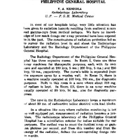 Evaluation of radiation hazards in the Philippine General Hospital
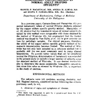 Hematocrit and mean corpuscular hemoglobin concentration in normal student Filipino students
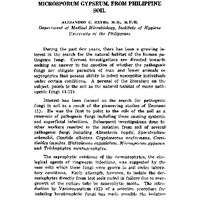 Isolation of the pathogenic fungus, microsporum gypseum, from Philippine soil
 Laboratory studies on the influenza epidemic of 1958
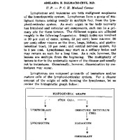 Lymphomas and leukemias - pathological and histogenetic aspects
 Notes on sanitary facilities in the Philippines
 Occupational diseases of the fisherman
 Primary carcinoma of the liver and its association with cirrhosis: a review of 54 deaths
 Radioactive fallout in the Manila Area and its public health significance
 Seminar
 Serum sodium and potassium levels with dietary evaluation
 Studies on body temperature of normal Filipino children
 Studies on escherichia coli serotypes in animals
 The changing concepts of health and the University
 Acta Medica Philippina
 A pattern of approach in opthalmological clinical conferences
 Burns - experiences with consecutive cases treated at the Philippine General Hospital
 Classification and transitions in chorionic tumors
 Nursery neonatal morbidity and mortality in the Philippine General Hospital
 Simplified methods of determining true sugar in blood
 The serum phospholipid levels and total cholesterol: phosopholipid ratios of apparently normal adult Filipino students
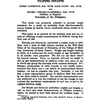 Weights of apparently well Filipino infants